141
Radiol Bras. 2011 Mai/Jun;44(3):141–146
Contribution of ultrasonography to the diagnosis of chronic
hepatitis C histopathological changes, with emphasis on
hepatic steatosis – Part I
*
Contribuição da ultrassonografia para o diagnóstico das alterações histopatológicas presentes na hepatite C crônica, com ênfase na esteatose hepática – Parte I
Marcia Wang Matsuoka1, Ilka Regina Souza de Oliveira2, Azzo Widman3, Arnaldo Zanoto4, Sérgio Keidi Kodaira5, Leonardo Ellery Marinho6, Wilson Jacob Filho7, Giovanni Guido Cerri8
Objective: To evaluate the role of ultrasonography in the assessment of histopathological changes in patients with chronic hepatitis C, with emphasis on hepatic steatosis. Materials and Methods: Liver ultrasonography results were compared with histopathological findings of liver biopsy of 192 patients with chronic hepatitis C virus infection. All the US examinations followed a single protocol, analyzing the following aspects: echogenicity, echotexture and attenuation. The patients sample was divided into two groups as follows: patients with sonographic changes and patients with no sonographic changes. Sonographic findings of both groups were compared with histopathological findings after liver biopsy. Results: Statistically significant intergroup differences were observed just regarding architectural changes grades 0 and 3 and hepatic steatosis. Attenuation was the sonographic criterion that was best correlated with hepatic steatosis. Conclusion: The results of the present study demonstrate that, in patients with chronic hepatitis C, ultrasonography has limitations in the characterization of histopathological changes, with an intermediate rate of agreement with the diagnosis of hepatic steatosis. Considering the specificity of 77.9% and the negative predictive value of 95.5%, the authors highlight the capacity of the method to demonstrate the probability of absence of hepatic steatosis. Keywords: Ultrasonography; Chronic hepatitis C virus infection; Hepatic steatosis; Liver biopsy.
Objetivo: Avaliar a contribuição da ultrassonografia no estudo das alterações histopatológicas encontradas na hepa-tite crônica pelo vírus C, com ênfase para a esteatose hepática. Materiais e Métodos: Foram comparados os resul-tados dos exames ultrassonográficos do fígado de 192 pacientes portadores de hepatite crônica pelo vírus C, com os achados histopatológicos dos fragmentos obtidos por biópsia hepática. Todos os exames ultrassonográficos obedece-ram a um mesmo protocolo, sendo analisados os seguintes critérios: ecogenicidade, ecotextura e atenuação. Os pacientes foram agrupados considerando-se os com alterações ultrassonográficas e os sem alterações ultrassonográ-ficas, sendo comparados com as alterações histopatológicas presentes. Resultados: Entre as alterações histopato-lógicas presentes, apenas os graus 0 e 3 de alteração arquitetural e a esteatose hepática apresentaram diferença estatística significante entre os dois grupos. Dentre os critérios ultrassonográficos avaliados, a atenuação foi o que apresentou melhor correlação com a esteatose hepática. Conclusão: Os resultados do trabalho demonstraram que, em pacientes com hepatite crônica pelo vírus C, a ultrassonografia apresentou limitações à caracterização das altera-ções histopatológicas, apresentando concordância regular com o diagnóstico de esteatose hepática. Destaca-se a capacidade do método em mostrar a probabilidade de inexistência de esteatose hepática, tendo em vista a especifi-cidade de 77,9% e o valor preditivo negativo de 95,5%.
Unitermos: Ultrassonografia; Hepatite C crônica; Esteatose hepática; Biópsia hepática. Abstract
Resumo
* Study developed at Instituto de Radiologia (InRad) do Hos-pital das Clínicas (HC) da Faculdade de Medicina da Universi-dade de São Paulo (FMUSP), São Paulo, SP, Brazil.
1. PhD, MD, Physician Assistant at Instituto da Criança (ICr) do Hospital das Clínicas da Faculdade de Medicina da Universi-dade de São Paulo (HC-FMUSP), São Paulo, SP, Brazil.
2. PhD, MD, Docent at Department of Radiology, Faculdade de Medicina da Universidade de São Paulo (FMUSP), São Paulo, SP, Brazil.
3. PhD, MD, Supervising Physician Assistant at Division of Digestive System Surgery II, Hospital das Clínicas da Faculdade de Medicina da Universidade de São Paulo (HC-FMUSP), São Paulo, SP, Brazil.
Matsuoka MW, Oliveira IRS, Widman A, Zanoto A, Kodaira SK, Marinho LE, Jacob Filho W, Cerri GG. Contribution of ultrasonography to the diagnosis of chronic hepatitis C histopathological changes, with emphasis on hepatic steatosis – Part I. Radiol Bras. 2011 Mai/ Jun;44(3):141–146.
4. Senior Assistant Professor, Division of Surgical Technique and Experimental Surgery, Department of Surgery, Faculdade de Medicina da Universidade de São Paulo (FMUSP), São Paulo, SP, Brazil.
5. PhD, MD, Physician Assistant at Instituto de Radiologia (In-Rad), Hospital das Clínicas da Faculdade de Medicina da Uni-versidade de São Paulo (HC-FMUSP), São Paulo, SP, Brazil.
6. MD, Specialist in Imaging Diagnosis of the Center of Com-puted Tomography at Hospital Samaritano, São Paulo, SP, Bra-zil.
7. Titular Professor, Director for the Unit of Geriatrics, Hospi-tal das Clínicas da Faculdade de Medicina da Universidade de São Paulo (HC-FMUSP), São Paulo, SP, Brazil.
8. Titular Professor, Department of Radiology, Faculdade de Medicina da Universidade de São Paulo (FMUSP), General Di-rector, Instituto do Câncer do Estado de São Paulo (Icesp), São Paulo, SP, Brazil.
Mailing Address: Dra. Marcia Wang Matsuoka. Avenida En-genheiro Luiz Gomes Cardim Sangirardi, 770, ap. 101, Vila Ma-riana. São Paulo, SP, Brazil, 04112-080. E-mail: mwmatsuoka@ yahoo.com.br
INTRODUCTION
Ultrasonography is an imaging method of first choice because of its noninvasive-ness, wide availability and for practicity of application(1–3). In the liver, the following
aspects are evaluated: biometry, shape, contours, intrahepatic vessels distribution, and the echographic characteristics of the parenchyma: echotexture, echogenicity and attenuation.
At ultrasonography, the normal hepatic parenchyma presents homogeneous echotexture and intermediate echogenicity, usually iso- or hyperechogenic in relation to the renal cortex and hypoechogenic in relation to the splenic tissue. The attenua-tion of the acoustic beam is subtle, allow-ing a good identification of intrahepatic vessels and of the diaphragm in the poste-rior region of the liver(4–6).
In inflammatory liver diseases, the sonographic findings vary according to the stage of the disease, being usually normal at the acute stage. At the chronic stage, with the onset of cirrhosis, irregularities in the hepatic contours, as well as changes in the characteristics of parenchymal tissues may be observed(7,8).
Among the different types of hepatitis, the one caused by the C virus has been object of clinical and sonographic investi-gation, given its high prevalence in the population as a whole(9–11).
Such a disorder that affects 170 million people around the world, may present a variable progression: 30% of the patients will develop mild and stable chronic hepa-titis for decades; 40% will present differ-ent degrees of fibrosis without the devel-opment of cirrhosis along the disease pro-gression; and 30% will present a severe progression of the disease, with develop-ment of cirrhosis(12). Among the patients
who develop cirrhosis, 20% will present complications caused by portal hyperten-sion with the disease progreshyperten-sion, and 1% to 7% may develop hepatocellular carci-noma(10,11,13,14).
Histopathological signs indicative of hepatitis C include architectural changes in the hepatic lobe, necroinflammation com-ponents and hepatic steatosis (HS), defined as a frequently reversible metabolic disor-der characterized by fatty liver infiltration
resulting from accumulation of triglycer-ides in within the hepatocytes(7,15).
In patients with hepatitis C, necropsy studies have demonstrated HS incidence in 31% to 72% of cases, and liver biopsies have demonstrated the presence of HS in approximately 50% of the cases(16).
How-ever, the role of HS in the onset, physiopa-thology and progression of the hepatic dis-ease is not well established yet, with the relationship between HS, necroinflamma-tory activity and development of fibrosis being described(17–20).
Hepatic biopsy is considered as the “gold standard” for the diagnosis and stag-ing of hepatitis C and HS, and is also rel-evant for therapeutic guidance in such dis-orders(11,21,22).
The evaluation of histopathological as-pects of hepatitis C and HS can be per-formed by means of ultrasonography in a limited manner. As regards HS, both the echographic characteristics of the paren-chyma and the liver biometry are usually taken into consideration.
Considering the echographic character-istics of the hepatic parenchyma, the diag-nosis of HS is based on a gradual increase in echogenicity and in attenuation of the acoustic beam. Textural changes, when present, are subtle and of minor relevance. However, such signs may also be present in cases of fibrosis and/or hepatic inflammatory processes, thus limiting the performance of the sonographic method for the diagnosis of HS(7,23–35). Additionally,
the analysis of echographic aspects of the hepatic parenchyma presents a subjective component, obtaining conflicting results with respect to the specific value of such method for the diagnosis of HS, with sen-sitivity rates ranging between 55% and 95%(25,26,36).
The present study was developed with a view on the controversial results of the HS diagnosis by ultrasonography. Initially, the results obtained exclusively by means of the analysis of echographic changes of the hepatic parenchyma will be presented (Part I). Sequentially, the results obtained by means of the joint analysis of the above mentioned echographic criteria and the bio-metric data regarding both the liver and the subcutaneous fat layer at the right hypo-chondrium will be presented (Part II).
MATERIALS AND METHODS
The present study was approved by the Committee for Ethics in Research (CAP-Pesq) of the Board of Clinical Directors of Hospital das Clínicas da Faculdade de Medicina da Universidade de São Paulo (HC-FMUSP), São Paulo, SP, Brazil.
The study sample comprised 192 con-secutive patients with clinical and labora-tory diagnosis of exclusively chronic C virus hepatitis referred to the Day Hospi-tal of the institution for US-guided liver biopsy in the period between July of 2002 and May of 2003.
In the studied population, the arithmetic mean age and respective standard deviation was 43.22 ± 13.21 years. According to the Kolmogorov-Smirnov test, the age distri-bution was normal, with sample homoge-neity being observed, allowing the com-parison between groups.
With the purpose of correlating the sonographic findings and histopathological changes in the liver in chronic hepatitis C, the patients were divided into two groups: a) patients with sonographic changes; b) patients without sonographic changes.
The means and standard deviations of ages in the two groups were, respectively, 45.03 ± 12.57 and 42.42 ± 13.46 years, with no statistically significant difference between both groups (unpaired t = 1.27; p
= 0.207050).
Among the 192 patients, 91 (47.4%) were men with 43.05 ± 12.95 years of age and 101 (52.6%) were women with 43.38 ± 13.51 years of age. There was no statis-tically significant difference between the proportion of both genders in the two groups (χ2c = 0.02; p = 0.88454) and
be-tween the age means (unpaired t = 0.17; p
= 0.86693), demonstrating sample homo-geneity and thus allowing the comparison between them.
Sonographic studies and liver biopsies were performed, each one by a same spe-cialist.
In the present study, HS was sonogra-phically classified as follows:
– moderate: hepatic parenchyma with moderate increase in echogenicity and sound beam attenuation, with moderate decrease in the visualization of the dia-phragm and intrahepatic vascularization;
– severe: hepatic parenchyma with a great in echogenicity and sound beam at-tenuation, with a marked or complete loss of the visualization of the diaphragm and of the intrahepatic vascularization(5,7);
The liver parenchyma echotexture was also evaluated, being classified as homo-geneous or heterohomo-geneous.
The group of patients with altered sonographic pattern of the liver paren-chyma was defined by the simultaneous presence of at least two of the analyzed parameters:
a) increased echogenicity (subtle, mod-erate or marked);
b) heterogeneous echotexture; c) increased attenuation of the acoustic beam (subtle, moderate or marked).
Previously to the liver biopsy, the pa-tients were submitted to ultrasonography with the purpose of localizing the best site for puncture.
Ultrasonography studies were per-formed in a Toshiba equipment model SSA-240A® (Toshiba; Tokyo, Japan) with
a 3.5 MHz convex transducer.
The study protocol was the following: a) total gain: adjusted in such a way that the liquid contents of the gallbladder and the blood in the inferior vena cava were non-echogenic. Gain curve adjusted to the neutral position;
b) patient positioning: horizontal dorsal decubitus;
c) transducer scan performed on the right hypochondrium in the longitudinal, transverse and oblique planes.
The patients with characteristics of fo-cal steatosis were not included in the study sample.
The liver biopsy specimens were ana-lyzed according to the criteria of the 1999 Brazilian Pathology Consensus of Socie-dade Brasileira de Patologia (Brazilian Pa-thology Society). Such Consensus evalu-ates chronic hepatitis by means of the stag-ing of present architectural changes, grades the activity of the ongoing condition by means of the necroinflammatory activity evaluation, and describes concomitant changes such as HS, lymphoid infiltrate,
ductal aggression and iron deposition. Moreover, according to the classification adopted by the Brazilian Pathology Con-sensus of 2002, the hepatic involvement by the C virus is classified as reactive liver, chronic hepatitis itself, and cirrhosis(15).
In order to allow the statistical analysis, the groups corresponding to the necroin-flammatory activity (portal activity, peri-portal activity and parenchymal activity) usually graded from 0 to 4 separately, were subdivided according to the sum of the grading for each one of such factors attrib-uted by pathological anatomy, establishing 13 subgroups (0 to 12). Subsequently, such subgroups were rearranged into four groups, in order to allow the comparison of necroinflammatory activity, HS and archi-tectural changes according to a single scale. The presence of HS was also graded as 0, 1, 2, 3, and 4, corresponding to increas-ing intensity of infiltration of fat particles in the liver, from mild to severe.
For the purpose of statistical analysis, 5% (α = 0.05) was adopted as significance level.
RESULTS
Among the 192 patients, 59 (30.7%) presented an altered sonographic pattern, and 133 (69.3%), presented unaltered sonographic pattern.
Histopathological patterns
The groups with and without sono-graphic changes were compared as regards histopathological patterns (architectural changes, necroinflammatory activity and HS). Statistically significant differences were observed between such groups for architectural changes and HS, and not for necroinflammation (Mann-Whitney non-parametric test).
1. Architectural changes (structural)
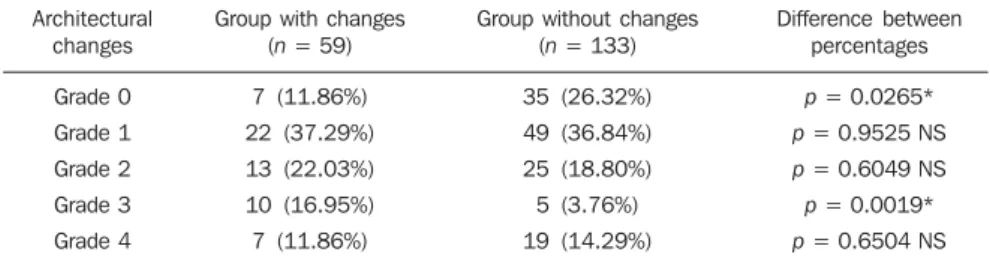
The quantification of the structural his-topathological changes in the groups with and without sonographic changes is shown on Table 1.
The chi-square test (χ2) was performed for analysis of joint variation in proportions (0, 1, 2, 3, 4) between the two groups (χ 2
= 13.40; p = 0.00949*).
Proportions for each quantification be-tween the groups with and without sono-graphic changes were analyzed by the test of difference between proportions, with a statistically significant difference observed at grade 0 (normal) and grade 3 (pre-cirrho-sis).
No difference was observed in the quan-tification 1 and 2 and also on 4, which cor-responds to cirrhosis.
2. Necroinflammation
The means of necroinflammatory changes were analyzed, with no statistically significant difference in the quantification of necroinflammatory activity being found between the groups with and without sonographic changes (Mann-Whitney non-parametric test).
3. Steatosis
Of the 192 patients, 81 (42.2%) pre-sented steatosis at the histopathological study, while 111 (57.8%) did not.
The present casuistry demonstrated a re-duced number of patients classified at each one of the HS grades (0, 1, 2, 3 and 4).
The results demonstrated a predomi-nance of mild grade HS in both patient groups (with and without sonographic changes), an HS was prevalent in the group without sonographic changes, with an es-timated prevalence of 95.5%.
Considering the above mentioned re-sults, which characterize the difficulty of
Table 1 Architectural changes (according to the 1999 Brazilian Pathology Consensus, of the Brazilian Pathology Society).
Architectural changes
Grade 0
Grade 1
Grade 2
Grade 3
Grade 4
Group with changes (n = 59)
7 (11.86%)
22 (37.29%)
13 (22.03%)
10 (16.95%)
7 (11.86%)
Group without changes (n = 133)
35 (26.32%)
49 (36.84%)
25 (18.80%)
5 (3.76%)
19 (14.29%)
Difference between percentages
p = 0.0265*
p = 0.9525 NS
p = 0.6049 NS
p = 0.0019*
p = 0.6504 NS
the method for diagnosing mild grade HS, and, with the objective of allowing the sta-tistical analysis, the different HS grades re-sulting from the histopathological analyses of the present casuistry were divided into two subgroups: I) grades 0 and 1; II) grades 2, 3 and 4 (Table 2).
Thus, as presence of HS in grades 2, 3, and 4 (moderate and severe) is considered, the analysis of the sample demonstrates a statistically significant difference between the groups with and without sonographic changes, with such difference being greater in the group with changes.
A statistically significant difference was observed between the HS proportions in the groups with and without sonographic changes and between the HS proportions in subgroups I and II (χ2c = 35.23; p =
0.00001*), with sensitivity of 79.3%, specificity of 77.9%, positive predictive value of 39.0%, negative predictive value of 95.5%, rate of agreement of 78.1% and
Cohen kappa coefficient (κ) = 0.4015, with a regular agreement.
Sonographic parameters
Among the three sonographic param-eters (echotexture, echogenicity and acous-tic beam attenuation), attenuation
pre-sented greater correlation with HS, as com-pared with the joint analysis of the same pa-rameters by means of the non-parametric Spearman’s rank correlation (Table 3).
Figures 1, 2 and 3 illustrate the differ-ent HS grades observed at ultrasonogra-phy.
Table 2 Hepatic steatosis groups.
Groups
With changes (n = 59) Without changes (n = 133)
Steatosis (HC)
Subgroup I
36 (61%) 127 (95.5%)
Subgroup II
23 (39%) 6 (4.5%)
p
p = 0.0185*
p = 0.00001*
HC, histological change. * Significant.
Table 3 Correlation between steatosis and sonographic parameters for diagnosis of changes at ultrasonography.
Sonographic parameter
Echotexture Echogenicity Attenuation
Spearman correlation (R)
–0.11 0.29 0.49
p
0.119704 NS 0.000037* 0.0000001*
* Significant; NS, non-significant.
Figure 1. Mild hepatic steatosis: ultrasonography of the right hepatic lobe with signs of subtle increase in echogenicity and attenuation of the acoustic beam; homogeneous texture.
Figure 2. Moderate hepatic steatosis: ultrasonography of the right hepatic lobe with moderate increase in echogenicity and attenuation of the acoustic beam; finely heterogeneous texture.
Figure 3. Severe hepatic steatosis: ultrasonography of the right hepatic lobe with marked increase in echogenicity and attenuation of the acoustic beam; impaired texture evaluation.
Figure 1 Figure 2
DISCUSSION
The literature has presented conflicting references with respect to the correlation between sonographic findings and histo-pathological findings of cirrhosis and HS. The development of the present study was favored by the possibility of compar-ing sonographic findcompar-ings with histopatho-logical findings, during US-guided percu-taneous liver biopsy, in a population of hepatitis C patients.
Between the two established patient groups (patients with and without sono-graphic changes), a statistically significant difference was observed between the archi-tectural changes and HS, and non-statis-tically significant difference as regards necroinflammatory activity. In the literature review, no study was found reporting sepa-rate correlation between such method and necroinflammation.
As architectural changes are considered, the patients group with sonographic changes and the group without sonographic changes presented statistically significant differences in fibrosis grades 0 and 3. No statistically significant difference was ob-served in grades 1, 2, and 4 of architectural changes. A comprehensive comparison between such results and those reported in the literature could not be undertaken be-cause of the absence of other systematized studies on sonographic and histopathologi-cal correlation with respect to architectural changes and their different grades. How-ever, considering exclusively grade 4 archi-tectural changes (cirrhosis), the obtained results are in agreement with data reported in the literature, with respect to the non-existence of statistically significant sono-graphic difference in this grade of architec-tural change(23,25,26,29,34,35,37–39).
Thus, the present study demonstrated that the characterization of different grades of architectural changes of the hepatic pa-renchyma poses limitations to ultrasonog-raphy.
As regards HS, the histopathological analysis demonstrated its presence in 42.2% of the patients, in agreement with data in the literature that report its presence in approximately 50% of the cases of chronic hepatitis C(19,20,39).
A statistically significant difference was observed between the groups with and
without sonographic changes, exclusively regarding the proportion of moderate and severe HS that was greater in the group with sonographic changes, demonstrating the capability of the method in identifying such steatosis grades (grades 2, 3 and 4).
The difficulty of ultrasonography in di-agnosing mild grade HS was demonstrated in the present study by the predominance of the mild grade HS both in the group with sonographic changes as well as in the group without sonographic changes.
The results in the present study are in agreement with other authors who report the high sensitivity of ultrasonography in the diagnosis of moderate and severe grades HS, and reduced sensitivity for the mild grades(7,24–33). However, other studies
report that it is not possible to diagnose HS by means of ultrasonography, differently from the obtained results(25,32,35).
It is important to highlight that, among the evaluated sonographic criteria, acous-tic beam attenuation was the criterion that presented the highest correlation with HS. Echogenicity, on its turn, presented corre-lation with HS, although less significantly, while echotexture did not present any cor-relation with HS (Table 3).
In the literature, data related to echo-genicity in the diagnosis of HS are conflict-ing. Some authors associate the increase in echogenicity with the diagnosis of this dis-order(40,41), while others affirm that there is correlation between hyperechogenicity and fibrosis or necroinflammation(32,35). As
re-gards attenuation, some authors consider that such parameter would be a good crite-rion for the diagnosis of HS(22,38,42).
Finally, it is observed that the result of the utilization of ultrasonography in the di-agnosis of HS may vary according to the performance of each one of the utilized pa-rameters: echotexture, echogenicity and attenuation. In the present study, attenua-tion was the most relevant parameter in the diagnosis of HS.
CONCLUSIONS
The results of the present study demon-strated that, considering the histopathologi-cal changes that are present in hepatitis C, ultrasonography was not capable of differ-entiating necroinflammation, and pre-sented limitations in the diagnosis of
archi-tectural changes. As regards HS, the statis-tical analysis demonstrated a regular agree-ment with the presence of HS observed at the histopathological analysis. The capabil-ity of the method to demonstrate the prob-ability of inexistence of HS should be high-lighted, considering the specificity of 77.9% and, mainly, the negative predictive value of 95.5%.
REFERENCES
1. Meire H, Cosgovre D, Dewbury K, et al. Abdomi-nal and general utrasound. 2nd ed. London: Churchill Livingstone; 2001.
2. Kodaira SK. Física. In: Cerri GG, editor. Ultra-sonografia abdominal. Rio de Janeiro, RJ: Revin-ter; 2002. p. 1–30.
3. Joy D, Thava VR, Scott BB. Diagnosis of fatty liver disease: is biopsy necessary? Eur J Gastro-enterol Hepatol. 2003;15:539–43.
4. Palmer PE. Manual de diagnóstico em ultra-so-nografia. Rio de Janeiro, RJ: Revinter; 1999. 5. Withers CE, Wilson SR. O fígado. In: Rumack
CM. Tratado de ultra-sonografia diagnóstica. Rio de Janeiro, RJ: Guanabara Koogan; 1998. p.73– 130.
6. Rodrigues MB, Amaro Jr E, Kodaira SK. Anato-mia e ultra-sonografia do abdome. In: Cerri GG, editor. Ultra-sonografia abdominal. Rio de Ja-neiro, RJ: Revinter; 2002. p. 31–53.
7. Machado MM, Rosa AC, Cerri GG. Doenças he-páticas difusas, hipertensão portal e transplante de fígado. In: Cerri GG, editor. Ultra-sonografia abdominal. Rio de Janeiro, RJ: Revinter; 2002. p. 55–124.
8. Ralls PW, Jeffrey RB Jr, Kane RA, et al. Ultra-sonography. Gastroenterol Clin North Am. 2002; 31:801–25.
9. Seeff LB. The natural history of chronic hepatitis C virus infection. Clin Liver Dis. 1997;1:587–602. 10. Poynard T, Ratziu V, Benmanov Y, et al. Fibrosis in patients with chronic hepatitis C: detection and significance. Semin Liver Dis. 2000;20:47–55. 11. Dove LM. A general approach to the management
of chronic hepatitis C. Gastroenterol Clin North Am. 2004;33:463–77.
12. Seeff LB. Natural history of hepatitis C. In: Schiff ER, Hoofnagle JH, editors. Update on viral hepa-titis. AASLD postgraduate course 2000. p. 112– 8.
13. Jármay K, Karácsony G, Ozsvár Z, et al. Assess-ment of histological features in chronic hepatitis C. Hepatogastroenterology. 2002;49:239–43. 14. Zheng RQ, Wang QH, Lu MD, et al. Liver
fibro-sis in chronic viral hepatitis: an ultrasonographic study. World J Gastroenterol. 2003;9:2484–9. 15. Gayotto LC, Comitê SBP/SBH. Visão histórica e
consenso nacional sobre a classificação das he-patites crônicas. GED. 2002;19:137–40. 16. Yoon EJ, Hu KQ. Hepatitis C virus (HCV)
infec-tion and hepatic steatosis. Int J Med Sci. 2006;3: 53–6.
18. Patton HM, Patel K, Behling C, et al. The impact of steatosis on disease progression and early and sustained treatment response in chronic hepati-tis C patients. J Hepatol. 2004;40:484–90. 19. Wyatt J, Baker H, Prasad P, et al. Steatosis and
fibrosis in patients with chronic hepatitis C. J Clin Pathol. 2004;57:402–6.
20. Negro F. Mechanisms and significance of liver steatosis in hepatitis C virus infection. World J Gastroenterol. 2006;12:6756–65.
21. Dao T, Lecointe I, Galateau F, et al. Contribution of liver biopsy and serology of hepatitis C virus to the diagnosis of a moderate and prolonged el-evation of aminotransferases. Gastroenterol Clin Biol. 1993;17:37–43.
22. Fontana RJ, Lok AS. Noninvasive monitoring of patients with chronic hepatitis C. Hepatology. 2002;36(5 Suppl 1):S57–64.
23. Foster KJ, Dewbury KC, Griffith AH, et al. The accuracy of ultrasound in the detection of fatty in-filtration of the liver. Br J Radiol. 1980;53:440–2. 24. Joseph AE, Dewbury KC, McGuire PG. Ultra-sound in the detection of chronic liver disease (the “bright liver”). Br J Radiol. 1979;52:184–8. 25. Celle G, Savarino V, Picciotto A, et al. Is hepatic
ultrassonography a valid alternative tool to liver biopsy? Report on 507 cases studied with both techniques. Dig Dis Sci. 1988;33:467–71. 26. Needleman L, Kurtz AB, Rifkin MD, et al.
Sonog-raphy of diffuse benign liver disease: accuracy of pattern recognition and grading. AJR Am J Roentgenol. 1986;146:1011–5.
27. Zwiebel WJ. Sonographic diagnosis of diffuse liver disease. Semin Ultrasound CT MR. 1995; 16:8–15.
28. Andrade RJ, García-Escaño MD. Hepatic steato-sis. Med Clin (Barc). 2000;114:574–6. 29. Ros PR, Mortele KJ. Diffuse liver disease. Clin
Liver Dis. 2002;6:181–201.
30. Hamaguchi M, Kojima T, Itoh Y, et al. The sever-ity of ultrasonographic findings in nonalcoholic fatty liver disease reflects the metabolic syndrome and visceral fat accumulation. Am J Gastroen-terol. 2007;102:2708–15.
31. Hirche TO, Ignee A, Hirche H, et al. Evaluation of hepatic steatosis by ultrasound in patients with chronic hepatitis C virus infection. Liver Int. 2007;27:748–57.
32. Perez NE, Siddiqui FA, Mutchnick MG, et al. Ultrasound diagnosis of fatty liver in patients with chronic liver disease: a retrospective observational study. J Clin Gastroenterol. 2007;41:624–9. 33. van Werven JR, Marsman HA, Nederveen AJ, et
al. Assessment of hepatic steatosis in patients undergoing liver resection: comparison of US, CT, T1-weighted dual-echo MR imaging, and point-resolved 1H MR spectroscopy. Radiology. 2010;256:159–68.
34. Taylor KJ, Riely CA, Hammers L, et al. Quanti-tative US attenuation in normal liver and in pa-tients with diffuse liver disease: importance of fat. Radiology. 1986;160:65–71.
35. Hepburn MJ, Vos JA, Fillman EP, et al. The ac-curacy of the report of hepatic steatosis on
ultra-sonography in patients infected with hepatitis C in a clinical setting: a retrospective observational study. BMC Gastroenterol. 2005;5:14. 36. Lupsor M, Badea R. Imaging diagnosis and
quan-tification of hepatic steatosis: is it an accepted alternative to needle biopsy? Rom J Gastroen-terol. 2005;14:419–25.
37. Kutcher R, Smith GS, Sen F, et al. Comparison of sonograms and liver histologic findings in pa-tients with chronic hepatitis C virus infection. J Ultrasound Med. 1998;17:321–5.
38. Lu ZF, Zagzebski JA, Lee FT. Ultrasound back-scatter and attenuation in human liver with dif-fuse disease. Ultrasound Med Biol. 1999;25: 1047–54.
39. Matos CA, Perez RM, Pacheco MS, et al. Steato-sis in chronic hepatitis C: relationship to the vi-rus and host risk factors. J Gastroenterol Hepatol. 2006;21:1236–9.
40. Tchelepi H, Ralls PW, Radin R, et al. Sonography of diffuse liver disease. J Ultrasound Med. 2002; 21:1023–32.
41. Palmentieri B, de Sio I, La Mura V, et al. The role of bright liver echo pattern on ultrasound B-mode examination in the diagnosis of liver steatosis. Dig Liver Dis. 2006;38:485–9.