RBCDH
RBCDH
Licence Creative Commom CC
1 Universidade de Trás-os--Montes e Alto Douro; Centro de Investigação em Desporto, Saúde e Desenvolvimento Humano. Vila Real. Portugal
2 Universidade de Fortaleza; Departamento de Fisioterapia. Fortaleza, CE. Brasil.
Received: 18 September 2013 Accepted: 17 October 2013
The inluence of positional release therapy
on the myofascial tension of the upper
trapezius muscle
A inluência da terapia de libertação posicional sobre a
tensão miofascial do músculo trapézio
Francisco José Saavedra1
Maria Teresa Cordeiro1,2
José Vilaça Alves1
Helder Miguel Fernandes1
Victor Machado Reis1
Daniela Gardano Bucharles Mont’Alverne2
Abstract – he objective of this study was to analyze the inluence of positional release therapy (PRT) on the myofascial tension of the upper trapezius muscle with an active myofascial trigger point (TrP). We studied 30 subjects (18 men and 12 women), mean age 34.5 ± 9.4 years, with an active TrP in the upper trapezius muscle on one side. A search for TrPs was performed bilaterally and the points were considered to be active when both local and referred pain evoked by manual palpation reproduced a deep aching and burning pain. he patients were evaluated under three conditions: (a) resting baseline, (b) concentric contraction and (c) isometric contraction, before and ater treatment with PRT, regarding the following parameters: (i) pain intensity during manual palpation (visual analogue pain scale) and (ii) upper trapezius muscle electromyographic (EMG) signals. A signiicant decrease in painful symptoms from 5.3 ± 1.9 to 2.8 ± 1.8 (p < 0.001) was observed ater treatment. here were no signiicant diferences in EMG signals during resting baseline and in the presence of concentric contraction ater the PRT session. It was concluded that PRT may be an efective treatment for pain relief and to reduce resting baseline EMG signals in the upper trapezius muscle with a TrP, suggesting that its use as an alternative or an adjunct to other therapies. he efectiveness of this type of treatment should be conirmed by further clinical studies.
Key words: Electromyography; Myofascial pain syndromes; Trigger points.
Positional release therapy Saavedra et al.
INTRODUCTION
Myofascial pain syndrome (MPS) is deined as a musculoskeletal pain condi-tion characterized by local and referred pain perceived as deep and aching, and by the presence of myofascial trigger points (TrPs) in any part of the body1.
A TrP is a hyperirritability spot in skeletal muscle or its fascia, located in palpable taut bands, which can be active or latent1,2. Active TrPs are
deined as those provoking spontaneous pain, thus being responsible for MPS. Latent TrPs have all the other characteristics of TrPs (taut band, local twitch response, and possibly referred pain on compression)1,3. Treatment
options for TrPs include trigger point injections, dry-needling, stretching exercise, massage therapy, and positional release therapy (PRT)2,4.
PRT is a technique in which muscles are placed in the position of great-est comfort, causing normalization of muscle hypertonicity and fascial ten-sion, a reduction in joint hypomobility, increased circulation and reduced swelling, decreased pain, and increased muscle strength4,5.
MPS is a form of musculoskeletal pain and, therefore, most of the available data pertain to musculoskeletal pain in general, which is currently reported to afect approximately 85% of the population at some point dur-ing their lives6. MPS represents the major cause of this pain, and the mean
prevalence of this condition among middle-aged adults (30–60 years) is reported to be 37% in men and 65% in women7. his prevalence reaches
85% in older adults (>65 years)8. hus, on the basis of the demographics
of aging, MPS can potentially become an increasingly important problem in the general population in years to come.
Several studies have reported a reduction in MPS symptoms ater the management of TrPs by diferent procedures9-12, but the efectiveness of PRT
in the improvement of patients with MPS remains unclear. he objective of the present study was to analyze the inluence of PRT on the myofascial tension of the upper trapezius muscle with an active TrP.
METHODOLOGICAL PROCEDURES
trapezius muscle by a physiotherapist with more than 8 years of experience in the diagnosis of TrPs. he diagnosis was made according to the criteria described by Simons et al.1: (1) presence of a palpable taut band within a
skeletal muscle, (2) presence of a hypersensitive tender spot in the taut band, (3) local twitch response elicited by snapping palpation of the taut band, and (4) reproduction of the typical referred pain pattern of the TrP in response to compression.
he following parameters were assessed in the patients before and af-ter PRT: (i) pain intensity during manual palpation using visual analogue pain scale13,14, and (ii) upper trapezius muscle electromyographic (EMG)
signals. he EMG signals of the upper trapezius muscle were acquired during three muscle function tests (baseline, concentric contraction, and maximal isometric voluntary contraction - MIVC).
To assess the EMG signals, participants were placed on a normal chair without back support and asked to relax their upper and lower arm. he resting baseline test was performed over a period of one minute to determine the silent parameters of the EMG signal, with or without TrP. For the meas-urements of the active elevation movement of the shoulder, the participants were asked to perform 10 elevations (3’’) controlled by an auditory signal, while EMG data were captured continuously. In addition to the elevation movement (concentric contraction), MIVC was acquired for 5’’ with 3’’ of rest between contractions. he participants were asked to perform three sets of three repetitions with 60’’ of rest between each set. he subject’s positions to obtain the MIVC were based on the guidelines of the Surface Electromyography for Non-Invasive Assessment of Muscles (SENIAM)15,16.
he EMG electrodes were positioned over the upper trapezius muscle according to SENIAM guidelines15,16. Myoelectric signals were sampled at
2000 Hz in the single diferential mode through a four-channel EMG sys-tem (Miotec 400® EMG Syssys-tem) using disposable Ag/AgCI circular bipolar electrodes (Medi-Trace 200 series, Kendall-LTP). he electrodes measuring 10 mm in diameter were coated with an adhesive conducting gel and were positioned on the skin covering the muscles with a center-to-center inter-electrode distance of 25 mm17. he signal was pre-ampliied 10 times at the
electrode location and sent to the ampliier (frequency range: 20–450 Hz18;
signal-to-noise ratio: 3 IV RMS; CMMR: 110 dB), which had a gain factor of 50, achieving a gain of 1000 for the EMG signal. he signal was sent to a 12-bit analog-to-digital converter (DT 3200, AMTI, USA) for subsequent mathematical analysis. For electrode placement, the skin was abraded with an alcohol-soaked gauze at the ixation sites in order to reduce impedance19.
Positional release therapy Saavedra et al.
site of the muscle ater full-wave rectiication and iltering through a zero lag 4th order Butterworth low-pass ilter with a cut-of frequency of 5 Hz.
he patients received PRT as described by D’Ambrogio et al.20. While the
patient was lying in the supine position, the therapist placed the trapezius muscle in a speciic position as follows: the patient’s head was lexed laterally toward the TrP and his/her shoulder was abducted to approximately 90º. In that position, the therapist monitored the TrP with her index inger and main-tained that position until release was felt. his could take from 5 to 20 min20.
Data were analyzed statistically using the SPSS® 17.0 sotware (SPSS,
Inc., Chicago, IL). Results are reported as the mean and standard deviation. he Shapiro-Wilk test showed a normal distribution of quantitative data (p
> 0.05). he diferences between the EMG signals were assessed with the
t-test for independent samples. Apaired sample t-test was used to assess the diferences in the EMG signals of active TrPs within referred pain intensity. Efect size was assessed using Cohen’s d (standardized mean diferences)21.
Taking into account the cut-of established by Cohen, the efect size can be small (∼0.2), medium (∼0.5), or large (∼0.8). he Pearson (r) test was used for the analysis of correlation between baseline and referred pain. Statistical analysis was conducted at the 95% conidence level, with the level of signiicance set at p < 0.05.
RESULTS
A total of 30 volunteers were evaluated; 12 of these patients were right-handed (40%) and 18 were let-handed (60%). he number of active TrPs was larger on the right side (n = 26; 86.7%) than on the let side (n = 4; 13.3%). No correla-tion between the dominant hand and TrP side was detected (p = 0.13). Table 1 shows the distribution of the dominant and TrP sides in the patients studied.
Table 1. Presentation of the dominant and trigger point (TrP) side [absolute frequency (n), relative frequency (%) and level of signiicance (p)].
Dominant side
Total (%) p right left
TrP side right 12 14 26 (86.7) 0.130
left 0 4 4 (13.3)
Total (%) 12 (40) 18 (60) 30 (100)
Prior to the intervention, the patients had a mean of 3.63 ± 2.86 mV in the basal condition on the active TrP side, and a mean of 2.34 ± 1.41 mV on the non-active myofascial trigger point (nTrP) side. Comparison of the two sides before the intervention revealed a signiicant diference (p = 0.02) (Figure 1). Ater the intervention, the side with a TrP showed a 23.15% reduction (2.79 ± 1.41 mV) in the EMG signal which, however, was not signiicant (p = 0.09; Cohen’s d = 0.4; r = 0.2).
not signiicant (p = 0.3).During MIVC (%), before the intervention a mean value of 19.65 ± 11.26 mV was observed on the TrP side and a mean value of 18.77 ± 16.79 mV on the healthy side (nTrP), with no signiicant difer-ence between sides (p = 0.8). Ater intervention, the side with a TrP showed a 9.27% decrease in the EMG signal (17.83 ± 10.68 mV) which, however, was not signiicant (p = 0.3; Cohen’s d = 0.2; r= 0.1). On the healthy side (nTrP), there was a 13% reduction in MVIC (1663 ± 9.05 mV), which was not statistically signiicant (p = 0.4; Cohen’s d = 0.2; r = 0.1). he EMG signals of the upper trapezius muscle acquired during the three muscle function tests (baseline, concentric contraction, and MVIC) are shown in Table 2.
Figure 1. Mean EMG signals during resting baseline, with comparison of the two sides before and after application of positional release therapy (B-TrP: baseline with a trigger point; B-nTrP: baseline without a trigger point). * Statistically signiicant diference (p = 0.02).
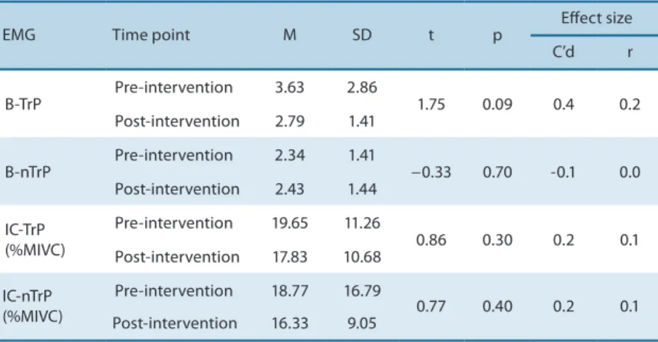
Table 2. EMG data before and after the application of positional release therapy [mean (M), standard deviation (SD), comparison of mean values (t) and level of signiicance (p). Efect size is expressed as Cohen’s d (C’d) and efect-size correlation (r)].
EMG Time point M SD t p
Efect size C’d r
B-TrP
Pre-intervention 3.63 2.86
1.75 0.09 0.4 0.2 Post-intervention 2.79 1.41
B-nTrP
Pre-intervention 2.34 1.41
−0.33 0.70 -0.1 0.0 Post-intervention 2.43 1.44
IC-TrP (%MIVC)
Pre-intervention 19.65 11.26
0.86 0.30 0.2 0.1 Post-intervention 17.83 10.68
IC-nTrP (%MIVC)
Pre-intervention 18.77 16.79
0.77 0.40 0.2 0.1 Post-intervention 16.33 9.05
B-TrP: baseline with a trigger point; B-nTrP: baseline without a trigger point; IC-TrP: isotonic contraction with a trigger point; IC-nTrP: isotonic contraction without a trigger point; IC-TrP (%MIVC): isotonic contraction with a trigger point for the percentage of maximal isometric voluntary contraction; IC-nTrP (%MIVC): isotonic contraction without a trigger point for the percentage of maximal isometric voluntary contraction.
Positional release therapy Saavedra et al.
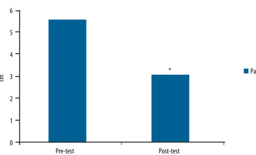
Figure 2. Mean pain intensity (visual analogue pain scale) before and after the application of positional release therapy. * Statistically signiicant diference (p < 0.001).
Ater intervention, on the TrP side, no correlations were found between resting baseline value and pain intensity (r = 0.1 and p = 0.5), or between pain intensity and isotonic contraction (r = 0.04 and p = 0.8).
DISCUSSION
Trigger points play a basic role in many chronic pain syndromes2.
Ac-cording to Harden et al.22, TrPs are associated with end-plate disorder
and increased release of acetylcholine, which result in local ischemia and sensitization of nociceptors. An increased release of inlammatory chemical substances such as histamine, prostaglandins, bradykinin and serotonin is observed at the TrP site22,23. hese substances can afect the membrane
of polymodal nociceptive receptors and cause peripheral sensitization, causing central sensitization and chronic pain23.
he present study showed that the EMG signal of the muscle with TrPs generates a greater change in the electrical signal when compared with nTrP muscle in the resting position (baseline). his result supports the use of EMG for the diagnosis of pain syndromes in order to identify the
TrPs24,25. Travell and Simons1 hypothesized that acetylcholine is constantly
released in the formation of TrPs, thus activating the release of Ca2+ in the
sarcoplasmic reticulum. hus, the presence of free Ca2+ leads to a constant
interaction of myoilaments and maintains muscle contraction, even in the absence of a voluntary action potential. his fact is in contrast to the EMG activity recorded in our study, in which an increase in the electrical signal was observed on the active TrP side.
We observed that the baseline EMG signal was lower than that ob-served during MIVC. We perceived that muscle with an active TrP did not inluence the reduction in strength during isotonic muscular concentric activity. he literature shows divergent results, with some studies report-ing a decrease in muscle activity26,27, others showing an increase in muscle
activity associated with pain intensity28,29, and still others30 inding no
signiicant diferences between the side with TrP and nTrP during active work, in agreement with the present results.
In the present study, all indicators showed small to medium size efects among patients. he mean pain intensity of the upper trapezius muscle with an active TrP was considered moderate; however, ater applying PRT, the tra-pezius muscle exhibited a low level of pain and this reduction was statistically signiicant. In PRT, the muscles are placed in the greatest comfort position. he resulting tissue relaxation improves vascular circulation and removes chemical mediators of inlammation2. hus, PRT may eliminate the
periph-eral and central sensitization. his technique may also directly reduce central sensitization by a damping efect on the facilitated segment in the spinal cord2.
We should recognize some limitations of the study. First, the sample size was small and therefore future studies with a greater number of pa-tients are recommended. Second, since active TrPs are not found oten in healthy controls, in the present study we only included patients. he reason was that we wanted to investigate referred pain areas in active TrPs in a patient population. Future studies should include a larger sample with and without active TrPs to permit better generalization of the present results.
CONCLUSIONS
he authors propose that PRT may be an efective treatment for pain relief, reducing the resting baseline EMG signals in the upper trapezius muscle with a TrP. his suggests that this technique may be used as an alternative or an adjunct to other therapies. he efectiveness of this type of treatment should be conirmed in further clinical studies.
REFERENCES
1. Simons DG, Travell JG, Simons LS.. Travell & Simons’ myofascial pain and dys-function: Upper half of body. 2nd ed. Vol. 1. Baltimore: Williams & Wilkins; 1999. 2. Ross EL. Pain management. Elsevier Health Sciences; 2004.
3. Gerwin RD. Classiication, epidemiology, and natural history of myofascial pain syndrome. Curr Pain Headache Rep 2001;5(5):412-20.
4. D’Ambrogio KJ, Roth GB, Robertson J, Halperin S, Wiley M. Positional release therapy: assessment and treatment of musculoskeletal dysfunction. Mosby, 1997. 5. Castro FM, Gomes RCV, Salomão JR, Abdom APV. A efetividade da Terapia de
Liberação Posicional em pacientes portadores de disfunção temporomandibular. Rev Odontol Univ Cid Sao Paulo 2006;18(1):67-74.
Positional release therapy Saavedra et al.
7. Drewes AM, Jennum P. Epidemiology of myofascial pain, low back pain, morning stifness and sleep-related complaints in the general population. J Muscoskel Pain 1995;3(1):121.
8. Podichetty VK, Mazanec DJ, Biscup RS. Chronic non-malignant musculoskel-etal pain in older adults: clinical issues and opioid intervention. Postgrad Med J 2003;79(937):627-33.
9. Moraska A, Chandler C. Changes in clinical parameters in patients with tension-type headache following massage therapy: a pilot study. J Man Manip Ther 2008;16(2):106-12.
10. Venancio RA, Alencar JR F, Zamperini C. Botulinum toxin, lidocaine, and dry needling injections in patients with myofascial pain and headaches. Cranio 2009;27(1):46-53.
11. Von Stülpnagel C, Reilich P, Straube A, Schäfer J, Blaschek A, Lee SH, et al. Myo-fascial trigger points in children with tension-type headache: a new diagnostic and therapeutic option. J Child Neurol 2009;24(4):406-9.
12. Mohamadi M, Ali G, Abbas R. Tension-type-headache treated by positional release therapy: a case report. Man her 2012;17(5):456-8.
13. Ervilha UF, Arendt-Nielsen L, Duarte M, Graven-Nielsen T. Efect of load level and muscle pain intensity on the motor control of elbow-lexion movements. Eur J Appl Physiol 2004;92(1-2):168-75.
14. Svensson P, Graven-Nielsen T, Matre D, Arendt-Nielsen L. Experimental muscle pain does not cause long-lasting increases in resting electromyographic activity. Muscle Nerve 1998;21(11):1382-9.
15. SENIAM (Surface Electromyography for the Non-Invasive Assessment of Muscles). European recommendations for surface electromyography: results of the Seniam project. Enschede (he Netherlands): Seniam/Biomed II/ European Union; 2005. Available from: <http://www.seniam.org/trapeziusdescendens.html> [2013 Sep 22]. 16. Hermens HJ, Freriks B, Disselhorst-Klug C, Rau G. Development of recommenda-tions for SEMG sensors and sensor placement procedures. J Electromyogr Kinesiol 2000;10(5):361-74.
17. Sacco IC, Gomes AA, Otuzi ME, Pripas D, Onodera AN. A method for better positioning bipolar electrodes for lower limb EMG recordings during dynamic contractions. J Neurosci Methods 2009;180:133-7.
18. Clancy EA, Morin EL, Merletti R. Sampling, noise-reduction and amplitude estima-tion issues in surface electromyography. J Electromyogr Kinesiol 2002;12(1):1-16. 19. Farina D. Interpretation of the surface electromyogram in dynamic contractions.
Exerc Sport Sci Rev 2006;34(3):121-7.
20. Davidof R. Trigger points and myofascial pain: toward understanding how they afect headaches. Cephalalgia 1998;18:436-48.
21. Cohen J. Quantitative Methods in Psycholog y. Psychological Bulletin 1992;112(1):155-9.
22. Harden RN, Cottrill J, Gagnon CM, Smitherman TA, Weinland SR, Tann B, et al. Botulinum toxin A in the treatment of chronic tension-type headache with cervi-cal myofascial trigger points: a randomized, double-blind, placebo controlled pilot study. Headache 2009;49(5):732-43.
23. Hwang M, Kang YK, Kim DH. Referred pain pattern of the pronator quadratus muscle. Pain 2005;116(3): 238-42.
24. Kuan TS, Hsieh YL, Chen JT, Chen JT, Yen WC, Hong CZ. he miofascial trigger point region: correlation between the degree of irritability and the prevalence of endplate noise. Am J Phys Med Rehabil 2007; 86(3):183-9.
Corresponding author
Francisco José Saavedra Universidade de Trás-os-Montes & Alto Douro
Parque Desportivo da UTAD, Apartado 1013
5001-801 Vila Real. Portugal E-mail: [email protected]
2006; 26:64-73.
27. Oberg T, Sandsjo L, Kadefors R, Larsson S. Electromyographic changes in work-related myalgia of the trapezius muscle. Eur J Appl Physiol Occup Physiol 1992;65(3):251-257.
28. Hubbard DR, Berkof GM. Trigger point show spontaneous needle EMG activity. Spine 1993;18(13):1803-7.
29. Farina D, Leclerc F, Arendt-Nielsen L, Buttelli O, Madeleine P. he change in spatial distribution of upper trapezius muscle activity is correlated to contraction duration. J Electromyogr Kinesiol 2008;18(1):16-25.
![Table 1. Presentation of the dominant and trigger point (TrP) side [absolute frequency (n), relative frequency (%) and level of signiicance (p)].](https://thumb-eu.123doks.com/thumbv2/123dok_br/14948781.504767/4.850.105.575.751.857/presentation-dominant-trigger-absolute-frequency-relative-frequency-signiicance.webp)