Hospital São José / Complexo Hospitalar Santa Casa de Porto Alegre (HSJ/CHSC), Porto Alegre RS, Brazil:1N e u ro s u rgeon at
HSJ-CHSC;2Medical - Residents in Neuro s u rg e ry at HSJ/CHSC;3Medical Student - Universidade Federal de Santa Maria, RS (UFSM); 4Medical - Resident in Pathology at CHSC; 5Professor of Pathology at the Medical School, Fundação Faculdade Federal de Ciências
Médicas de Porto Alegre (FFFCMPA);6P rofessor of Neuro s u rg e ry at FFFCMPA and Universidade Federal do Rio Grande do Sul (UFRGS);
Chief of the Neurosurgery Service at HSJ-CHSC.
Received 14 September 2005, received in final form 26 December 2005. Accepted 23 February 2006.
D r. Arthur de Azambuja Pereira Filho - Av. Prof. Oscar Pereira 3008 - 91710-000 Porto Alegre RS - Brasil. E-mail: art h u r p e re i r a f i l h o @ g m a i l . c o m
PRIMARY MENINGEAL BURKITT-TYPE
LYMPHOMA PRESENTING AS THE FIRST
CLINICAL MANIFESTATION OF ACQUIRED
IMMUNODEFICIENCY SYNDROME
Pedro Luís Gobbato
1, Arthur de Azambuja Pereira Filho
2, Gustavo de David
2,
Mário de Barros Faria
2, Felipe de David
3, Pedro Bandeira Aleixo
4,
Marinez Bizarro Barra
5, Nelson Pires Ferreira
6ABSTRACT - The purpose of this study is to re p o rt a rare case of primary meningeal high grade Burkitt-type lymphoma presenting as the first clinical manifestation of acquired immunodeficiency syndrome. A 3 8 - y e a r-old Caucasian man, with a negative past medical history, sought treatment after experiencing glob-al headache for five days. CT-Scan reveglob-aled a right fro n t - t e m p o ro-parietglob-al hyperdense subdurglob-al expansive mass. A craniotomy was performed and a hard white subdural was microsurgically dissected. Some hours after the surg e ry, the patient developed hemispheric cerebral edema and intracranial hypertension syn-d rome. Decompressive craniotomy was perf o rmesyn-d ansyn-d the patient hasyn-d an excellent re c o v e ry. Scre e n i n g blood tests diagnosed human immunodeficiency virus infection. Further investigation ruled out systemic diseases. Eleven days after the initial surgery, the patient developed an acute respiratory failure and sep-sis, dieing on that day. Pathological studies diagnosed Burkitt-type lymphoma.
KEY WORDS: Burkitt-type lymphoma, meningeal neoplasm, acquired immunodeficiency syndrome.
Linfoma de Burkitt primitivo da meninge como primeira manifestação clínica da síndrome da imunodeficiência adquirida
RESUMO - O objetivo desse estudo é relatar um caso de linfoma de Burkitt de alto grau primitivo da meninge, que se apresentou como primeira manifestação clínica da síndrome de imunodeficiência adquiri-da. Um homem branco, de 38 anos, previamente hígido, referia cefaléia holocraniana há cinco dias. A TC de crânio evidenciou coleção hiperdensa subdural na região fro n t o - t e m p o ro-parietal direita. Após cranio-tomia fronto-temporal direita, um tumor branco e rígido de localização subdural foi micro c i ru rg i c a m e n t e ressecado. Algumas horas após, o paciente apresentou edema cerebral hemisférico e hipertensão intracra-niana, tendo sido submetido à craniotomia descompressiva com excelente melhora clínica. Testes soro l ó g i-cos evidenciaram infecção por vírus da imunodeficiência humana. Investigações complementares afastaram outras doenças sistêmicas. Onze dias após a primeira ciru rgia, o paciente apresentou insuficiência re s p i-ratória aguda e sepse, evoluindo para o óbito. Análise histopatológica evidenciou linfoma de Burkitt.
PALAVRAS-CHAVE: linfoma de Burkitt, tumor meningeal, síndrome da imunodeficiência adquirida.
t u m o r, often located deeply in the cerebral hemi-s p h e rehemi-s with a predilection for periventricular brain tissue3. A meningeal location has been estimated in
a round 7 - 10% of all cases, with a poor outcome not i m p roved by aggressive systemic therapy7. The
major-ity of these patients normally presents with seizure s , headache, cranial nerves deficit, hemiparesis or arm P r i m a ry central nervous system (CNS) lymphoma
is defined as non-Hodgkin’s lymphoma arising with-in the CNS and confwith-ined to it at the time of diagno-sis1,2. It is a rare tumor, representing less than 2% of
all primary brain tumors in some series3 - 6. Primary
s y n d rome (AIDS) patients it re p resents the most fre-quent brain tumor8 , 9. A high prevalence is also
ob-s e rved in renal and cardiac tranob-splantob-s, patientob-s with IgA deficiency, or Wiskott-Aldrich syndro m e1 0 , 1 1. A
number of clinical re p o rts suggest an increasing inci-dence of this tumor over the past decades12-14,
how-ever there is no published data about it as the first clinical manifestation of AIDS.
The purpose of this study is to re p o rt and discuss a rare case of primary meningeal high grade Burkitt-type lymphoma, presenting as the first clinical man-ifestation of AIDS.
CASE
A 38-year-old Caucasian man, with a negative past med-ical history, sought treatment after experiencing global headache of moderate intensity for five days. There were no other complaints. The patient’s neurological examina-tion was intact. Computed tomography (CT-scan) re v e a l e d a right fro n t - t e m p o ro-parietal hyperdense subdural expan-sive mass, with a 1cm midline brain shift, suggesting an acute subdural hematoma (Fig1).
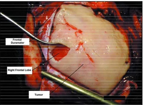
A right fro n t - t e m p o ro-parietal craniotomy was per-f o rmed. A hard white subdural mass with extension to f rontal, temporal and parietal spaces was micro s u rg i c a l l y dissected (gross-total removal) (Fig 2). A few hours after the surg e ry, the patient developed right hemispheric cere-bral edema and intracranial hypertension syndrome. De-c o m p ressive De-craniotomy was perf o rmed and the patient had an excellent re c o v e ry. Screening blood tests (2 sam-ples) diagnosed human immunodeficiency virus (HIV) infec-tion some days after the surgeries. This fact was not known neither by the patient nor by his family. Further comple-m e n t a ry radiological and hecomple-matological investigation ru l e d out systemic diseases. Eleven days after the initial surg e ry, the patient developed an acute re s p i r a t o ry failure and sep-sis by a Pneumocistis carinni infection. The patient died on that day.
Pathological studies– At microscopy the biopsy was characterized by a diffuse monotonous infiltrate of medi-um sized cells, with a slight molding pattern. The distinct s t a rry-sky pattern caused by interspersed tingible-body m a c rophages was present. The cells showed round nuclei, with coarse chromatin and basophilic nucleoli, with a notable rim of basophilic cytoplasm. Kary o rrhectic debris and mitotic figures were frequent. Immunohistochemically, the tumor e x p ressed positivity for B cell markers CD20 and CD79a. CD10 and Bcl-6 were also positive. The pro l i f e r a-tive index measured by the Ki-67 was very high bearing 100%. The negativity to Bcl-2 also helped in the diff e re n-tial diagnosis with diffuse large B cell lymphoma. These f e a t u res are compatible with high grade Burkitt-type lym-phoma (Fig 3).
lymphomas consist of secondary involvement of the brain, spinal cord, or covering membranes by nodal or extranodal (rather than CNS) lymphomas1 6. Primary
CNS lymphoma is by definition an extranodal lym-phoma arising in the central nervous system in the absence of systemic disease3. These tumors are
prima-rily the non-Hodgkin type, and are high grade and of B-cell lineage, with large cell morphological charac-teristics, although nearly all other types of malignant lymphoma of the CNS are on re c o rd1 6.
P r i m a ry CNS lymphoma has a much higher inci-dence in patients with established immune deficient status, especially in AIDS patients3. In its early stages,
human immunodeficiency virus infection has no symptoms or causes only a flu-like illness with many of the following symptoms: fever, sore throat, rash, nausea and vomiting, diarrhea, fatigue, swollen lymph nodes, muscle aches, headaches and joint pain. When the number of CD4 cells drops significantly, the patient develops AIDS and opportunistic infec-tions may overcome. Some of the germs that can cause these include Candida fungus, cryptococcosis, c y t o m e g a l o v i rus, herpes simplex virus, Mycobacterium
Fig 2. Transoperative photography showing the meningeal Burkitt-type lymphoma with subdural extension.
primary meningeal CNS lymphoma.
Several questions remain re g a rding the optimal management of patients with primary CNS lym-p h o m a2 1 , 2 3 , 2 4. Surg e ry traditionally plays little or no
role in the treatment of primary central nervous sys-tem lymphoma due to its infiltrative behavior and often multicentric appearance. Bataille et al.2 5p
re-sented a re t rospective multicentric-based analysis of 248 cases, 51% of them with surgical resection. They concluded that partial surgical resection was an unfa-vorable prognostic factor. However, no specific infor-mation was given about extent of resection and selec-tion criteria. In this respect most re p o rts give infor-mation re g a rding survival re g a rdless of the multiplic-ity of the lesion and the extent of resection26,27. The
results of surgical treatment are often based on a high percentage of multicentric lesions or partial re-s e c t i o n2 8. This makes it difficult to determine the
im-pact of surg e ry on survival. Although stere o t a c t i c biopsy appears to be indicated in multicentric cases, t h e re may be a subset of patients with well circ u m-scribed and surgically accessible lesions that could benefit from surgical resection3.
P r i m a ry CNS lymphoma is clearly a both chemo-sensitive and radiochemo-sensitive tumor. The current ther-apeutic recommendations place chemotherapy as the first line of tre a t m e n t2 9 , 3 0: chemotherapy alone is used
for patients older than 60 years, radiotherapy being re s e rved to patients with residual tumor after chemo-therapy. The combined treatment of chemotherapy followed by radiotherapy is apllied to the patients younger than 60 years. Some studies have demon-strated that methotrexate remains the most impor-tant single agent effective in primary CNS lymphoma, because of its good blood brain barrier penetration and its tumoricidic activity2 0. In the present case, it
was not possible to offer complementary tre a t m e n t besides aggressive surg e ry, because of the poor out-come of our patient.
In conclusion, although a threefold increase in the incidence of primary CNS lymphomas has been re p o rted re c e n t l y, primary meningeal high grade Burkitt-type lymphoma is still a very rare tumor. The peculiarities of this case are the lesion`s topography, its radiological characteristic and the fact that the p r i m a rymeningeal high grade Burkitt-type lympho-ma presented as the first clinical lympho-manifestation of AIDS.
phoma, as brain lymphoma . Primary meningeal Burkitt-type lymphoma as the first manifestation of AIDS has not been reported previously.
The incidence of primary central nervous system lymphomas among immunocompetent and immuno-c o m p romised patients has been on the rise in the past three decades3 , 1 8 , 1 9. This rise can be part i a l l y
attributed to the prolonged survival of AIDS patients and the extensive use of immunosuppressive thera-py in organ transplantation and autoimmune dis-ease3,6.
Most authors have found that primary CNS lym-phoma is a disease of late middle age3 , 2 0 , 2 1but some
series describe a diff e rent age distribution, with the majority of patients being in the seventh, eighth or even in the ninth decades of life1 2. In the pre s e n t
case, the patient was in the third decade of life, which is an atypical age for this disease according to the lit-erature.
A preoperative diagnosis of primary CNS lym-phoma can be made with a high degree of cert a i n-ty if the lesion is hyperdense on CT-scan, is invasive to the surrounding parenchyma, shows homogeneous enhancement, and has broad contact with ependy-ma and/or leptomeninges1 6 , 2 2. In a large number of
cases, Lee et al.2 2evaluated CT-scan findings in some
brain lymphomas and have correlated these findings with the tumors` pathological features. These authors have clearly discussed that homogeneous enhance-ment of primary CNS lymphoma on CT scanning is a common phenomenon, but that peripheral enhance-ment may be rarely seen, especially in AIDS re l a t e d lymphomas. They found central necrosis in the high-grade lymphomas with peripheral enhancement; none of those cases were the Burkitt type.
In the present case, it was observed an abnorm a l radiological imaging in contrast with those usual findings described above: the CTscan revealed a fro n t -t e m p o ro-parie-tal hyperdense subdural expansive mass, which suggested an acute subdural hematoma, needing urgent neurosurgical intervention.
Dubuisson et al.1 2re p o rted a series where the vast
REFERENCES
1. Jahnke K, Thiel E, Schilling A, et al. Low grade primary central nerv-ous system lymphoma in immunocompetent patients. Br J Haematol 2005;128:616-624.
2. Plasmwilm L, Herrlinger U, Korfel A, et al. Primary central nervous system (CNS) lymphoma in immunocompetent patients. Ann Hematol 2002;81:415-423.
3. Bellinzona M, Roser F, Ostertag H, Gaab RM, Saini M. Surgical re m o v a l of primary central nervous system lymphomas (PCNSL) presenting as space occupying lesions: a series of 33 cases. EJSO 2005; 31: 100-105. 4. A b rey LE, DeAngelis LM, Yahalom J. Long-term survival in primary
CNS lymphoma. J Clin Oncol 1998;16:859-863.
5. Mead GM, Bleehen NM, Gregor A, et al. A medical re s e a rch council randomized trial in patients with primary cerebral non-Hodgkin lym-phoma: cerebral radiotherapy with and without cyclophosphamide, d o x o rubicin, vincristine, and predinisone chemotherapy. Cancer 2000; 89:1359-1370.
6. C a m i l l e r i - B roet S, Martin A, Moreau A, et al. Primary central nervous system lymphomas in 72 immunocompetent patients: pathologic find-ings and clinical correlations Groupe Ouest Est d`etude des Leucenies et A u t res Maladies du Sang (GOELAMS). Am J Clin Pathol 1998;11 0 : 607-612.
7. Lachance DH, O`Neill BP, Macdonald DR. Primary leptomeningeal lymphoma: report of 9 cases, diagnosis with immunocytochemical analysis, and review of the literature. Neurology 1991;41:95-100. 8. Ciacci JD, Tellez C, VonRoenn J, Levy RM. Lymphoma of the central
nervous system in AIDS. Semin Neurol 1999;19:213-221.
9. Schabet M. Epidemiology of primary CNS lymphoma. J Neuro o n c o l 1999;43:199-201.
10. Boder E. Ataxia-telangiectasia: some historic, clinical and pathologic observations. Birth Defects Orig Artic Ser 1975;11:255-270.
11. B u r k h a rdt D, Schipper HI, Kaboth U, Felgenhauer K. IgA p ro d u c i n g primary intracerebral lymphoma. J Neurol Neuro s u rg Psychiatry 1992;55:623-625.
12. Dubuisson A, Kaschten B, Lénelle J, et al. Primary central nervous sys-tem lymphoma: report of 32 cases and review of the literature. Clin Neurol Neurosurg 2004;107:55-63.
13. Corn BW, Marcus SM, Topham A, Hauck W, Curran WJ. Will primary central nervous system lymphoma be the most frequent brain tumor diagnosed in the year 2000? Cancer 1997;79:2409-2413.
14. Eby NL, Gru fferman S, Flannelly CM, Schold SC, Vogel FS, Burger PC. I n c reasing evidence of primary brain lymphoma in the US. Cancer 1988;62:2461-2465.
15. Menniti A, Moschettoni L, Liccardo G, Lunardi P. Low-grade primary meningeal lymphoma: case report and review of the literature . Neurosurg Rev 2005;28:229-233.
16. Monabati A, Rakei M, Kumar P, Taghipoor M, Rahimi A. Primary Burkkit lymphoma of the brain in an immunocompetent patient. J Neurosurg 2002;96:1127-1129.
17. Bennett JC, Plum F. Cecil Tratado de Medicina Interna, 20ª Ed. Rio de Janeiro: Editora Guanabara Koogan S.A., 1997:2027-2088.
18. Bacellar H, Munoz A, Miller EM, et al. Temporal trends in the incidence of HIV- 1 - related neurologic diseases: multicenter AIDS cohort study, 1985-1992. Neurology 1994;44:1892-1900.
19. Hao D, DiFrancesco LM, Brasher PM, et al. Is primary CNS lymphoma really becoming more common? A population-based study of incidence, clinicopathological features and outcomes in Alberta from 1975 to 1996. Ann Oncol 1999;10:65-70.
20. Batchelor T, Carson K, O`neill A, et al. Treatment of primary CNS lym-phoma with methotrexate and deferred radiotherapy: a report of NABTT 96-07. J Clin Oncol 2003;21:1044-1049.
21. Socié G, Pipro t - C h a u ffat C, Schlienger M, et al. Primary lymphoma of the central nervous system. Cancer 1990;65:322-326.
22. Lee Y Y, Bruner JM, Van Tassel P, et al. Primary central nervous system lymphoma: CT and pathologic correlation. AJR 1986;147:747-752. 23. Bataille B. Traitment des lymphomas cérébraux primitives. Principes
généraux et resultants. Neurochirurgie 1997;43:372-375.
24. Shibamoto Y, Tsutsui K, Dodo Y, Yamabe H, Shima N, Abe M. Impro v e d survival rate in primary intracranial lymphoma treated by high-dose radiation and systemic vincristinedoxoru b i c i n c y c l o p h o s p h a m i d e -prednisolone chemotherapy. Cancer 1991;65:1907-1912.
25. Bataille B, Delwail V, Menet E, et al. Primary intracerebral malignant lymphoma: report of 248 cases. J Neurosurg 2000;92:261-266. 26. Henry JM, Heffner Jr RR, Dillard SH, Earle KM, Davis RL. Primary
malignant lymphomas of the central nervous system. Cancer 1974;34:1293-1302.
27. O`Neill BP, Illig JJ. Primary central nervous system lymphoma. Mayo Clin Proc 1989; 64:1005- 1020.
28. Hayakawa T, Takakura K, Abe H, et al. Primary central nervous sys-tem lymphoma in Japan: a re t rospective, cooperative study by CNS-Lymphoma Study Group in Japan. J Neurooncol 1994;19:197-215. 29. De Angelis LM. Primary central nervous system lymphoma. Curr Opin
Neurol 1999;12:687-691.