Andrea Kanako Yamazaki(a) Cacio Moura-Netto(b)
Ricardo Julio Cabrales Salgado(c) Brigida Monica Kleine(d)
Igor Prokopowitsch(e)
(a) PhD student; (d)MSc student; (e)PhD, Professor – Department of Endodontics, School of Dentistry, University of São Paulo, São Paulo, SP, Brazil.
(b) PhD, Professor, Department of Endodontics, Paulista University (UNIP), São Paulo, SP, Brazil.
(c) PhD, Professor of Endodontics – São Paulo Dental Association (APCD), São Caetano do Sul, SP, Brazil.
Corresponding author:
Cacio Moura Netto
R. Prof. Lineu Prestes, 2227 – Depto de Dentística
Cidade Universitária São Paulo - SP - Brazil CEP: 05508-000 E-mail: [email protected]
Received for publication on Apr 17, 2009 Accepted for publication on Nov 09, 2009
Ex vivo
analysis of root canal cleaning
using Endo-PTC associated to NaOCl
and different irrigant solutions
Abstract: The aim of this study was to assess qualitatively, by means of SEM images, the cleaning of the dentin walls of root canals after chemi-cal-surgical preparation using Endo-PTC cream with 0.5% and 1% so-dium hypochlorite and different inal irrigating solutions. Seventy-two single-rooted human teeth were divided into eight groups and prepared using Endo-PTC cream with sodium hypochlorite (NaOCl) at different concentrations, and irrigated with NaOCl at different concentrations. Fi-nal irrigation was performed with either EDTA-T or EDTA-C. The best results were obtained with Group 1, followed by Groups 5, 2, 7, 8, 3, 6 and 4. We can conclude that the use of 0.5% NaOCl during instrumen-tation and inal lush of the root canals was more eficient in cleaning than was 1% sodium hypochlorite. EDTA-T was more eficient in remov-ing smear layer than EDTA-C, and the cervical third presented better cleaning of the root canal walls than did the middle third, which showed cleaner dentin walls than the apical third.
Descriptors: Root canal therapy; Microscopy, electron, scanning; Root canal irrigants.
Introduction
Smear layer is formed during endodontic instrumentation by adher-ing to the instrument and beadher-ing compacted against canal walls, thereby covering dentin tubules’ entrances. The presence of the smear layer may
compromise the success of the endodontic therapy.1-3 Considering the
cleaning of root canal walls, large numbers of chemical substances and various combinations of them have been tested to seek the best protocol to bring together all the necessary requirements.
In addition to all the irrigating solutions, there is Endo-PTC cream which acts as a lubricant and thus reduces friction between the instru-ment and the root canal wall. This cream is a mixture of Tween 80 de-tergent, urea peroxide and carbowax as a lubricant. Because of the an-timicrobial action of Endo-PTC, when used along with 0.5% sodium hypochlorite it becomes effective even in the presence of organic
mate-rial.4-6 Due to its antibacterial and chemical properties, sodium
its cytotoxicity will also be greater when in contact with periapical tissues. In association with chelating agents, it results in cleaner root walls that are free
from smear layer.2,7-13
Based on the above, this study aimed to evalu-ate qualitatively, through SEM image analysis, the cleaning of the inner root canal walls as evidenced by the presence or not of smear layer, promoted by different irrigation protocols.
Material and Methods
After receiving approval from the Ethics Com-mittee of the Dental College of the University of São Paulo, 72 single-rooted human teeth were opened and emptied with #10 K iles (Dentsply Maillefer, Ballaigues, Switzerland) in the presence of 0.5% sodium hypochlorite (Fórmula e Ação, São Paulo, SP, Brazil). The working length of each tooth was established to be 1 mm short from the apical fora-men. The specimens were randomly divided into eight groups of nine teeth each. During instrumen-tation of the root canals, all apexes were covered with wax to allow the relux of the substances, simulating clinical conditions. The instrumentation was made up to a #35 ile and apical preparation with a #40 ile, using Endo-PTC cream (Fórmula e Ação, São Paulo, SP, Brazil) associated with 0.5 or 1% sodium hypochlorite (Fórmula e Ação, São Paulo, SP, Brazil) (Table 1). It is important to state that the sodium hypochlorite was renewed during instrumentation in order to maintain the efferves-cent reaction with Endo-PTC until all lubricant was
dissociated.
After preparation, the inal lush was done us-ing 20 ml of the irrigatus-ing solution correspondus-ing to each group (Table 1), either EDTA-T or EDTA-C (Fórmula e Ação, São Paulo, SP, Brazil). After in-troducing each 5 ml of irrigant solution, mechanical agitation was performed inside the root canal using a #15 K ile to enable greater contact between the ir-rigation solution and the root canal walls.
The crowns of all of the teeth were cut using diamond disks. Following this, a groove was made longitudinally on the buccal and lingual faces of the specimens, using a diamond-tipped steel disk, to a depth of approximately 1 mm. Then, the roots were split into two halves using a stainless steel blade and a small hammer. Only one of the two sections of each root was selected, the one with the better in-tegrity.
The teeth were then prepared for scanning elec-tron microscopy analysis (EDAX CDU Leap Detec-tor, Philips, the Nederlands) in their cervical, mid-dle and apical thirds (9, 6 and 2 mm from the apex, respectively), equidistant from the lateral walls, at 1000x magniication.
To evaluate these photomicrographs, the
Fo-toscore program (developed by Guerisoli,8 2002)
was utilized. Among the images obtained, three were selected as patterns for score’s grades. Four calibrated professors in Endodontics blindly as-sessed the photomicrographs. These images were di-vided according to scores for the degree of cleaning of the dentin wall as follows:
Group
Instrumentation Final flush
Endo-PTC NaOCl NaOCl EDTA-T EDTA-C
0.5% 1% 0.5% 1% 2.5%
G1 X X X X
G2 X X X X
G3 X X X X
G4 X X X X
G5 X X X X
G6 X X X X
G7 X X X X
Score 1 = dentin surface free of smear layer and with dentin tubules visible.
Score 2 = up to 50% of dentin surface with pres-ence of smear layer and with dentin tubules vis-ible.
Score 3 = more than 50% of dentin surface with presence of smear layer and without visible den-tin tubules.
The statistical analysis was made by means of the Kruskal-Wallis non-parametric test at a signii-cance level of 1%.
Results
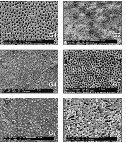
The qualitative assessment data on root wall cleaning that the examiners obtained were convert-ed into numerical values, using the scoring system adopted for this study, by the Fotoscore program. Figure 1 shows a sample of each group. Statistical
•
•
•
analysis showed that there was a signiicant differ-ence at the 1% level between the eight experimental groups. (Table 2)
The best results regarding the cleaning of the dentin walls of the root canals were obtained from Group 1, followed by Group 5, Group 2, Group 7, Group 8, Group 3, Group 6 and Group 4, respec-tively.
Discussion
A large number of studies have been conducted in the constant search for chemical substances, or combinations of such substances, or methods that would be capable effectively of removing this smear
layer.8,14
Among the substances selected for intermittent use, sodium hypochlorite is the one most commonly utilized. Because of its characteristics, which favor
disinfection of the root canal, this solution has been routinely studied at various concentrations in rela-tion to toxiicarela-tion and dissolurela-tion of the pulp tis-sue.4,15-17
Although sodium hypochlorite meets most of the basic requirements for an auxiliary substance to achieve sanitization of the root canal system, it does not meet all of them. Used alone, therefore, it is in-suficient to achieve this objective and needs to be used in combination with other chemical substances such that the characteristics of one solution
comple-ment the characteristics of the other.10,15
Gly-Oxide, RC-PREP and Endo-PTC are
among the substances available for continuous use. These have a creamy or gel-like consistency and they aid in lubricating the root walls thereby diminishing friction through suspension of the detritus resulting
from instrument use.4,10,13,15-19
Removal of the smear layer impregnated in the dentin walls of the root canals is also aided by the demineralizing and chelating action, respec-tively, of organic and inorganic acids such as citric acid and EDTA, with or without associated
deter-gents.2,7,8,20,21
Adding a detergent solution to the original Fór-mulation of EDTA has led to increased surface ten-sion for this substance, with the result that greater permeability of the dentin tubules and better clean-ing of the dentin walls of the root canals has thereby
tive detergent associated with EDTA in the EDTA-T solution, as well as Cetavlon, a cationic detergent present in EDTA-C.
The cleanest root dentin surfaces were seen in Group 1, in which Endo-PTC neutralized by 0.5% sodium hypochlorite and inal irrigation using 0.5% sodium hypochlorite and EDTA-T were utilized. Among the sodium hypochlorite concentrations uti-lized during the inal irrigation of the root canals (0.5%, 1% and 2.5%), the concentration of 0.5% presented the best result regarding dentin clean-ing. The lowest quantities of smear layer were ob-served in Groups 1, 5 and 2 (in decreasing order of cleaning), in which the instruments in the root canals were used along with Endo-PTC neutralized by 0.5% sodium hypochlorite. However, there were no statistically signiicant differences at the 1% level between Groups 1, 2 and 5. A slower reaction between 0.5% NaOCl and Endo-PTC in compari-son with 1% and 2.5% NaOCl could have led to a greater time for oxygen formation and, therefore, a better performance in smear layer removal.
There was a statistically signiicant difference at the 1% level between Group 2 and Group 5, in re-lation to the inal irrigation. EDTA-C was used in Group 2 and T was used in Group 5. EDTA-T presented better cleaning. EDTA-Tergensol, being an anionic detergent, is more effective for dealing with calcium ions and oily residues, which results in a better removal of smear layer.
The experimental group that presented the greatest quantity of smear layer on the root dentin surface was Group 4, in which Endo-PTC, neutral-ized by 1% sodium hypochlorite and inal irrigation with 0.5% sodium hypochlorite and EDTA-C, was utilized. However, Group 4 did not present a sta-tistically signiicant difference at the 1% level com-pared with Groups 3 and 6.
Considering the chelating agents utilized in this study, EDTA-T and EDTA-C, EDTA-T showed cleaner dentin walls than did EDTA-C. There was a statistically signiicant difference at the 1% level in this respect between Groups 1 and 2 and Groups 4 and 8. This statistical difference at the 1% level did not occur between Groups 6 and 7, although in the
Table 2 - Mean values for the score readings for the cervi-cal, middle and apical thirds in each experimental group.
Group Third Kruskal-Wallis*
cervical middle apical
G1 1.42 1.42 2.00 a,c
G2 1.92 1.83 2.67 a
G3 1.83 2.25 3.00 a,b
G4 2.17 2.25 3.00 b,d
G5 1.25 1.83 2.67 b,c,d
G6 1.67 2.58 2.92 d
G7 2.08 1.75 2.75 e
G8 2.25 2.17 2.58 e
cally signiicant difference at the 1% level.
As was observed in this study, the apical third is the most critical region with regard to the character-istic of greatest accumulation of smear layer com-ing from the chemical-surgical preparation. Efforts need to be concentrated on this, in order to achieve more eficient action by the auxiliary chemical sub-stance to combat microorganisms and clean the root
dentin.14
It is clear that it is important to carry out new studies on dentin cleaning of root canal walls, with the aim of improving the cleaning of these walls. Given that in modern Endodontics resinous illing cements are used, that penetrate into the dentin tu-bules and thereby cause greater sealing of the root canal system, the more complete cleaning of canal walls becomes a decisive factor in endodontic suc-cess.
Conclusion
The use of 0.5% sodium hypochlorite in asso-ciation with Endo-PTC cream during the chemi-cal-surgical preparation was shown to be more effective in dentin cleaning from root canal walls than was 1% sodium hypochlorite.
Among the sodium hypochlorite concentrations utilized during the inal irrigation of the root ca-nals (0.5%, 1% and 2.5%), the concentration of 0.5% presented the best result regarding dentin cleaning.
The inal irrigation performed using EDTA-T provided a better result with regard to removal of smear layer than did EDTA-C.
In relation to thirds of the root, the cervical third presented better cleaning of the dentin walls of the root canals than did the middle and apical thirds, while the middle third of the root showed cleaner dentin walls than did the apical third. 1.
2.
3.
4.
References
1. Hülsmann M, Heckendorff M, Lennon A. Chelating agents in root canal treatment: mode of action and indications for their use. Int Endod J. 2003 Dec;36(12):810-30.
2. Jeon IS, Spångberg LS, Yoon TC, Kazemi RB, Kum KY. Smear layer production by 3 rotary reamers with different cutting blade designs in straight root canals: a scanning electron mi-croscopic study. Oral Surg Oral Med Oral Pathol Oral Radiol Endod. 2003 Nov;96(5):601-7.
3. Lopes HP, Elias CN, Estrela C, Toniasso S. Mechanical stir-ring of smear layer removal: influence of the chelating agent (EDTA). Braz Endod J. 1996 Jan;1(1):52-5.
4. Okino LA, Siqueira EL, Santos M, Bombana AC, Figueiredo JA. Dissolution of pulp tissue by aqueous solution chlorhexi-dine digluconate and chlorhexichlorhexi-dine digluconate gel. Int Endod J. 2004 Jan;37(1):38-41.
5. Pérez-Heredia M, Ferrer-Luque CM, González-Rodríguez MP. The effectiveness of different acid irrigating solutions in root canal cleaning after hand and rotary instrumentation. J Endod. 2006 Oct;32(10):993-7.
6. Tanomaru Filho M, Leonardo MR, Silva LA, Aníbal FF, Faccioli LH. Inflammatory response to different endodontic irrigating solutions. Int Endod J. 2002 Sep;35(9):735-9 7. Goldberg F, Spielberg C. The effect of EDTAC and the
varia-tion of its working time analyzed with scanning electron mi-croscopy. Oral Surg Oral Med Oral Pathol. 1982 Jan;53(1):74-7.
8. Guerisoli DMZ. Estudo, por meio da microscopia eletrônica de varredura, da remoção de smear layer dos canais
radicula-res após aplicação de diferentes quelantes e do laser Er: YAG [Dissertação de Mestrado]. Ribeirão Preto: Faculdade de Odon-tologia da Universidade de São Paulo; 2002.
9. Marchesan MA, Arruda MP, Silva-Sousa YTC, Saquy PC, Pé-cora JD, Sousa-Neto MD. Morphometrical analysis of clean-ing capacity usclean-ing nickel-titanium rotary instrumentation associated with irrigating solutions in mesio-distal flattened root canals. J Appl Oral Sci. 2003 Jan-Mar; 11(1):55-9. 10. Ram Z. Effectiveness of root canal irrigation. Oral Surg Oral
Med Oral Pathol. 1977 Aug;44(2):306-12.
11. Torabinejad M, Walton RE. Principles and practice of endodon-tics. 2nd ed. Philadelphia: WB Saunders Company; 1996. 12. Vasconcelos BC, Luna-Cruz SM, De-Deus G, Moraes IG,
Maniglia-Fereira C, Gurgel-Filho ED. Cleaning ability of chlorhexidine gel and sodium hypochlorite associated or not with EDTA as root canal irrigants: a scanning electron mi-croscopy study. J Appl Oral Sci. 2007 Oct;15(5):387-91. 13. Niu W, Yoshioka T, Kobayashi C, Suda H. A scanning
elec-tron microscopic study of dentinal erosion by final irriga-tion with EDTA and NaOCl soluirriga-tions. Int Endod J. 2002 Nov;35(11):934-9.
14. Baker NA, Eleazer PD, Averbach RE, Seltzer S. Scanning electron microscopic study of the efficacy of various irrigating solutions. J Endod. 1975 Apr;1(4):127-35.
16. Peters OA, Barbakow F. Effects of irrigation on debris and smear layer on canal walls prepared by two rotary tech-niques: a scanning electron microscopic study. J Endod. 2000 Jan;26(1):6-10.
17. Mader CL, Baumgartner JC, Peters DD. Scanning electron microscopic investigation of the smeared layer on root canal walls. J Endod. 1984 Oct;10(10):477-83.
18. Sampaio JEC, Campos FP, Pilatti GL, Theodoro LH, Leite FRM. A scanning electron microscopy study of root surface smear layer removal after topical application of EDTA plus a detergent. J Appl Oral Sci. 2005 Jul-Sep;13(3):247-52.
19. Spanó JCE, Barbin EL, Santos TC, Guimarães LF, Pécora JD. Solvent Action of sodium hypochlorite on bovine pulp and physico-chemical properties of resulting liquid. Braz Dent J. 2001 Jul;12(3):154-7.
20. Marais JT. Cleaning efficacy of a new root canal irrigation solution: a preliminary evaluation. Int Endod J. 2000 Jul; 33(4):320-5.