The influence of ovariectomy, simvastatin
and sodium alendronate on alveolar
bone in rats
Influência da ovariectomia, da sinvastatina e do
alendronato sódico no osso alveolar em ratas
Abstract: Bisphosphonates are currently used in the treatment of many diseases involv-ing increased bone resorption such as osteoporosis. Statins have been widely used for the treatment of hypercholesterolemia and recent studies have shown that these drugs are also capable of stimulating bone formation. The purpose of this study was to evaluate the inluence of an estrogen deicient state and the effects of simvastatin and sodium alendro-nate therapies on alveolar bone in female rats. Fifty-four rats were either ovariectomized (OVX) or sham operated. A month later, the animals began to receive a daily dose of sim-vastatin (SIN - 25 mg/kg), sodium alendronate (ALN - 2 mg/kg) or water (control) orally. Thirty-ive days after the beginning of the treatment, the rats were sacriiced and their left hemimandibles were removed and radiographed using digital X-ray equipment. The alveolar radiographic density under the irst molar was determined with gray-level scal-ing and the values were submitted to analysis of variance (α = 5%). Ovariectomized rats gained more weight (mean ± standard deviation: 20.06 ± 6.68%) than did the sham oper-ated animals (12.13 ± 5.63%). Alveolar radiographic density values, expressed as gray lev-els, were lowest in the OVX-water group (183.49 ± 6.47), and differed signiicantly from those observed for the groups receiving alendronate (sham-ALN: 193.85 ± 3.81; OVX-ALN: 196.06 ± 5.11) and from those of the sham-water group (193.66 ± 4.36). Other comparisons between groups did not show signiicant differences. It was concluded that the ovariectomy reduced alveolar bone density and that alendronate was eficient for the treatment of this condition.
Descriptors: Alendronate; Models, animal; Osteoporosis; Ovariectomy; Simvastatin.
Resumo: Os bisfosfonatos são empregados atualmente para o tratamento de várias doen-ças caracterizadas pelo aumento da reabsorção óssea, como a osteoporose. As estatinas são amplamente utilizadas para redução de níveis elevados de colesterol e estudos recentes têm revelado sua ação anabólica no osso. O objetivo deste trabalho foi avaliar a inluência da deiciência estrogênica e dos tratamentos com sinvastatina ou alendronato sódico no osso alveolar em ratas. Cinqüenta e quatro ratas sofreram ovariectomia (OVX) ou cirurgia simulada (“sham”). Um mês após, os animais passaram a receber diariamente, via oral, 25 mg/kg de sinvastatina (SIN), 2 mg/kg de alendronato (ALN) ou água (controle). Trinta e cinco dias depois do início do tratamento os animais foram sacriicados, as hemimandí-bulas esquerdas removidas e radiografadas em aparelho de raios X digital. Foi calculada a densidade radiográica em tons de cinza da área de osso alveolar sob o primeiro molar mandibular e os valores foram submetidos a ANOVA, ao nível de 5%. Ratas ovariec-tomizadas ganharam mais peso (média ± desvio-padrão: 20,06 ± 6,68%) que as demais (12,13 ± 5,63%). Os valores de densidade radiográica, em tons de cinza, foram menores nos animais do grupo OVX-água (183,49 ± 6,47), signiicantemente diferentes daqueles observados nos grupos que receberam alendronato (“sham”-ALN: 193,85 ± 3,81; OVX-ALN: 196,06 ± 5,11) e no grupo “sham”-água (193,66 ± 4,36). Outras comparações entre grupos não revelaram diferenças estatísticas. Concluiu-se que a ovariectomia reduziu a densidade óssea alveolar e que o tratamento com alendronato sódico foi eiciente para o tratamento desta situação.
Descritores: Alendronato; Modelos animais; Osteoporose; Ovariectomia; Sinvastatina. Ana Lia Anbinder(a)
Fernanda de Almeida Prado(b)
Marcela de Almeida Prado(b)
Ivan Balducci(c)
Rosilene Fernandes da Rocha(d)
(a) PhD, Professor, Dentistry Department,
Taubaté University (UNITAU).
(b) DDSs;(c)PhD, Professor – Department of
Biosciences and Oral Diagnosis, School of Dentistry of São José dos Campos, São Paulo State University.
(d) MS, Professor, Department of Social
Dentistry and Pediatric Clinics, School of Dentistry of São José dos Campos, São Paulo State University.
Corresponding author:
Ana Lia Anbinder
R. Helena David Neme, n. 148, ap. 61, Jd. São Dimas
São José dos Campos - SP - Brazil CEP: 12245-310
E-mail: [email protected]
Introduction
Osteoporosis is deined worldwide as a systemic skeletal disease characterized by low bone density and microarchitectural deterioration of bone tis-sue, which leads to increased bone fragility and risk of fracture.6 The disease should be considered as a
public health problem due to its social, physical and economic impact. The majority of osteoporosis cas-es occur in post-menopause women, since they have an estrogen deiciency and this condition is associ-ated to a rapid increase of bone resorption.6
Bisphosphonates are drugs that inhibit bone re-sorption and that have been successfully used in the systemic treatment of osteoporosis, selectively acting on bone tissue and interfering with the action of os-teoclasts.18 As inhibitors of osteoclasts,
bisphospho-nates have been used with success in studies related to the inhibition of periodontal disease induced in animals without osteoporosis or other systemic dis-eases.16
Statins or 3-hydroxy-3-methyglutaryl coenzyme A (HMG-CoA) reductase inhibitors are drugs wide-ly used over the last decade for the reduction of el-evated blood cholesterol levels. Some recent studies have reported an action of these substances on bone formation through the stimulation of bone morpho-genetic protein-2 (BMP-2).13 In view of these
ind-ings, it has been suggested that statins, if selectively directed at bone, may have beneicial effects on the treatment of osteoporosis and fractures.13
Thus, the objective of the present study was to evaluate the inluence of the absence of ovarian hor-mones and the inluence of simvastatin or sodium alendronate treatment on alveolar bone in rats.
Material and Methods
Fifty-four adult female Wistar rats (Rattus
nor-vegicus) aged approximately 90 days were used. The
study was conducted according to the Ethical Princi-ples on Animal Experimentation adopted by the Bra-zilian College of Animal Experimentation (COBEA). At 3 months of age, the animals were divided randomly, using a table of random numbers, with one half being submitted to bilateral ovariectomy (OVX group) and the other half, to sham surgery for the simulation of surgical stress (SHAM group).
After anesthesia with a mixture of 13 mg/kg of 2% xylazine hydrochloride (Rompun - Bayer - São Pau-lo, SP, Brazil) and 33 mg/kg of ketamine base (Fran-cotar - Virbac - Roseira, SP, Brazil), and shaving of the lateral abdominal region, the skin and muscula-ture were incised longitudinally below the last rib, and the ovary was identiied and exposed. In the OVX group, hemostasis was performed by ligation of the upper part of the Fallopian tube with #4.0 silk suture and the ovary was excised together with surrounding fat, the oviduct and a small portion of the uterus. The muscle layer was then closed with absorbable #4.0 catgut and the skin, with #4.0 silk suture. In the SHAM group, exposure of the ovary was followed by replacement of the organs into the abdominal cavity and suture.
Thirty days after ovariectomy or sham surgery, the rats were again randomly subdivided, according to an oral administration of 25 mg/kg of simvastatin (Sinvascor - Baldacci - São Paulo, SP, Brazil) (SIN), 2 mg/kg of sodium alendronate (TDM/AL - 013 - Galena - Campinas, SP, Brazil) (ALN), or a corre-sponding volume of iltered water (W-control), daily, until the day of sacriice 35 days after the beginning of treatment. The anesthetized animals were sacri-iced by decapitation. The left hemimandibles were then removed and ixed in 10% formalin.
All animals were weighed on the day of ovariec-tomy or sham surgery (initial weight), after 30 days (day of the beginning of treatment) and after 65 days (day of sacriice), and the percental weight gain on days 30 and 65 was calculated. The specimens were radiographed with a digital dental X-ray apparatus (Gendex, Des Plaines, IL, USA) using the following parameters: 7 mA, 65 kVp, focus-object distance of 30 cm, and exposure time of 0.063 s. The hemiman-dible was placed upon the sensor of the apparatus in order to maintain the lingual and buccal cusps in a parallel position, thus permitting the overlapping of the radiographic images of the cusps and roots.
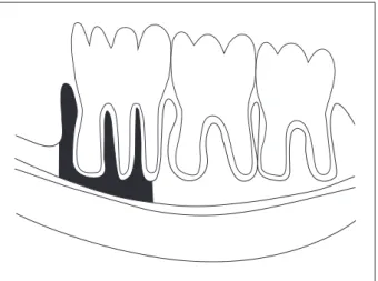
ligament of the roots of the irst molar and lower in-cisor, between the apex of the distal root of the irst molar and the alveolar bone mesial to this tooth. To standardize the mesial limit of the analyzed region, an imaginary vertical line was traced connecting the space of the periodontal ligament of the lower inci-sor and the point of highest convexity of the mesial alveolar bone as shown in Figure 1.
Three measurements were obtained at distinct times by a single examiner who was unaware of which group the animals belonged to. The mean of the three measurements obtained for each sample was used for statistical analysis. The coeficient of variation of the repeated measurements was used to determine intra-examiner calibration. A variation of less than 5% was considered as a good degree of intra-examiner reproducibility.12 If the values of
co-eficient of variation were higher than 5%, the mea-surements were repeated.
In the present study, for bone density data, the following two independent variables were analyzed:
ovarian hormones and drug, the irst consisting of two levels, presence (SHAM group) or absence (OVX group), and the second consisting of three levels, alendronate, simvastatin or water. For analy-sis of changes in animal weight, besides ovarian hor-mones and drug, the time factor was also studied at two levels (day 30 and day 65).
The data regarding body weight and alveolar ra-diographic density were submitted to descriptive and inferential statistics (analysis of variance - ANOVA and Tukey multiple comparisons test; α = 5%). Sta-tistical analysis was performed with the Statistica for Windows 5.5 (2000) (StatSoft Inc., Tulsa, OK, USA) and Statistix for Windows, version 8.0 (2003) software (Analytical Software - Tallahassee, FL, USA).
Results
For inferential statistical analysis of the data, statistical assumptions were evaluated before using the ANOVA model. The results indicated that the residuals were normally distributed. Uniformity was tested by plotting against predicted values, and none of the ANOVA assumptions was violated.
Body weight
The body weight gain data were summarized by means and standard deviation (Table 1).
Analysis of the percental body weight gain of the animals by repeated measures ANOVA showed no signiicant interaction effect among the three factors
(F(2;48) = 0.78; p = 0.4626), or between hormone and
time (F(1;48) = 0.41; p = 0.5258). Signiicant differ-ences were observed for the main effects hormones
(F(1;48) = 41.14; p < 0.001) and time (F(1;48) = 52.62;
p < 0.001), but not for the main effect drug
(F(2;48) = 2.05; p = 0.14). For the hormones factor,
Figure 1 - Schematic drawing of the alveolar bone region where radiographic density was evaluated.
Table 1 - Mean (± standard deviation) for the body weight gain data (%).
Ovarian Hormones
Water Alendronate Simvastatin
Day 30 Day 65 Day 30 Day 65 Day 30 Day 65
SHAM (presence) 9.57 ± 5.61 17.97 ± 5.83 7.88 ± 3.32 11.33 ± 5.56 10.63 ± 3.00 15.41 ± 3.44
OVX (absence) 17.84 ± 3.17 25.35 ± 5.03 20.01 ± 4.25 24.71 ± 5.71 15.36 ± 6.11 17.09 ± 8.71
its absence had a positive inluence on weight gain, with OVX animals showing a higher weight gain (mean ± standard deviation, 20.06 ± 6.68%) than SHAM animals (12.13 ± 5.63%). For the time fac-tor, the weight gain of the animals compared to the initial weight was higher on day 65 (18.64 ± 7.55%) than on day 30 (13.54 ± 6.16%).
Alveolar density
The coeficient of variation obtained for the three measurements of alveolar radiographic den-sity ranged from 0.097% to 2.439%. The data were submitted to descriptive statistics and means and standard deviation are shown in Table 2.
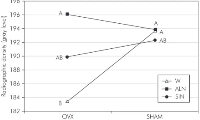
After ANOVA, signiicant differences were observed for the interaction effect (F(2;48) = 4.36; p = 0.0182). Considering the main effects, by plot-ting the means, there was a disordinal interaction for hormones (F(1;48) = 4.05; p = 0.0497) and for
drug (F(2;48) = 4.61; p = 0.0148); thus, these effects should not be analyzed separately. The effect of the drug factor depended on the level of the hormones factor (Graph 1). The Tukey test was applied for comparison of means between the experimental conditions. Alveolar radiographic density values, ex-pressed as gray levels, were lowest in the OVX-wa-ter group (183.49 ± 6.47), and differed signiicantly from those observed for the groups receiving alen-dronate (SHAM-ALN: 193.85 ± 3.81; OVX-ALN: 196.06 ± 5.11) and from those of the SHAM-water group (193.66 ± 4.36) (Graph 1). Other comparisons between means did not show signiicant differences.
Discussion
Menopausal women, deprived of the effects of estrogens and experiencing negative environmental factors such as sedentarism, show a higher tendency to gain weight. The increase of fat in these women might be related to a reduction in energy expendi-ture at rest and in physical activities, leading to a positive energy balance.14 Studies have shown that
the removal of estrogen is related to an increase of food intake and weight gain in rodents.3 Based
on the literature, in the present study the success of ovariectomy in reducing ovarian hormone lev-els could be demonstrated by changes in the body weight of the animals. The time factor also resulted in a signiicant increase of body weight, indicating the growth of the animals during the study period despite their maturity and irrespective of their hor-monal situation.
Conlicting results regarding the inluence of the absence of ovarian hormones on alveolar bone loss have been reported in both human and animal stud-ies. In the present study, among animals which re-ceived water, lower bone density was observed in the ovariectomized group. Observation periods of 3
Ovarian Hormones
Drug
Water Alendronate Simvastatin Row
SHAM (presence) 193.66 ± 4.36 193.85 ± 3.81 192.34 ± 7.95 193.28 ± 5.50
OVX (absence) 183.49 ± 6.47 196.06 ± 5.11 189.86 ± 8.80 189.80 ± 8.49
Column 188.58 ± 7.49 194.96 ± 4.52 191.10 ± 8.23 –
Graph 1 - Mean radiographic density (gray level) for the interaction between ovarian hormones (ovariectomized-OVX; sham operated-SHAM) and drug (simvastatin-SIN; alendronate-ALN; water-W). The same letters indicate the absence of a significant difference. The lowest value was found in group OVX-W (indicated as letter B), and it differed significantly from those observed for the groups OVX-ALN, SHAM-ALN and SHAM-W (indicated as letter A).
A
A A
AB
AB
B
R
adio
graphic
dens
ity
(gray
lev
el)
198
196
194
192
190
188
186
184
182
OVX SHAM
ALN SIN W
months or longer have generally been employed in the evaluation of the effects of ovariectomy on the lower jaw,24 but Tanaka et al.20 (2002) reported a reduction
in the interradicular septum of the irst lower mo-lar after 60 days. On the other hand, some authors did not observe maxillary or periodontal alterations resulting from ovariectomy. According to Teóilo et al.21 (2003), estrogen deiciency was not suficient to
promote maxillary osteoporosis in rats 11 weeks af-ter ovariectomy. A combination of calcium and estro-gen deiciency may accelerate the onset of osteoporo-sis signs in the maxillae of experimental animals.21
The relationship between the use of statins and improvement of bone quality reported in the litera-ture is still controversial. Some animal studies have reported positive effects of statins on bone tissue, increasing bone formation and reducing the effects of osteoporosis,13 stimulating fracture19 and bone
defect healing when applied to the site of injury,23
and increasing bone density.1 If the anabolic effect
of statins is considered to be real, one possible ex-planation of this effect would be that small GTPas-es, prenylated by products of the mevalonate meta-bolic pathway, negatively regulate the expression of BMP-2. Inhibition of this pathway, preventing the prenylation and function of GTPases, may stimulate the expression of BMP-2, causing the proliferation and differentiation of osteoblasts and, consequently, bone formation.17
Our results are compatible with those reported by Von Stechow et al.22 (2003) and Inoue et al.8 (2003)
who did not observe changes in bone tissue, after ovariectomy, or in bone healing with the adminis-tration of simvastatin. Junqueira et al.9 (2002),
how-ever, observed improvement of bone defect healing in ovariectomized rats with the use of simvastatin. The differences between the results of these studies might be attributed to variations in methodology; however, the controversy continues and further in-vestigations are needed to clarify it.
The divergence between results is even greater in clinical and epidemiological studies conducted on humans in whom it is dificult to standardize and control variables. While some investigators reported an increase in bone density4 and a reduction in
frac-ture risk,2 others7,10 did not ind these beneits, and
our results agree with theirs. The high hepatoselec-tivity of the drug used might be one of the reasons for its ineficacy observed in these studies. The dose tested, based on Mundy et al.13 (1999), did not
im-prove alveolar bone tissue. Further studies are neces-sary to formulate doses and modes of administration that optimize the effect of statins on bone without reducing its beneicial cardiovascular effects.
In the present study, higher radiographic densi-ty was observed in animals receiving alendronate, especially those submitted to ovariectomy, suggest-ing that the beneicial effects of alendronate on the treatment of systemic osteoporosis may extend to alveolar bone. Bisphosphonates administered sys-temically have been shown to be effective in the re-duction of alveolar bone loss induced by periodontal disease in animal16 and in human11 studies,
show-ing that their complementary use in the treatment of periodontal disease seems promising. However, there are recent reports in literature of a possible association between intravenously administered bisphosphonates – prescribed for the treatment of hypercalcemia of malignancy due to multiple my-eloma bone lesions or bone metastases –, or chronic oral bisphosphonate use in patients with osteoporo-sis or Paget’s disease, and bone necroosteoporo-sis in the man-dible or maxilla.5 Some authors have studied the
successful use of topical bisphosphonates in peri-odontal defects in patients15 and this may be a good
alternative for the use of these drugs. Most of the knowledge about the effects of bisphosphonates in the stomatognathic system comes from animal and
in vitro studies. Therefore, more research is needed
before these medicaments can be freely used in den-tal clinics.
Conclusion
Based on the methods used, we concluded that ovariectomy reduced alveolar density in rats and that sodium alendronate was effective in the treat-ment of this condition.
Acknowledgments
References
1. Anbinder AL, Balducci I, Rocha RF, Carvalho YR. Influência da sinvastatina na densidade óssea em tíbias de ratos. RPG Rev Pós-Grad. 2002;9(4):331-6.
2. Chan KA, Andrade SE, Boles M, Buist DS, Chase GA, Dona-hue JG et al. Inhibitors of hydroxymethylglutaryl-coenzyme A reductase and risk of fracture among older women. Lancet. 2000;355(9222):2185-8.
3. Chen Y, Heiman ML. Increased weight gain after ovariec-tomy is not a consequence of leptin resistance. Am J Physiol Endocrinol Metab. 2001;280:E315-22.
4. Chung YS, Lee MD, Lee SK, Kim HM, Fitzpatrick LA. HMG-CoA reductase inhibitors increase BMD in type 2 diabetes mel-litus patients. J Clin Endocrinol Metab. 2000;85(3):1137-42. 5. Estefania Fresco R, Ponte Fernandez R, Aguirre Urizar JM.
Bisphosphonates and oral pathology II. Osteonecrosis of the jaws: review of the literature before 2005. Med Oral Patol Oral Cir Bucal. 2006;11(6):E456-61.
6. Genant HK, Cooper C, Poor R, Reid I, Ehrlich G, Kanis J et al. Interim report and recommendations of the World Health Organization task-force for osteoporosis. Osteoporos Int. 1999;10(4):259-64.
7. Hsia J, Morse M, Levin V. Effect of simvastatin on bone markers in osteopenic women: a placebo-controlled, dose-ranging trial [ISRCTN85429598]. BMC Musculoskelet Dis-ord. 2002;3:7. Available from: http://www.biomedcentral. com/1471-2474/3/7.
8. Inoue TM, Anbinder AL, Balducci I, Carvalho YR. Influência da sinvastatina, administrada por via subcutânea, na reparação óssea em tíbia de rato. RPG Rev Pós-Grad. 2003;10(2):113-9. 9. Junqueira JC, Mancini MN, Carvalho Y, Anbinder AL, Bal-ducci I, Rocha RF. Effects of simvastatin on bone regenera-tion in the mandibles of ovariectomized rats and on blood cholesterol level. J Oral Sci. 2002;44(3/4):117-24.
10. Lacroix AZ, Cauley JA, Pettinger M, Hsia J, Bauer DC, Mc-Gowan J et al. Statin use, clinical fracture, and bone density in postmenopausal women: results from the women’s health ini-tiative observational study. Ann Intern Med. 2003;139(2):97-104.
11. Lane N, Armitage CG, Loomer P, Hsieh S, Majumdar S, Wang HY et al. Bisphosphonate therapy improves the out-come of conventional periodontal treatment: results of a 12-month, randomized, placebo-controlled study. J Periodontol. 2005;76(7):1113-22.
12. Leichter JW, Pack ARC, Kardos TB. A comparison of stereo-logical and computer-assisted histomorphometric analysis as tools for histological quantification in regenerative studies. J Periodontol Res. 1998;33(2):99-104.
13. Mundy G, Garret R, Harris S, Chan J, Chen D, Rossini G et al. Stimulation of bone formation in vitro and in rodents by statins. Science. 1999; 286(5446):1946-9.
14. Poehlman ET. Menopause, energy expenditure, and body com-position. Acta Obstet Gynecol Scand. 2002;81(7):603-11. 15. Reddy GT, Kumar TM, Veena. Formulation and evaluation of
alendronate sodium gel for the treatment of bone resorptive lesions in periodontitis. Drug Deliv. 2005;12(4):217-22. 16. Reddy MS, Weatherford TW 3rd, Smith CA, West BD, Jeffcoat
MK, Jacks TM. Alendronate treatment of naturally-occurring periodontitis in beagle dogs. J Periodontol. 1995;66(3):211-7. 17. Rogers MJ. Statins: lower lipids and better bones? Nat Med.
2000;6(1):21-3.
18. Russel R, Rogers MJ, Frith JC, Luckman SP, Coxon FP, Ben-ford HL et al. The pharmacology of bisphosphonates and new insights into their mechanisms of action. J Bone Miner Res. 1999;14(2):53-65.
19. Skoglund B, Forslund C, Aspenberg P. Simvastatin improves fracture healing in mice. J Bone Miner Res. 2002;17(11):2004-8.
20. Tanaka, M, Ejiri S, Toyooka E, Kohno S, Ozawa H. Effects of ovariectomy on trabecular structures of rat alveolar bone. J Periodontol Res. 2002;37(2):161-5.
21. Teófilo JM, Azevedo AC, Petenusci SO, Mazaro R, Lamano-Carvalho TC. Comparison between two experimental pro-tocols to promote osteoporosis in the maxilla and proximal tibia of female rats. Pesqui Odontol Bras. 2003;17(4):302-6. 22. Von Stechow D, Fish S, Yahalom D, Bab I, Chorev M, Muller
R et al. Does simvastatin stimulate bone formation in vivo? BMC Musculoskelet Disord. 2003;4:8. Available from: http:// www.biomedcentral.com/1471-2474/4/8.
23. Wong RWK, Rabie ABM. Statin collagen grafts used to repair defects in the parietal bone of rabbits. Br J Oral Maxillofac Surg. 2003;41(4):244-8.