Boniek Castillo Dutra Borges(a) Fabrício Lopes da Rocha Pereira(b) Roberta Caroline Bruschi Alonso(c) Rodivan Braz(d)
Marcos Antônio Japiassú Resende Montes(d)
Isauremi Vieira de Assunção Pinheiro(b)
Alex José Souza dos Santos(b)
(a) Department of Dentistry, School of Dentistry, Potiguar University (Laureate International Universities), Natal, RN, Brazil.
(b) Department of Dentistry, School of Dentistry, Federal University of Rio Grande do Norte, Natal, RN, Brazil.
(c) Department of Dentistry, School of Dentistry, Bandeirante University of São Paulo (UNIBAN), São Paulo, SP, Brazil. (d) Department of Restorative Dentistry,
Pernambuco School of Dentistry, University of Pernambuco, Camaragibe, PE, Brazil.
Corresponding author: Boniek Castillo Dutra Borges E-mail: [email protected]
Received for publication on Oct 25, 2011 Accepted for publication on Feb 14, 2012
Impact of adhesive and photoactivation method
on sealant integrity and polymer network
formation
Abstract: We evaluated the inluence of photoactivation method and hydropho -bic resin (HR) application on the marginal and internal adaptation, hardness
(KHN), and crosslink density (CLD) of a resin-based issure sealant. Model issures were created in bovine enamel fragments (n = 10) and sealed using
one of the following protocols: no adhesive system + photoactivation of the sealant using continuous light (CL), no adhesive system + photoactivation of the sealant using the soft-start method (SS), HR + CL, or HR + SS. Marginal and internal gaps and KHN were assessed after storage in water for 24 h. The CLD was indirectly assessed by repeating the KHN measurement after 24 h of immersion in 100% ethanol. There was no difference among the samples with regard to marginal or internal adaptation. The KHN and CLD were similar for samples cured using either photoactivation method. Use of a hydrophobic resin
prior to placement of issure sealants and curing the sealant using the soft-start method may not provide any positive inluence on integrity or crosslink den -sity.
Descriptors: Hardness Tests; Dental Marginal Adaptation; Polymerization.
Introduction
Dental decay remains a common disease of the oral cavity.1 The eficacy
of issure sealants on the primary and secondary prevention of caries disease
has been well demonstrated.2,3 However, their eficacy relies directly on their
ability to thoroughly ill pits and issures and remain intact and bonded to the
enamel surface for a lifetime.4,5 Therefore, factors which may affect marginal
adaptation of the sealant must be studied.
A well-adapted sealant and a strong polymeric network are required for sealant effectiveness.6 Although some authors have suggested that application
of adhesive systems can improve marginal adaptation and retention of issure
sealants,7 others have argued that this procedure offers no beneits to sealant
retention or marginal integrity.8 Complete retention of sealant material in the
issures depends on adhesion to enamel,9 and the inluence of adhesive sys
-tems may be predicted by analyzing the supericial and internal margins of issure sealants.
In an attempt to decrease stresses during polymerization of resin-based materials, alternative photoactivation methods such as soft-starting have been proposed. These methods modify the polymerization kinetics by modulating the power density during photoactivation.10 The soft-start modulated
photoac-tivation method was reported to be effective in reducing contraction stress and
Declaration of Interests: The authors certify that they have no commercial or associative
interest that represents a conlict of interest in
improving the strength of the bonded interface without compromising the polymerization quality of the restor-ative composite.11
Although resin-based issure sealants possess low elastic moduli to facilitate low during polymerization,
the occlusal surface can present a higher C factor, lead-ing to the development of elevated shrinkage stress.9
Photoactivation of issure sealants using soft-start meth -ods might decrease polymerization stresses, improving marginal adaptation. However, the literature is unclear
concerning the beneits of soft-start photoactivation on
maintaining marginal integrity or the effect on polymer
network formation of issure sealants.
Using a novel issure model, we aimed to evaluate
the use of an adhesive system before sealant placement as well as soft-start modulated photoactivation on
su-pericial and internal adaptation, sealant hardness, and crosslink density. Our hypothesis was that supericial
and internal marginal adaptation of the sealant would be improved by the adhesive system and by modulated soft-start photoactivation without decreasing sealant hardness or crosslink density.
Methodology
Sample preparation
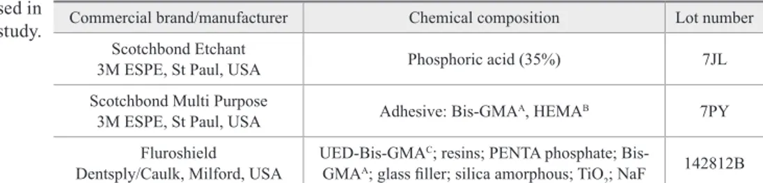
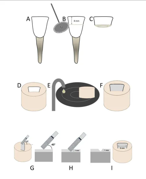
Commercial brand names, chemical compositions, and lot numbers for the materials used in this study are listed in Table 1. Representative examples of the sample preparation stages are depicted in Figures 1A-I. Forty bovine incisors were used to prepare the samples (Figure 1A). The crowns were sectioned in the mesio-distal direction using double-faced diamond disks (KG Sorensen, Barueri, Brazil) (Figure 1B) in an attempt to obtain 8 mm-long enamel/dentin fragments (Figure 1C). These fragments were then embedded in polystyrene resin (Figure 1D) and the enamel surface was ground on
a water-cooled mechanical polisher (Arotec, São Paulo, Brazil) using 320-, 600-, and 1200-grit silicon carbide (SiC) abrasive papers (Figures 1E and F). A cylindrical diamond bur (#1094, KG Sorensen, Barueri, Brazil) was mounted in a high-speed handpiece (Kavo, Joinville, Brazil), positioned at 45° to the enamel surface (Figure 1G), and used to generate a groove in the enamel (Figure
1H) under constant air-water cooling to produce issure
models measuring 1 mm deep × 8 mm long (Figure 1I).
To achieve a uniform issure size, the handpiece was
mounted on a cavity standardization device. The dia-mond bur was replaced after every 5th preparation. The
specimens were examined in a stereomicroscope (Carl
Zeiss, Manaus, Brazil) at 25× magniication to verify
whether the enamel remained on the lower surface. The materials were prepared according to the
manu-facturers’ recommendations. The issure was etched us -ing 35% phosphoric acid gel for 15 s, rinsed for 30 s and dried for 30 s with oil-free air. When applicable, a hydrophobic resin was applied after enamel etching and light cured for 10 s using the Ultra-Lume LED 5
(Ultra-dent, South Jordan, USA). The resin-based issure seal
-ant was placed in the issures, covered with a polyester
strip, and photoactivated according to the protocol
speci-ied for that sample group. The continuous light photo -activation method involved curing the material for 20 s at 800 mW/cm2 (following the manufacturer’s
instruc-tions), resulting in a total energy of 16 J. The soft-start photoactivation method was standardized with an initial light exposure of 10 s at 150 mW/cm2, followed by 18 s
at 800 mW/cm2 (total energy 16 J).
The specimens were stored in distilled water at 37°C
for 24 h and then inished and polished in a water-cooled
mechanical polisher (Arotec, São Paulo, Brazil) using 600- and 1200-grit SiC sandpaper.
Commercial brand/manufacturer Chemical composition Lot number
Scotchbond Etchant
3M ESPE, St Paul, USA Phosphoric acid (35%) 7JL
Scotchbond Multi Purpose
3M ESPE, St Paul, USA Adhesive: Bis-GMA
A, HEMAB 7PY
Fluroshield Dentsply/Caulk, Milford, USA
UED-Bis-GMAC; resins; PENTA phosphate; Bis-GMAA; glass iller; silica amorphous; TiO
2; NaF
142812B
A: bisphenol-A diglycidyl methacrylate; B: hydroxyethyl methacrylate; C: urethane modified Bis-GMA dimeth-acrylate.
Marginal adaptation analysis
A 1.0% acid red propylene glycol solution (Caries Detector, Kuraray, Osaka, Japan) was applied at the restoration margins for 5 s. After staining, the speci-mens were rinsed in tap water and gently blown dry.
The issure margins were evaluated using a Leica MZ6
stereomicroscope (Leica Microsystems, Heerbrugg,
Switzerland) at 16× magniication. A digital image of
each specimen was obtained at this stage. The lengths
of the dye-stained gaps along the issure margins were
measured (in millimeters) using the Image tool software version 2.0 (University of Texas Health and Science Center, San Antonio, USA). The length of the marginal gap was calculated as a percentage of the entire margin
length. Samples were also scored according to the pres-ence (score 1) or abspres-ence (score 0) of gaps.
Internal adaptation analysis
Three slices from sealed issures (1-mm thick) were
cut in the mesio-distal direction using an Isomet 1000 machine (Buehler, São Paulo, Brazil). Caries Detector solution was applied to the internal interfaces, and the same procedures described previously were performed for the evaluation of internal adaptation. The internal gap percentages of the slices were averaged to obtain a mean for each specimen. The samples were also scored according to the presence (score 1) or absence (score 0) of gaps.
Knoop hardness test (KHN)
After analyzing the internal margins, the KHN was evaluated for two slices. An initial hardness (MHi) read-ing was obtained on the top surface of each specimen using a Knoop hardness tester (HMV-2T E, Shimadzu Corporation, Tokyo, Japan, 50 g load for 15 s). A total
of ive Knoop measurements were performed on the top
surface of each specimen: one at the center and the other four at a distance of approximately 200 µm from the
central location. The average of the ive values was used
to represent the KHN value of each specimen.
Crosslink density (CLD)
After analysis of KHN, all specimens were im-mersed in absolute ethanol (100%) at room temperature. The CLD was indirectly estimated based on the per-centage decrease in hardness (%HD) that occurred as a result of ethanol exposure.12 After immersion for 24 h
a second hardness reading was obtained (MHf). Five Knoop measurements were performed on the top sur-face of each specimen as previously described. The re-sults were tabulated and the %HD was calculated using the following equation:
%HD = 100 − [(MHf × 100) / MHi)],
where MHf represents the inal KHN value (after ethanol storage) and MHi represents the initial KHN value (before ethanol storage).
Statistical analysis
Marginal and internal adaptations were determined from the percentage of gaps (quantitative data) and the presence or absence of gaps (qualitative data). To com-pare the quantitative data, the Kruskall-Wallis test was performed, while the Mann-Whitney test was used to analyze the qualitative data. KHN and %HD were evaluated by means of Analysis of Variance (ANOVA)
and Tukey’s test. The level of signiicance was set at
P < 0.05. The Assistat Beta 7.5 software (Federal Uni-versity of Campina Grande, Campina Grande, Brazil) was used to perform all tests.
Results
Supericial marginal adaptation
There were no differences between groups in terms
of percentage of supericial marginal gaps or presence
of gaps. The median gaps and gap scores related to
su-pericial marginal adaptation are listed in Table 2.
Internal marginal adaptation
There were no differences between groups either in percentage of internal marginal gaps or scale rank. The
median gaps and gap scores related to supericial mar -ginal adaptation are listed in Table 3.
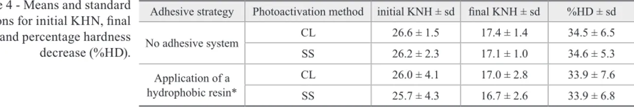
HKN and CLD
There were no differences between groups in either initial KNH or %HD (CLD). Means and standard
de-viations for initial KHN (before ethanol storage), inal
KHN (after ethanol storage), and percentage hardness decrease are contained in Table 4.
Discussion
An important parameter for evaluating the clinical success of sealant materials is the marginal adaptation. Many studies13,14 have used human third molars to
evalu-ate the integrity of issure sealants. However, the issure
morphology of the human third molar is highly variable,
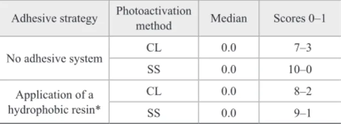
Table 2 - Median gaps (%) and gap scores related to supericial marginal adaptation.
Adhesive strategy Photoactivation
method Median Scores 0–1
No adhesive system CL 0.0 7–3
SS 0.0 10–0
Application of a hydrophobic resin*
CL 0.0 8–2
SS 0.0 9–1
*Hydrophobic resin of Scothbond Multipurpose Plus. CL: continuous light; SS: soft-start.
Table 3 - Median gaps and gap scores related to internal mar-ginal adaptation.
Adhesive strategy Photoactivation
method Median Scores 0 – 1
No adhesive system CL 0.0 18–2
SS 0.0 18–2
Application of a hydrophobic resin*
CL 0.0 19–1
SS 0.0 19–1
and does not permit standardization of the C-factor. For
this reason we employed an artiicial issure model to provide similar conditions while avoiding the inluence
of the C-factor.
Clinically, sealants are applied to issures with in -tact, aprismatic enamel instead of the ground, prismatic enamel15 present in the issure model used in this in
-vestigation. However, some authors16,17 have reported
no difference in the adhesion of etch-and-rinse adhe-sive systems to ground or unground enamel. The use of ground, prismatic tissue in this study most likely did
not inluence the results since an etch-and-rinse adhe -sive system was utilized. The preparation of simulated
issures in bovine enamel to evaluate the integrity of issure sealants has the advantage of standardizing the C-factor of the issure using an acceptable substitute for
human teeth.
In the present investigation the marginal and
inter-nal gap occurrence was assessed by staining the issure
margins with a 1.0% acid red propylene glycol solution. This technique has been effectively used to evaluate gap formation, but not microleakage.18-21 A lat, inished, and
polished specimen is required to perform this technique
without bias, and the samples were lattened, inished,
and polished using abrasive paper even though this pro-cedure is not commonly performed in vivo.
No beneits were achieved in terms of early sealant
integrity by applying a hydrophobic resin or curing the
issure sealant using the soft-start method, although this
photoactivation method did not decreased hardness or CLD. Therefore, our hypothesis was partially validated. As the marginal and internal integrity was assessed 24 hours after polymerization, the presence of gaps could be attributed exclusively to polymerization shrinkage. The monomer conversion resulted in similar stresses
during photoactivation of the resin-based issure seal
-ant. This might have been facilitated by the low charac -teristics of the material providing better accommodation
in the illed substrate due to its low viscosity. On the
other hand, if the samples had been subjected to aging conditions simulating an oral environment, it is likely that some differences might be observed. Further
stud-ies must be conducted to evaluate the long-term eficacy
of the soft-start method.
Applying a hydrophobic resin before sealant place-ment did not improve sealant integrity, possibly
be-cause sealants have reduced surface tension and low
characteristics that promote satisfactory adhesion to the enamel after acid etching. Although application of an adhesive system prior to the sealant has demonstrated a positive effect on micro leakage,21 several criticisms of
this method have appeared due to its low reliability in
predicting the true iniltration of substances.22 Tracers
used in microleakage investigations are generally not suitable for testing the marginal seal of restorations in
vitro, as the molecules are so small that they penetrate
through tiny and invisible paths along the substrate / re-storative material interface. Evaluating the marginal integrity as performed in this work represents a more realistic method.23
Considering the absence of negative effects for mod-ulated photoactivation methods such as the soft-start method on select physical properties of resin-based ma-terials, the results of this study corroborate those found elsewhere with respect to degree of conversion24 and
crosslink density.25 In fact, composite materials
submit-ted to equivalent radiant exposure irrespective of the curing method possess similar degrees of conversion.24
Although other photoactivation methods such as the pulse-delay can decrease the crosslink density of poly-meric networks, the soft-start method did not reduce the level of crosslinking.25 It must be pointed out that
insuf-icient crosslinking of the polymer matrix may make
resin-based materials more sensitive to the plasticizing effects of exogenous substances containing chemicals such as acids, bases, salts, alcohols, and oxygen that
en-Adhesive strategy Photoactivation method initial KNH ± sd inal KNH ± sd %HD ± sd
No adhesive system CL 26.6 ± 1.5 17.4 ± 1.4 34.5 ± 6.5
SS 26.2 ± 2.3 17.1 ± 1.0 34.6 ± 5.3
Application of a hydrophobic resin*
CL 26.0 ± 4.1 17.0 ± 2.8 33.9 ± 7.6
SS 25.7 ± 4.3 16.7 ± 2.6 33.9 ± 6.8
*Hydrophobic resin of Scothbond Multipurpose Plus adhesive system. CL: continuous light; SS: soft-start. Table 4 - Means and standard
ter the oral environment during eating and drinking25
and may have a deleterious effect on the polymer
net-work and compromise its clinical eficacy.
Conclusion
The use of a modulated photoactivation method such as the soft-start and a hydrophobic resin before seal-ant placement did not provide any early improvement
in marginal or internal adaptation of the pit and issure
sealant tested. Hardness and crosslink density were not affected by the photoactivation method.
Acknowledgements
The authors thank KG Sorensen (Barueri, Brazil) for providing some of the materials used in this study.
References
1. Piovesan C, Mendes FM, Antunes JL, Ardenghi TM. Inequalities in the distribution of dental caries among 12-year-old Brazilian schoolchildren. Braz Oral Res. 2011 Jan-Feb;25(1):69-75. 2. Ahovuo-Saloranta A, Hiiri A, Nordblad A, Mäkelä M,
Worthing-ton HV. Pit and fissure sealants for preventing dental decay in the permanent teeth of children and adolescents. Cochrane Database Syst Rev. 2008 Oct 8;(4):CD001830.
3. Borges BC, Campos GP, Silveira AD, Lima KC, Pinheiro IV. Ef-ficacy of a pit and fissure sealant on arresting dentin non-cavitated caries: a 1-year follow-up randomized and controlled clinical trial. Am J Dent. 2010 Dec;23(6):311-6.
4. Hevinga MA, Opdam NJ, Bronkhorst EM, Truin GJ, Huysmans MC. Long-term performance of resin based fissure sealants placed in a general dental practice. J Dent. 2010 Jan;38(1):23-8. 5. Griffin SO, Oong E, Kohn W, Vidakovic B, Gooch BF. The
effec-tiveness of sealants in managing caries lesions. J Dent Res. 2008 Feb;87(2):169-74.
6. Borges BC, Bezerra GV, Mesquita JA, Pereira MR, Aguiar FH, Santos AJ, Pinheiro IV. Effect of irradiation times on the polym-erization depth of contemporary fissure sealants with different opacities. Braz Oral Res. 2011 Apr;25(2):135-42.
7. Feigal RJ, Quelhas I. Clinical trial of a self-etching adhesive for sealant application: success at 24 months with Prompt L-Pop. Am J Dent. 2003 Aug;16(4):249-51.
8. Mascarenhas AK, Nazar H, Al-Mutawaa S, Soparkar P. Effective-ness of primer and bond in sealant retention and caries prevention. Pediatr Dent. 2008 Jan-Feb;30(1):25-8.
9. Glavina D, Vranic DN, Milosevic SA, Bergman V, Majstorovic M, Skrinjaric I. Soft-start polymerization of fissure sealant: retention after three years. Coll Antropol. 2007 Dec;31(4):1089-92. 10. Alonso RC, Correr GM, Cunha LG, De Moraes Souto Pantoja CA,
Puppin-Rontani RM, Sinhoreti MA. Modulated photoactivation methods: effect on marginal and internal gap formation of restora-tions using different restorative composites. J Biomed Mater Res B Appl Biomater. 2007 Aug;82(2):346-51.
11. Cunha LG, Alonso RC, Pfeifer CS, Correr-Sobrinho L, Ferracane JL, Sinhoreti MA. Modulated photoactivation methods: Influence on contraction stress, degree of conversion and push-out bond strength of composite restoratives. J Dent. 2007 Apr;35(4):318-24. 12. Schneider LF, Moraes RR, Cavalcante LM, Sinhoreti MA, Correr-Sobrinho L, Consani S. Cross-link density evaluation through softening tests: effect of ethanol concentration. Dent Mater. 2008 Feb;24(2):199-203.
13. Braz AK, Aguiar CM, Gomes AS. Evaluation of the integrity of dental sealants by optical coherence tomography. Dent Mater. 2011 Apr;27(4):e60-4.
14. Perdigao J, Sezinando A, Gomes G. In vitro sealing potential of a self-adhesive pit and fissure sealant. Quintessence Int. 2011 May;42(5):e65-73
15. Papacchini F, Cury AH, Goracci C, Chieffi N, Tay FR, Polimeni A, et al. Noninvasive pit and fissure sealing: microtensile bond
strength to intact bovine enamel of different pit and fissure sealants in a simplified fissure model. J Adhes Dent. 2006 Dec;8(6):375-80. 16. Loguercio AD, Moura SK, Pellizzaro A, Dal-Bianco K, Patzlaff
RT, Grande RH, et al. Durability of enamel bonding using two-step self-etch systems on ground and unground enamel. Oper Dent. 2008 Jan-Feb;33(1):79-88.
17. Perdigão J, Gomes G, Gondo R, Fundingsland JW. In vitro bond-ing performance of all-in-one adhesives. Part I--microtensile bond strengths. J Adhes Dent. 2006 Dec;8(6):367-73.
18. Yoshikawa T, Burrow MF, Tagami J. A light curing method for improving marginal sealing and cavity wall adaptation of resin composite restorations. Dent Mater. 2001 Jul;17(4):359-66. 19. Alonso RC, Correr GM, Cunha LG, Borges AFS, Puppin-Rontani
RM, Sinhoreti MA. Dye staining test: an alternative method for assessing gap formation in composite restorations. Acta Odontol Scand. 2006 Jun;64(3):141-5.
20. Correr GM, Alonso RC, Puppin-Rontani RM, Correr-Sobrinho L, Sinhoreti MA. Marginal and internal adaptation of composite restorations using a resin liner on deproteinized substrate. Acta Odontol Scand. 2005 Aug;63(4):227-32.
21. Asselin ME, Fortin D, Sitbon Y, Rompré PH. Marginal microle-akage of a sealant applied to permanent enamel: evaluation of 3 application protocols. Pediatr Dent. 2008;30(1):29-33.
23. Heintze S, Forjanic M, Cavalleri A. Microleakage of Class II resto-rations with different tracers--comparison with SEM quantitative analysis. J Adhes Dent. 2008 Aug;10(4):259-67.
24. Cunha LG, Alonso RC, Pfeifer CS, Correr-Sobrinho L, Ferra-cane JL, Sinhoreti MA. Contraction stress and physical properties development of a resin-based composite irradiated using modu-lated curing methods at two C-factor levels. Dent Mater. 2008 Mar;24(3):392-8.