0103 - 5053 $6.00+0.00
Review
* e-mail: [email protected]
Chemical Similarity and Biological Activities
Hugo Kubinyi
University of Heidelberg, Germany and BASF AG, Ludwigshafen, Germany
“Moléculas similares exercem atividades farmacológicas similares”. Baseados neste conceito, os(as) químicos(as) medicinais modificam as estruturas de compostos biologicamente ativos. Mudanças bioisostéricas de átomos e grupos podem ser usadas em inibidores enzimáticos, agonistas e antagonistas, dentre outros princípios ativos. Entretanto, estruturas químicas similares e dissimilares não são facilmente definidas de forma objetiva. Surpreendentemente, várias relações entre a estrutura e a atividade demonstram que compostos similares podem ter atividades e ações biológicas muito diferentes. Modificações químicas simples de alguns ligantes podem resultar em efeitos inesperados, incluindo novos modos de ação. Enantiômeros ópticos também apresentam diferenças em suas atividades biológicas.
“Similar molecules exert similar biological activities”. Since long, medicinal chemists use this concept to modify the structures of biologically active compounds. Bioisosteric replacements of atoms and groups pave the way from various lead structures to therapeutically useful enzyme inhibitors, receptor agonists and antagonists, and other active principles. However, similarity and diversity of chemical structures cannot be defined in an objective manner. Several surprising structure-activity relationships demonstrate that chemically similar compounds may have significantly different biological actions and activities. Some protein ligands exert unexpected new binding modes, after only minor chemical modification. Of course, even optical enantiomers most often have different biological activities.
Keywords: chemical similarity, similarity vs. activity, structure-activity relationships, agonists and antagonists, hydrogen bonds, optical enantiomers
1. Similarity and Dissimilarity of Molecules
“Similar molecules exert similar biological activities”.
Since long, medicinal chemists used this concept to modify
the structures of biologically active compounds.
1-6Bioisosteric replacements of atoms and groups pave the
way from lead structures to therapeutically useful enzyme
inhibitors, receptor agonists and antagonists, and other
active principles.
7-11Several surprising structure-activity
relationships demonstrate that chemically similar
compounds may have significantly different biological
actions and activities. Of course, even optical enantiomers
most often have different biological activities.
Correspondingly, a sophisticated consideration of
“chemical similarity” and “chemical diversity” is a waste
of time. Similarity and diversity of molecules depend on
the 3D structure and binding site properties of the
biological target, not on any artificial parameters.
The principle of bioisosteric replacement of functional
groups serves as a successful optimization strategy.
5-11Its
systematic application has resulted in a broad variety of
therapeutically used drugs, many of them finally having
the desired combination of favorable properties. In their
attempts to optimize lead structures, medicinal chemists
intuitively follow the principles of evolution. The
biological activity, in later stages a selectivity index or
some other biological property, serve as the “fitness
function” for the “survival” of certain structural entities.
However, several surprising structure-activity relationships
demonstrate how difficult rational drug design can be and
to which extent structure-based and computer-aided
approaches can support this process.
5,12-21Similarity and diversity of chemical structures cannot
be defined in an objective manner (Figure 1).
6-11,22-24There
is no doubt that for maximum affinity a ligand of a
biological macromolecule has to fit the binding pocket
geometrically (Figure 2).
25On the other hand, for recognition by the binding site
it does not matter whether a certain acceptor atom of the
ligand is either nitrogen or oxygen. An illustrative example
for this equivalence are two series of scytalone dehydratase
inhibitors, salicylamides and quinazolines, which exert
the same type of interactions with the protein. Also the
important role of water can be seen: several conserved
water molecules mediate the contact between the ligand
and the enzyme. In many cases, the replacement of such
water molecules reduces binding affinity but a significant
enhancement of affinity is observed for the scytalone
dehydratase inhibitors, if one water molecule is replaced
by a cyano substituent - its nitrogen atom mimics the
oxygen atom of the water molecule (Figure 4).
292. Different Mechanisms of Action of Similar
Molecules
Several well-known examples of different modes of
action of closely related analogs can be found in medicinal
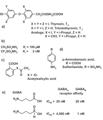
Figure 1. Bioisosteric replacement of atoms or groups can a) retain biological activity, b) significantly enhance biological activity, c) destroy biological activity, d) turn a substrate into an inhibitor, or e) modify selectivity. a) Thyroxin, T4 (X = Y = Z = I), is the less active depot form of the natural hormone, T3 (X = Y = I); a monoisopropyl analog (X = I, Y = isopropyl) of T3 is slightly more active than the natural hormone; even an analog without any halogen atoms (X = Me, Y = isopropyl) has some thyromimetic activity.22 b)
Trifluoromethanesulfonamide is 50,000 times more active as a carboanhydrase inhibitor than the methanesulfonamide.23 c) An
ex-change of the ester oxygen atom in acetylsalicylic acids to -CH2- or -NH- destroys biological activity because these analogs cannot any longer transfer an acetyl residue to cyclooxygenase. d) Sulfanil-amide is an antimetabolite of p-aminobenzoic acid; it acts as a false substrate and therefore inhibits bacterial dihydrofolate biosynthesis. e) GABA (γ-aminobutyric acid) binds with equal affinities to GABAA and GABAB receptors; its analog γ-aminopropylphosphonous acid is a highly selective GABAB receptor ligand.24
Figure 3. Peptide antibiotics, like vancomycin, are the last weapons against multiresistant bacterial pathogens. However, in the last years, several vancomycin-resistant strains were observed. One mecha-nism of resistance development is shown here: the natural substrate for bacterial cell wall biosynthesis, Lys-D-Ala-D-Ala, is converted to Lys-D-Ala-D-Lac (exchange of NH against O), leading to the loss of one hydrogen bond in its complex with vancomycin. The conse-quence is a 1,000-fold lower sensitivity of such strains against van-comycin.27,28
chemistry and pharmacology textbooks
.
1,2,4,30Norepinephrine, epinephrine and isoproterenol are
adrenergic agonists. However, in going from R = H to R =
CH
3and R = isopropyl, the mechanism of action gradually
changes from a more or less specific
α
-adrenergic agonism
to a pure
β
-adrenergic agonism. If the two hydroxyl groups
of isoproterenol are exchanged against two chlorine atoms,
the
β
-adrenergic antagonist dichloroisoproterenol (DCI)
results; in fact, this compound was the first
β
-blocker. Some
other receptor antagonists,
e.g.
the histamine, dopamine and
morphine receptor antagonists, are lipophilic analogs of
the corresponding agonists (Figure 5). Several peptide
receptor antagonists differ from the corresponding agonists
only by minor changes in their alkyl groups (Figure 6).
31-34A well-known example of different modes of action of
closely related analogs are the anti-allergic agent
promethazine, the neuroleptic drug chlorpromazine, and
the antidepressants imipramine and desipramine. Despite
their very similar chemical structures, promethazine acts
mainly as a histamine H
1antagonist, chlorpromazine is a
dopamine antagonist, imipramine is an unspecific
norepinephrine and serotonin uptake inhibitor, and
desipramine is a norepinephrine-specific uptake inhibitor
(Figure 7). Steroid hormones, like the estrogens, the
gestagens, the male sex hormones and anabolics, provide
another striking example of different biological effects of
chemically closely related analogs (Figure 8).
Several therapeutically used drugs bind to more than
one receptor and are correspondingly termed
“promiscuous” ligands. However, whether a certain
(balanced) unspecific mode of action is advantageous for
therapy or not remains still uncertain.
Figure 6. Even minor changes in the alkyl groups of some peptidomimetic angiotensin receptor antagonists convert these com-pounds into agonists, some of them with modified selectivity.31-34
Figure 5. Norepinephrine, epinephrine and isoproterenol differ only by the presence and size of an N-alkyl group. Nevertheless, they exert very different biological effects. Norepinephrine is an α -adrenergic agonist, epinephrine is a mixed α, β-adrenergic agonist, and isoproterenol is a selective β-adrenergic agonist. The exchange of the phenolic hydroxyl groups of isoproterenol against chlorine atoms produced the very first β-blocker. In morphine, the exchange of the N-methyl group against an allyl group converts the agonist to an antagonist.
3. The Biological Activity of a Ligand Depends
on its Flexibility
Other common structural modifications in the
optimization of a lead structure are the dissection of rings
or the rigidification of flexible molecules. Molecules with
several rotatable bonds may adopt many different
geometries, some of them being favorable because of low
internal energies, others being less favorable because of
van der Waals or electrostatic repulsion between
nonbonded atoms or groups. If different conformations of
such molecules are “frozen” by closing rings between
certain atoms, either one of two very different consequences
results. If the frozen conformation differs from the bioactive
conformation of the flexible lead or if the added atoms
interfere with the binding, biological activity will be more
or less destroyed. If the ring closure stabilizes the bioactive
conformation, usually a significant increase in biological
activity results.
An example of rigid analogs with remarkable selectivity
differences are chemically closely related integrin ligands
(Figure 10).
36-424. Biological Potencies of Similar Molecules
For compounds with comparable threedimensional
structures, most often similar analogs have also similar
biological activities. That this needs not always be the
case can be demonstrated by a comparison of three series
of thermolysin inhibitors. Analogs with X = -NH- and
-CH
2- are about 1,000-times more potent than the X =
-O-analogs. The explanation for this effect can be easily
derived from the 3D structure of thermolysin. If X is an
-NH- group, a hydrogen bond is formed between this group
and the oxygen atom of an alanine carbonyl group. In the
-O- analog, this hydrogen bond cannot be formed; in
addition, an electrostatic repulsion between the two
oxygen atoms results. The biological activity of the -CH
2-analog has been predicted to be comparable to the
-NH-analog and to be much higher than for the -O- -NH-analog. This
was later confirmed by the synthesis of these inhibitors
(Figure 11; cf. Figure 3).
43-47Figure 8. Steroid hormones are another example of chemically related compounds with strikingly different actions. These orally active, synthetic analogs of natural sex hormones are estrogens, gestagens (the female sex hormones), androgens (male sex hor-mone) and anabolics.
analogy, promethazine is an H1 antagonist, acting as an antiallergic drug, chlorpromazine is a dopamine antagonist for the treatment of schizophrenia, and imipramine and desipramine are neurotransmit-ter uptake inhibitors that are used in the therapy of depression.
Figure 9. The concept of the program CAVEAT35 is based on the
Figure 11. Certain peptide analogs are inhibitors of the bacterial zinc protease thermolysin. Whereas the X = -NH- analogs are potent inhibitors, the X = -O- analogs are less potent by about 3 orders of magnitude (cf. Figure 3). This difference was explained by the fact that -NH- forms a hydrogen bond with the >C=O group of Ala-113, which cannot be formed by the -O- analog; in addition, there is a repulsive effect of the two electronegative oxygen atoms. From this structure-activity relationship it was predicted that the -CH2- analogs should be as active as the -NH- analogs. Despite the fact that these analogs cannot form a hydrogen bond to the enzyme, there is no negative effect of desolvation of this part of the ligand, as observed for the -NH- and -O- analogs.43-47
Figure 10. Integrins are a series of different cell surface receptors that mediate cell-cell association (e.g. platelet aggregation) by bind-ing oligofunctional proteins that contain the same bindbind-ing motif: Arg-Gly-Asp (the so-called RGD motif) in different conformations. Small cyclic peptides and peptidomimetics containing this motif are high affinity integrin ligands; dependent on their (frozen) confor-mation they bind more or less selectively to only a certain integrin. Research at SmithKline Beecham led to the compounds SB 214 857 and SB 223 245, which are highly selective for the fibrinogen receptor (α2β3 integrin) and the vitronectin receptor (αvβ3 integrin), respectively, despite their close chemical similarity.36-42
A similar but less pronounced effect is observed for
thrombin inhibitors; in this case the nonbonded contact
between the -X- group of the ligand and the carbonyl group
of Gly 216 in the binding site of thrombin is responsible
for the structure-activity relationship. If, on the other hand,
a carbonyl group of the inhibitors, which forms a hydrogen
bond with the -NH- group of Gly 216, is replaced by -CH
2-,
affinity is reduced by some orders of magnitude; only in
one case a small reduction of affinity is observed (Figure
12).
48-51There are many examples in literature where the
introduction of an -OH group into a ligand either causes
an increase or a decrease of biological affinities. From a
theoretical point of view, this is not surprising. If the new
-OH group forms hydrogen bonds with polar groups at the
binding site (either as a hydrogen bond donor or as an
acceptor), the net free energy depends on the balance
between the desolvation energies of the water shells at the
surfaces of the ligand and the binding site, as compared to
the energy of the formed hydrogen bond (or bonds) and
the entropy gain by the release of some water molecules.
Certain tightly bound water molecules in the binding cavity
of a protein (usually seen in the X-ray structures), e.g. those
which form more than two hydrogen bonds to the protein,
are not easily removed. The attempt to introduce an -OH
group into the ligand, to replace such a water molecule,
must necessarily fail.
An example where this is not the case and where
significantly enhanced binding affinities result after the
introduction of such a hydroxyl group, are cytidine and
adenosine deaminase inhibitors, which are capable to add
a water molecule in the binding site, to mimic a transition
state (Figure 13).
52,53In this special case the resulting
affinity differences are 7 to 8 orders of magnitude!
5. Biological Potencies
vs
. Similar Biological
Targets
Figure 12. Typical thrombin inhibitors, a) X = NH, form β-sheet type hydrogen bonds with Gly-216. If X = -NH- is exchanged against -O- or -CH2-, a structure-activity relationship is observed that is similar but less pronounced as in the thermolysin inhibitors (Figure 11). b) An even smaller effect is observed, if the amino group is removed. Reduction of the interacting carbonyl group to a -CH2- group reduces affinities by factors of about 400 (c), 2,000 (d), 10,000 (e), and 4 (f). The small activity difference in the latter compound pair can be explained by the fact that, even after reduc-tion, the attached nitrogen atom remains neutral whereas it becomes a basic nitrogen in the other cases.48-51
Figure 13. The natural product zebularin is a highly potent inhibi-tor of cytidine deaminase because it perfectly mimics the transition state of the enzymatic reaction after addition of a conserved water molecule within the binding site. 3,4-Dihydrozebularine can nei-ther add nor replace this water molecule. Correspondingly, this com-pound is much less active; two hydrogen atoms make an activity difference of more than seven orders of magnitude.52,53
Figure 14. Remikiren, a renin inhibitor, shows different potency against the renins of rats and dogs, as compared to monkeys and humans. Such differences are caused by minor amino acid sequence differences and the resulting different binding site geometries of these homologous renins.54,55
progress in gene technology it is now possible to produce
human proteins in sufficient quantities to establish test
models. Thus, their biological activity in humans can be
forecasted from investigations at the molecular level.
Thiorphan and its
retro-inverso
peptide,
retro
-thiorphan, are inhibitors of the structurally related zinc
proteases thermolysin and NEP 24.11. Although the
affinities of the ligand pair differ, from enzyme to enzyme,
by three orders of magnitude, there are no significant
activity differences between them. Thus, they may be
considered to be “similar”, which was also confirmed by
the X-ray structure analyses of their thermolysin
complexes. On the other hand, their activities against yet
another related zinc protease, angiotensin converting
enzyme (ACE), are significantly different; with respect to
this enzyme the analogs are “dissimilar” (Figure 16).
57,58Corresponding problems are also observed in
predictions for toxicity in humans.
2,3,7,8-Tetrachlorodioxin is highly toxic to several species,
e.g
.
guinea pigs and mink. It is much less toxic for mice, rats,
hamsters, rabbits, dogs, and monkeys. If one extrapolates
from the monkey, dioxin should be relatively “harmless”
for humans, at least if only acute toxicity is considered.
However, the significantly different toxicity
vs
. the closely
related species guinea pig and hamster puts a caveat on
too simple and straightforward extrapolations (Figure 17).
596. Chirality and Biological Activities
Due to the chiral nature of amino acids (except glycine),
drug binding sites of proteins are asymmetric. In the past,
the different actions of enantiomers of chiral molecules on
enzymes and receptors were often neglected. For economic
reasons, racemates of synthetic drugs were used in therapy.
Today, researchers and drug companies are more aware of
the different effects of enantiomers and diastereomers, in
their biological activities (Figure 18) as well in their
pharmacokinetics.
60-77Figure 16. Thiorphan and retro-thiorphan differ in the direction of the amide group of both molecules. Nevertheless, they are equally potent against the zinc protease thermolysin. X-ray structure deter-mination of the complexes confirms that both molecules display equivalent interactions. Although being much more potent, they also show identical activities against another zink protease, NEP 24.11 (originally called enkephalinase). However, their activities differ significantly against a third zinc protease, the angiotensin-converting enzyme (ACE), which gives evidence that chemical and biological similarities cannot be defined in an objective manner -they depend on the structure of the biological target.57,58
Figure 18. Butaclamol is just one example that the eudismic ratio, the ratio of affinities of (+)- and (-) enantiomers, differs from receptor to receptor, for the same compound.68 The (S)-(-) form of the calcium
channel ligand Bay K 8644 is an agonist (stabilizing the open calcium channel), whereas the (R)-(+) form is a weak antagonist, a calcium channel blocker (stabilizing the closed channel).73,74 Corresponding
differences are observed for a CCK1 ligand, where one diastereomer is an agonist, whereas its enantiomer is an antagonist.75-77
Figure 17. 2,3,7,8-Tetrachlorodioxin (the so-called “Seveso dioxin”) shows significantly different acute toxicities in different species.59
7. Conclusions
An overreliance in target-independent similarity
indices has to be questioned, because of the dependence
of “similarity” on the biological macromolecule to which
the analogs bind. Sophisticated investigations on the
“dissimilarity” of chemical databases are most often futile.
Similar compounds may have very different actions and
different molecules can be very similar in their biological
activities. Considering the examples presented in this
paper and the many more cases in literature, one has to
admit that we are far from a deeper understanding of the
details which underlie the observed structure-activity
relationships. Applying the results from one series of
analogs to another, one may arrive at completely wrong
conclusions.
Acknowledgements
This publication reviews the content of two lectures,
presented at the 3
rdWorkshop on Chemical Structure and
Biological Activity: Perspectives in QSAR, Instituto de
participate in these symposia. The financial contributions
of Deutscher Akademischer Austauschdienst (DAAD),
Bonn, Germany, Fundação de Amparo à Pesquisa do Estado
de São Paulo (FAPESP), São Paulo, Brazil, and the congress
organisations in São Paulo and Caxambu, Brazil, are
gratefully acknowledged.
References
1. Hansch, C.; Sammes, P. G.; Taylor, J. B. eds.; Comprehensive Medicinal Chemistry, Pergamon Press: Oxford, 1990. 2. Wolff, M. E. ed.; Burger’s Medicinal Chemistry, 5th ed., John
Wiley & Sons: New York, 1995.
3. Dean, P. M. ed.; Molecular Similarity in Drug Design, Blackie Academic & Professional: London, 1995.
4. Wermuth, C. G. ed.; The Practice of Medicinal Chemistry, Academic Press: London, 1996.
5. Böhm, H.-J.; Klebe, G.; Kubinyi, H.; Wirkstoffdesign. Der Weg zum Arzneimittel, Spektrum Akademischer Verlag: Heidelberg, 1996.
6. Kubinyi, H. In 3D QSAR in Drug Design. Volume II. Ligand-Protein Interactions and Molecular Similarity; Kubinyi, H.; Folkers, G.; Martin, Y. C. eds., Kluwer/ESCOM: Dordrecht, 1998, pp. 225-252; also published at Persp. Drug Design Discov.1998, 9-11, 225.
7. Hansch, C.; Bioisosterism, Intra-Science Chem. Rept. 1974, 8, 17.
8. Thornber, C. W.; Chem. Soc. Rev.1979, 8, 563. 9. Lipinski, C. A.; Annu. Rep. Med. Chem. 1986, 21, 283. 10. Burger, A.; Prog. Drug. Res.1991, 37, 287.
11. Patani, G. A., LaVoie, E. J.; Chem. Rev. 1996, 96, 3147. 12. Müller, K. ed.; Persp. Drug Discov. Design 1995, 3,1. 13. Bohacek, R. S.; McMartin, C.; Guida, W. C.; Med. Res. Rev.,
1996, 16, 3.
14. Veerapandian, P. ed.; Structure-Based Drug Design, Marcel Dekker: New York, 1997.
15. Babine, R. E.; Bender, S. L.; Chem. Rev. 1997, 97, 1359. 16. Gubernator, K.; Böhm, H.-J., Structure-Based Ligand
De-sign, Methods and Principles in Medicinal Chemistry, Vol. 6, Mannhold, R.; Kubinyi, H.; Timmerman, H. eds., Wiley-VCH: Weinheim, 1998.
17. Borchardt, R. T.; Freidinger, R. M.; Sawyer, T. K.; Smith, P. L. Figure 19. Because of the chiral nature of our sensoric receptors,
the enantiomers of limonene and carvone differ in their typical odor.78 For two diastereomers of the wine lactone, the odor
eds., Integration of Pharmaceutical Discovery and Develop-ment. Case Histories, Pharmaceutical Biotechnology, Volume 11, Plenum Press : New York, 1998.
18. Kubinyi, H.; Curr. Opin. Drug Discov. Dev. 1998, 1, 4. 19. Kubinyi, H.; Curr. Opin. Drug Discov. Dev. 1998, 1, 16. 20. Klebe, G.; J. Mol. Med. 2000, 78, 269
21. Leung, D.; Abbenante, G.; Fairlie, D. P.; J. Med. Chem. 2000,
43, 305.
22. Dietrich, S. W.; Bolger, M. B.; Kollman, P. A.; Jorgensen, E. C.; J. Med. Chem. 1977, 20, 863.
23. Maren, T. H.; Conroy, C. W.; J. Biol. Chem. 1993, 268, 26233. 24. Froestl, W.; Furet, P.; Hall, R. G.; Mickel, S. J.; Strub, D.; von Sprecher, G.; Baumann, P. A.; Bernasconi, R.; Brugger, F.; Felner, A.; Gentsch, C.; Hauser, K.; Jaekel, J.; Karlsson, G.; Krebs, K.; Maître, L.; Marescaux, C.; Moser, P.; Pozza, M. F.; Rihs, G.; Schmutz, M.; Steinmann, M. W.; van Riezen, H.; Vassout, A.; Mondadori, C.; Olpe, H.-R.; Waldmeier, P. C.; Bittiger, H. In Perspectives in Medicinal Chemistry; Testa, B. Kyburz, E.; Fuhrer, W.; Giger, R. eds., Verlag Helvetica Chimica Acta: Basel and VCH: Weinheim, 1993, p. 259.
25. Cramer, F.; Freist, W.; Acc. Chem. Res. 1987, 20, 79. 26. Kubinyi, H. In Pharmacokinetic Optimization in Drug
Re-search. Biological, Physicochemical, and Computational Strat-egies; Testa, B.; van de Waterbeemd, H.; Folkers, G.; Guy, R. eds.; Helvetica Chimica Acta and Wiley-VCH: Zürich, 2001, p. 513.
27. Loll, P. J.; Kaplan, J.; Selinsky, B. S., Axelsen, P. H.; J. Med. Chem.1999, 42, 4714.
28. Walsh, C. T.; Fisher, S. L.; Park, I.-S.; Prahalad, M.; Wu, Z.;
Curr. Biol. 1996, 3, 21.
29. Chen, J. M.; Xu, S. L.; Wawrzak, Z.; Basarab, G. S.; Jordan, D. B.; Biochemistry 1998, 37, 17735.
30. Emmett, J. C. In Membranes and Receptors, Volume 3 of Com-prehensive Medicinal Chemistry. The Rational Design, Mecha-nistic Study and Therapeutic Application of Chemical Com-pounds; Hansch, C., Sammes, P. G.; Taylor, J., eds., Pergamon Press: Oxford, 1990.
31. Underwood, D. J.; Strader, C. D.; Rivero, R.; Patchett, A. A.; Greenlee, W.; Prendergast, K.; Chem. Biol.1994, 1, 211. 32. Perlman, S.; Costa-Neto, C. M.; Miyakawa, A. A.; Schambye,
H. T.; Hjorth, S. A.; Paiva, A. C. M.; Rivero, R. A.; Greenlee, W. J.; Schwartz, T. W.; Mol. Pharmacol. 1997, 51, 301. 33. Beeley, N. R. A.; Drug Discov. Today 2000, 5, 354. 34. Ooms, F.; Curr. Med. Chem. 2000, 7, 141.
35. Lauri, G.; Bartlett, P. A.; J. Comput.-Aided Mol. Design 1994,
8, 51.
36. Haubner, R.; Finsinger, D.; Kessler, H.; Angew. Chem. Int. Ed. Engl. 1997, 36, 1375.
37. Aumailley, M.; Gurrath, M.; Müller, G.; Calvete, J.; Timpl, R.; Kessler, H.; FEBS Letters 1991, 291, 50.
38. Ku, T. W.; Ali, F. E.; Barton, L. S.; Bean, J. W.; Bondinell, W.
E.; Burgess, J. L.; Callahan, J. F.; Calvo, R. R.; Chen, L.; Eggleston, D. S.; Gleason, J. G.; Huffman, W. F.; Hwang, S. M.; Jakas, D. R.; Kharash, C. B.; Keenan, R. M.; Kopple, K. D.; Miller, W. M.; Newlander, K. A.; Nichols, A.; Parker, M. F.; Peishoff, C. E.; Samanen, J. M.; Uzinskas, I.; Venslavsky, J. W.; J. Am. Chem. Soc. 1993, 115, 8861.
39. Pfaff, M.; Tangemann, K.; Müller, B.; Gurrath, M.; Müller, G.; Kessler, H.; Timpl, R.; Engel, J.; J. Biol. Chem. 1994, 269, 20233.
40. Mousa, S. A.; Cheresh, D. A.; Drug Discov. Today1997, 2, 187.
41. Samanen, J. M.; Ali, F. E.; Barton, L. S.; Bondinell, W. E.; Burgess, J. L.; Callahan, J. F.; Calvo, R. R.; Chen, W.; Chen, L.; Erhard, K.; Feuerstein, G.; Heys, R.; Hwang, S. M.; Jakas, D. R.; Keenan, R. M.; Ku, T. W.; Kwon, C.; Lee, C. P.; Miller, W. H.; Newlander, K. A.; Nichols, A.; Parker, M.; Peishoff, C. E.; Rhodes, G.; Ross, S.; Shu, A.; Simpson, R.; Takata, D.; Yellin, T. O.; Uzsinskas, I.; Venslavsky, J. W.; Yuan, C. K.; Huffman, W. F.; J. Med. Chem. 1996, 39, 4867.
42. Keenan, R. M.; Miller, W. H.; Kwon, C.; Ali, F. E.; Callahan, J. F.; Calvo, R. R.; Hwang, S. M.; Kopple, K. D.; Peishoff, C. E.; Samanen, J. M.; Wong, A. S.; Yuan, C. K.; Huffman, W. F.; J. Med. Chem.1997, 40, 2289.
43. Bartlett, P. A.; Marlowe, C. K.; Science 1987, 235, 569. 44. Tronrud, D. E.; Holden, H. M.; Matthews, B. W.; Science
1987, 235, 571.
45. Bash, P. A.; Singh, U. C.; Brown, F. K.; Langridge, R.; Kollman, P. A.; Science 1987, 235, 574.
46. Merz, K. M.; Kollman, P. A.; J. Am. Chem. Soc.1989, 111, 5649.
47. Morgan, B. P.; Scholtz, J. M.; Ballinger, M. D.; Zipkin, I. D.; Bartlett, P. A.; J. Am. Chem. Soc. 1991, 113, 297.
48. Shuman, R. T.; Rothenberger, R. B.; Campbell, C. S.; Smith, G. F.; Gifford-Moore, D. S.; Gesellchen, P. D.; Chemistry and Biology (Proceedings of the 12th American Peptide Sympo-sium,Cambridge, MA, USA, 1991), Smith, J. A.; Rivier, J. E., ed., ESCOM Science Publishers B.V.: Leiden, 1992, p. 801. 49. Balasubramanian, N.; St. Laurent, D. R.; Federici, M. E.;
Meanwell, N. A.; Wright, J. J.; Schumacher, W. A.; Seiler, S. M.; J. Med. Chem. 1993, 36, 300.
50. Klein, S. I.; Dener, J. M.; Molino, B. F.; Gardner, C. J.; D’Alisa, R.; Dunwiddie, C. T.; Kasiewski, C.; Leadley, R. J.; Bioorg. Med. Chem. Lett. 1996, 6, 2225.
51. Obst, U.; Banner, D. W.; Weber, L.; Diederich, F.; Chem. Biol.
1997, 4, 287.
52. Wolfenden, R.; Kati, W. M.; Acc. Chem. Res. 1991, 24, 209. 53. Xiang, S.; Short, S. A.; Wolfenden, R.; Carter Jr., C. W.;
Bio-chemistry 1995, 34, 4516.
54. Clozel, J.-P.; Fischli, W.; Arzneim.-Forsch. (Drug. Res.) 1993,
43, 260.
60. Ariëns, E. J.; Soudijn, W.; Timmermans, P. B. M. W. M.;
Stereochemistry and Biological Activity of Drugs, Blackwell Scientific Publishers: Oxford, 1983.
61. Smith, D. F. ed.; CRC Handbook of Stereoisomers: Therapeu-tic Drugs, CRC Press: Boca Raton, Florida, 1989.
62. Holmstedt, B.; Frank, H.; Testa, B.; Chirality and Biological Activity, Alan R. Liss, Inc.: New York, 1990.
63. Brown, C. ed.; Chirality in Drug Design and Synthesis, Aca-demic Press: London, 1990.
64. Roth, H. J.; Müller, C. E.; Folkers, G.; Stereochemie & Arzneistoffe: Grundlagen - Betrachtungen - Auswirkungen, Wissenschaftliche Verlagsgesellschaft: Stuttgart, 1998. 65. Lehmann F., P. A.; Rodriguez de Miranda, J. F.; Ariëns, E. J.;
Prog. Drug Res.1976, 20, 101.
66. Ariëns, E. J.; Eur. J. Clin. Pharmacol. 1984, 26, 663.
73. Schramm, M.; Thomas, G.; Towart, R.; Franckowiak, G.;
Nature1983, 303, 535.
74. Franckowiak, G.; Bechem, M.; Schramm, M.; Thomas, G.;
Eur. J. Pharmacol.1985, 114, 223.
75. de Tullio, P.; Delarge, J.; Pirotte, B.; Curr. Med. Chem. 1999,
6, 433.
76. Hughes, J.; Dockray, G. J.; Hill, D.; Garcia, L.; Pritchard, M. C.; Forster, E.; Toescu, E.; Woodruff, G.; Horwell, D. C.;
Regul. Pept.1996, 65, 15.
77. Beinborn, M.; Quinn, S. M.; Kopin, A. S.; J. Biol. Chem.
1998, 273, 14146.
78. Friedman, L.; Miller, J. G.; Science 1971, 172, 1044. 79. Guth, H.; Helv. Chim. Acta 1996, 79, 1559.