Short Report
S
J. Braz. Chem. Soc., Vol. 23, No. 5, 977-983, 2012.Printed in Brazil - ©2012 Sociedade Brasileira de Química 0103 - 5053 $6.00+0.00
*e-mail: [email protected], [email protected]
Cytotoxic Sesquiterpene Lactones and other Constituents of
Centaurea omphalotricha
El Hadj Kolli,a Francisco León,*,b Fadila Benayache,c Sara Estévez,d José Quintana,d
Francisco Estévez,d Ignacio Brouard,b Jaime Bermejoband Samir Benayache*,a
aLaboratoire de Valorisation des Ressources Naturelles et Synthèse de Substances Bioactives, Equipe Associée à l’A.N.D.R.S. and cLaboratoire de Phytochimie et Analyses Physico-Chimiques et
Biologiques, Université Mentouri, Route de Aïn El Bey, 25000 Constantine, Algeria
bInstituto de Productos Naturales y Agrobiología, CSIC, Av. Astrofísico Fco. Sánchez, 3, 38206 La Laguna, Tenerife, Spain
dDepartamento de Bioquímica, Unidad Asociada al CSIC, Facultad de Ciencias de la Salud, Universidad de Las Palmas de Gran Canaria, and Instituto Canario de Investigación del Cáncer,
Av. S. Cristóbal, 35016 Las Palmas de Gran Canaria, Gran Canaria, Spain
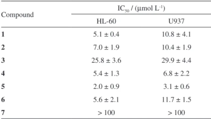
A investigação fitoquímica das partes aéreas de Centaurea omphalotricha levou ao isolamento de três lactonas sesquiterpênicas novas, 4’-acetilcinaropicrina, 4’-acetilcebelina F e 15-acetil desidromelitensina, juntamente com doze compostos conhecidos, sendo sete lactonas sesquiterpênicas, dois isoprenóides e três flavonóides. As estruturas dos novos compostos foram elucidadas por meio de RMN 1D e 2D, e espectrometria de massas, e por comparação com os dados descritos na literatura. O efeito de lactonas sesquiterpênicas sobre a viabilidade das células tumorais humanas, linhagens HL-60 e U937, também foi investigado e 3-acetilcinaropicrina e 4’-acetilcinaropicrina foram os compostos mais citotóxicos contra células de leucemia humana com valores de IC50 de 2,0 ± 0,9 e 5,1 ± 0,4 µmol L
-1, respectivamente.
Phytochemical research of the aerial parts of Centaurea omphalotricha led to the isolation of three new sesquiterpene lactones, 4’-acetyl cynaropicrin,4’-acetyl cebellin F and 15-acetyl dehydromelitensin, together with twelve known compounds, seven sesquiterpene lactones, two isoprenoids and three flavonoids. The structures of the new compounds were elucidated by means of extensive 1D and 2D NMR, and MS, and by comparison with reported data in the literature. The effect of sesquiterpene lactones on the viability of the human tumor cell lines HL-60 and U937 was also investigated and 3-acetyl cynaropicrin, and 4’-acetyl cynaropicrin were found to be the most cytotoxic compounds against human leukemia cells with an IC50 values of 2.0 ± 0.9 and 5.1 ± 0.4 µmol L-1, respectively.
Keywords:Centaurea, Asteraceae, sesquiterpene lactones, cytotoxic activity, HL-60
Introduction
The genus Centaurea (Asteraceae: Centaureinae) comprises more than 500 species, most of which grow around the Mediterranean and in Western Asia.1Centaurea
species have long been used for their biological properties, mainly as anti-inflammatory,2 antipyretic,3 cytotoxic,4
antibacterial,5 and antiproliferative agents.6 Phytochemical
investigations revealed that the compounds responsible for their pharmacological properties are flavonoids7 and
sesquiterpene lactones predominantly germacranolides,
eudesmanolides, elemanolides, and guaianolides.8 As a part
of our continuing search for novel, plant-derived anticancer chemotherapeutic agents and our systematic investigation of the composition of plants of this genus,4,9 we have
investigated the chemical constituents of the aerial parts of
Centaurea omphalotricha Coss. & Durieu ex Batt. et Trab., a species endemic to Algeria and Tunisia.10,11
and 15-acetyl dehydromelitensin (3), which are new in the literature, together with the seven sesquiterpene lactones: linichlorin B (4),12 3-acetyl cynaropicrin (5),13
8-(4-hydroxymethacrylate)-dehydromelitensin (6),14
15-acetyl melitensin (7),15 cynaropicrin (8),16
desacyl-cynaropicrin (9)16 and 8-hydroxy-11,13-dihydrozaluzanin
C (10);16 two isoprenoids: 3-hydroxy-5,6-epoxy-ionone,17
dehydrovomifoliol17 and three flavonoids: circimaritin,
apigenin and luteolin.18 The structures of the known
compounds were confirmed by comparison of their spectroscopic data (MS, 1H and 13C NMR) with literature
references. In this study we also demonstrate that the isolated sesquiterpene lactones 1-6 (Figure 1) induce cytotoxicity in human leukemia cell lines HL-60 and U937.
Experimental
General
Optical rotations were measured using a Perkin-Elmer model 343 polarimeter using CHCl3 as solvent. UV spectra
were recorded using a SHIMADZU model UV-1700 spectrophotometer. IR spectra were recorded as CHCl3
films on NaCl plates, using a Bruker model IFS-55 and Perkin-Elmer model FTIR-8400S spectrophotometer.
1H and 13C NMR spectra were obtained on a Bruker model
Avance 400 and AMX-500 spectrometer with standard pulse sequences, operating at 400 and 500 MHz for 1H and
100 and 125 MHz for 13C, respectively. CDCl
3 was used as
solvent and TMS as internal standard. EIMS were taken on a Micromass model Autospec (70 eV) spectrometer. HRESIMS was performed with a LCT Premier XE Micromass Waters spectrometer in positive ionization mode (Waters Corporation). Column chromatography (CC) was carried out with Si gel Fluka (cat. 60737) (40-63 µm), and column fractions were monitored by TLC (Si gel 60 F254,
0.2 mm, Macherey Nagel (cat. 818-333)) by detection with a spraying reagent (CH3COOH/H2O/H2SO4; 80:16:4)
followed by heating at 100 °C. Preparative TLC was carried out on Si gel 60 PF254 + 366 (20 × 20 cm, 1 mm thickness,
Analtech (cat. 02014)).
Plant material
Aerial parts of Centaurea omphalotricha (Coss. & Durieu ex Batt.) Willk. were collected from the Daya of Mogheul near Bechar in southwest Algeria (32.0192 N and 2.22 W) in April 2010 and identified by Professor M. Kabeche of University of Setif and M. Benabdelhakem from the National Agency of Preservation of Natural
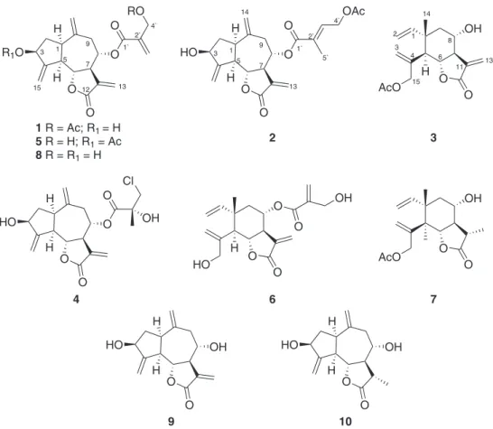
Figure 1. Structures of compounds 1-10.
O
R1O O
O RO
O
O
HO O
O
OAc
O
OH
O AcO
O
O
HO O
O
OH Cl
O
O
O HO
O O
OH OH
O AcO
O 1 R = Ac; R1 = H
5 R = H; R1 = Ac 8 R = R1 = H
2 3
4 6 7
1 3
5 7 9
13 1`
2` 4`
1 3
5 7 9
13 1`
2` 4`
5`
1
3
13 2
4
15 6
8 14
12
11
15
14
H
H
H
H H
H H
H
O
HO OH
O H
H
O
HO OH
O H
H
Resources of Bechar. A voucher specimen (COB N. 175-2010) has been deposited in the Herbarium of Constantine University.
Extraction and isolation
Air-dried aerial parts (2604 g) of C. omphalotricha
(Asteraceae) were powdered and macerated at room temperature with EtOH/H2O (80:20 v/v) for 48 h, three
times. After filtration, the filtrate was concentrated and suspended in H2O (800 mL). The residue was extracted
successively with CHCl3, EtOAc and n-butanol. The
organic phases were dried with Na2SO4, filtered using
common filter paper and concentrated in vacuum at 25 ºC to obtain the following extracts: CHCl3 (10.0 g), EtOAc (9.8 g)
and n-butanol (41.8 g). The CHCl3 extract was fractionated
by column chromatography (CC) (Si gel; petroleum ether/EtOAc with increasing polarity; 100 × 7 cm) to yield 25 fractions (1-25). Fraction 22 (petroleum ether/ EtOAc, 65:35 and 45:55; 1.3 g) was subjected to Si gel CC (CH2Cl2/MeOH; 100 × 1.5 cm) with increasing polarity to
give 8 subfractions. Subfraction 1 (CH2Cl2; 220 mg) was
purified by preparative TLC (n-hexane/Et2O, 2:4, three
elutions) to give dehydrovomifoliol (2.2 mg); subfraction 4 (CH2Cl2/MeOH, 96:4; 413.6 mg) was rechromatographed
on a Si gel column (CH2Cl2/acetone; 50 × 1.5 cm) with
increasing polarity to give 22 subfractions (sub1-sub 22). Sub 6 (CH2Cl2/acetone, 98:2; 8.2 mg) was purified by
preparative TLC (CH2Cl2/EtOAc/acetone; 50:10:4) to
afford compound 7 (4.2 mg); sub 9 (CH2Cl2/acetone, 96:4;
14.5 mg) was submitted to preparative TLC (Et2O/n-hexane,
4:1, three elutions) to give the new compound 3 (1.8 mg) and 5 (1.3 mg); sub 10 (CH2Cl2/acetone, 96:4; 16.9 mg)
gave after purification by preparative TLC (n-hexane/ Et2O, 1:5, four elutions) 3-hydroxy-5,6-epoxy-ionone
(3.8 mg); sub14 (CH2Cl2/acetone, 90:10; 16.3 mg) was
rechromatographed by preparative TLC (n-hexane/Et2O,
1:5, four elutions) to afford compound 4 (3 mg).
Fraction 23 (petroleum ether/EtOAc, 40:60; 720 mg) which was purified by Si gel TLC on preparative plates (CH2Cl2/MeOH/AcOH; 80:9:1, one elution) gave in
order of increasing polarity compound 9 (15.1 mg) and a mixture which was purified by TLC (CH2Cl2/MeOH/H2O,
10:1:0.1, one elution) yielding compound 8 (2.1 mg). Fraction 24 (petroleum ether/EtOAc 20:80; 1270 mg) was chromatographed on a Si gel CC (CH2Cl2/isopropanol;
100 × 1.5 cm) with increasing polarity to give 26 subfractions. Subfraction 8 (CH2Cl2/isopropanol, 96:4;
113.3 mg) was submitted to preparative TLC (n-hexane/ Et2O, 1:2 five elutions) to afford the new product 1
(1.3 mg) and a mixture of compounds which was purified
by preparative TLC (CH2Cl2/MeOH, 92:8, two elutions) to
yield the new product 2 (4 mg) and circimaritin (3.4 mg). Subfraction 9 (CH2Cl2/isopropanol, 96:4; 87.7 mg) was
submitted to preparative TLC (n-hexane/Et2O, 1:2, three
elutions) to yield 6 (4.6 mg). Subfraction 14 (CH2Cl2/
isopropanol, 96:4; 14 mg) was a pure compound 10. A part of the EtOAc extract (5.8 g) was chromatographed on a Si gel CC (CHCl3/acetone, 100 × 7 cm) with increasing
polarity to give 8 fractions. Fraction 8 (CHCl3/acetone,
50:50 to 100% acetone; 421.2 mg) was rechromatographed on a Si gel CC (CH2Cl2/EtOAc; 120 × 1.5 cm) with
increasing polarity to give 13 subfractions. Subfraction 5 (CH2Cl2/EtOAc, 80:20; 11 mg) afforded pure apigenin
(10.2 mg). Subfraction 7 (CH2Cl2/EtOAc, 75:25; 21.8 mg)
was rechromatographed by TLC using CH2Cl2/MeOH/H2O
(100:10:0.1) as mobile phase to yield luteolin (11.3 mg).
4’-Acetyl cynaropicrin (1)
Amorphous solid; [α]D25 = + 94 (c0.026, CHCl3); IR
(NaCl) νmax/cm
-1: 3478, 2939, 1755, 1740, 1729, 1642,
1450, 1373, 1267, 1152, 1048, 1031, 961; 1H NMR
(500 MHz, CDCl3) see Table 1;
13C NMR (125 MHz,
CDCl3) see Table 2; HRESIMS positive ion m/z 411.1428
[M + Na] + (Calc. for C
21H24O7Na, 411.1420).
4’-Acetyl cebellin F (2)
Amorphous solid; [α]D25 = + 81 (c0.018, CHCl3); IR
(NaCl) νmax/cm
-1: 3481, 2937, 1751, 1729, 1653, 1374,
1231, 1135, 1030, 910; 1H NMR (500 MHz, CDCl 3)
see Table 1; 13C NMR (CDCl
3, 125 MHz) see Table 2;
HRESIMS positive ion m/z 425.1578 [M + Na] + (Calc. for
C22H26O7Na, 425.1576).
15-Acetyl dehydromelitensin (3)
Amorphous solid; [α]D25 = + 60 (c0.009, CHCl3); IR
(NaCl) νmax/cm
-1: 3420, 2935, 1751, 1734, 1727, 1651,
1384, 1239, 1137, 1051, 910; 1H NMR (500 MHz, CDCl 3)
see Table 1; 13C NMR (125 MHz, CDCl
3) see Table 2;
HRESIMS positive ion m/z 329.1360 [M + Na] +, (Calc.
for C17H22O5Na, 329.1365).
Cell culture and cytotoxicity assays
The human leukemia HL-60 and U937 cells (DSMZ, German Collection of Microorganisms and Cell Cultures, Braunschweig, Germany) were grown in RPMI 1640 containing 2 mmol L-1 L-glutamine supplemented with
10% (v/v) heat-inactivated fetal bovine serum as previously described.19
Stock solutions of 25 mmol L-1 sesquiterpene lactones
dilutions were made in culture media just before use. In all experiments, the final concentration of DMSO did not exceed 0.4% (v/v), a concentration which is non toxic to the cells. The cytotoxicity of sesquiterpene lactones on human tumor cells was analyzed by colorimetric 3-[4,5-dimethylthiazol-2-yl-]2,5-diphenyl tetrazolium bromide (MTT) assay as previously described.20 Concentrations inducing a 50%
inhibition of cell growth (IC50) were determined graphically
using the curve fitting algorithm of the computer software Prism 4.0 (GraphPad). Values are means ± SE from at least three independent experiments, each performed in triplicate. The antitumor agent etoposide was used as a positive control in both HL-60 (IC50 = 0.4 ± 0.1 µmol L-1)
and U937 cells (IC50= 1.4 ± 0.3 µmol L -1).
Results and Discussion
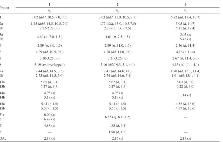
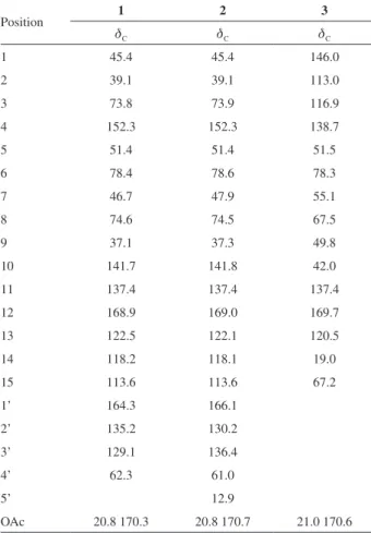
Compound 1 was obtained as an amorphous solid. HRESIMS experiments indicated the molecular formula C21H24O7 (calc. for [M + Na]
+ 411.1420; found 411.1428).
The IR spectra exhibited absorption bands for OH groups (3478 cm-1) and for carbonyl groups (1755 (α,β-unsaturated-
γ-lactone), 1740 and 1729 (ester carbonyls) cm-1). The
presence of these groups was confirmed by the 1H (Table 1)
and 13C NMR spectra (Table 2). The 1H NMR of 1 showed
the presence of eight olefinic methylene protons at dH 5.65
(d, 1H, J 3.1 Hz, H-13a), 6.27 (d, 1H, J 3.5 Hz , H-13b), 4.98 (s, 1H, H-14a), 5.19 (s, 1H, H-14b), 5.41 (t, 1H, J 1.5 Hz, H-15a), 5.53 (t, 1H, J 1.5 Hz, H-15b), 6.00 (s, 1H, H-3’a) and 6.49 (s, 1H, H-3’b), one acetyl group at dH 2.14 (s, 3H,
OAc), three oxygenated methines at dH 4.29 (dd, 1H, J 10.5,
9.0 Hz, H-6), 4.60 (tt, 1H, J 7.0, 1.5 Hz, H-3) and 5.19 (m, 1H, H-8) and one oxygenated methylene at dH 4.88 (s, 2H,
H2-4’).The connectivities were established by analysis of its
COSY spectrum. The 13C NMR (Table 2) and DEPT data
indicated the presence of one carbonyl group corresponding to a γ-lactone, two ester carbonyl groups, three aliphatic methylene, eight olefinic, one methyl (acetate), and six methine carbons. Both the 1H and 13C NMR spectral data of
compound 1 were close to those of cynaropicrin,16 with the
exception of an extra acetyl group in compound 1, which was assigned by a HMBC experiment. Thus, the correlation observed between the signal at dH 4.88 (s, 2H, H2-4’) and
dC 170.3 (OAc), allowed us to confirm the position of the
acetoxy group at the C-4’ position. Therefore, the structure of 1 was elucidated as 4’-acetyl cynaropicrin.
Compound 2 was obtained as an amorphous solid. The
1H NMR data (Table 1) of 2 were very similar to those
Table 1.1H NMR data of compounds 1-3 in CDCl
3 (d in ppm and J in Hz)
Proton 1 2 3
dH dH dH 1 3.02 (ddd, 10.5, 9.0, 7.5) 3.03 (ddd, 11.0, 10.5, 7.5) 5.82 (dd, 17.4, 10.7)
2a 2b
1.75 (ddd, 14.5, 10.5, 7.0) 2.22-2.27 (m)
1.77 (ddd, 13.0, 10.5,7.5) 2.28 (dt, 13.0, 7.5)
5.05 (d, 10.7) 5.11 (d, 17.4)
3a
3b 4.60 (tt, 7.0, 1.5 ) 4.61 (tt, 7.5, 1.5)
5.05 (s) 5.45 (s)
5 2.89 (tt, 9.0, 1.5) 2.89 (tt, 11.0, 1.5) 2.46 (d, 11.4)
6 4.29 (dd, 10.5, 9.0) 4.28 (dd, 11.0, 9.0) 4.16 (t, 11.4)
7 3.20-3.25 (m) 3.21-3.26 (m) 2.67 (tt, 11.4, 3.0)
8 5.19 (m, overlapped) 5.16 (ddd, 9.3, 5.1, 4.0) 4.15 (td, 11.4, 4.1)
9a 9b
2.44 (dd, 14.5, 3.5) 2.75 (dd, 14.5, 5.0)
2.41 (dd, 14.6, 4.0) 2.74 (dd, 14.6, 5.1)
1.70 (dd, 13.1, 11.4) 1.91 (dd, 13.1, 4.1)
13a 13b
5.65 (d, 3.1) 6.27 (d, 3.5)
5.62 (d, 3.1) 6.27 (d, 3.5)
6.03 (d, 3.0) 6.22 (d, 3.0)
14a 14b
4.98 (s) 5.19 (s)
4.98 (s)
5.19 (s) 1.14 (s)
15a 15b
5.41 (t, 1.5) 5.53 (t, 1.5)
5.41 (t, 1.5) 5.55 (t, 1.5)
4.52 (d, 13.6) 4.57 (d, 13.6)
3’a 3’b
6.00 (s)
6.49 (s) 6.85 (tq, 6.1, 1.2) —
4’ 4.88 (s) 4.83 (d, 6.1) —
5’ — 1.98 (d, 1.2) —
of 1, suggesting that both compounds are closely related in structure. The major variation is that compound 2 has a different acyl group. A detailed comparison of the 1H
(Table 1) and 13C NMR (Table 2) signals of 2 and 1 revealed
an extra carbon signal in 13C NMR, besides the presence
of a methyl group at dH 1.98 (d, 3H, J 1.2 Hz, H3-5’) and a
vinylic proton signal at dH 6.85 (tq, 1H, J 6.1, 1.2 Hz, H-3’)
in 1H NMR suggesting that the substituent group in 2 is the
4-acetoxy-2-methyl butenoyl. The relative stereochemistry of the double bond in the acyl group was determined as E
based on the observed correlation between the signals at
dH 4.83 and 1.98 in the ROESY spectrum. The combination
of all the above data and the HRESIMS experiment led us to assign the structure of 2 as 4’-acetyl cebellin F.
The HREIMS and 13C NMR data of 3 indicated the
molecular formula C17H22O5. The IR spectrum of this
compound showed the presence of hydroxyl groups (3420 cm-1), and carbonyl groups (1751, 1734, 1727 cm-1).
The 1H NMR (Table 1) spectrum of 3 exhibited the presence
of one methyl group at dH 1.14 (s, 3H, CH3-14), one acetyl
methyl group at 2.13 (s, 3H, OAc), one vinylic proton at 5.82 (dd, 1H, J 17.4, 10.7 Hz, H-1), and six olefinic
methylene protons at 5.05 (d, 1H, J 10.7 Hz, H-2a), 5.11 (d, 1H, J 17.4 Hz, H-2b), 5.05 (s, 1H, H-3a), 5.45 (s, 1H, H-3b), 6.03 (d, 1H, J 3.0 Hz, H-13a), 6.22 (d, 1H,
J 3.0 Hz, H-13b), typical of a 1,3,11(13)-elematrien-6,12-olide.21,22 The relationships between the proton signals in
3 were established from the 1H-1H COSY spectrum, which
disclosed the following connectivities: H-1 with H2-2, H-5
with H-6, H-13 with H-7, H-8 with H2-9. The
13C NMR
(Table 2) and DEPT spectral data indicated the presence of two carbonyl groups, corresponding to a γ-lactone, and an acetoxy group, six olefinic carbons, two methyl groups, two aliphatic methylene carbons, one of them oxygenated, four methine carbons, two of them oxygenated and one quaternary carbon. These assignments were similar to those of the known elemanolide dehydromelitensin.22 However,
a hydroxyl group was replaced by an acetoxy group, the location of this group in 3 being confirmed by the low shift position (+ 0.5 ppm) of the protons H2-15 at dH 4.52 (d,
1H, J 13.6 Hz, H-15a) and 4.57 (d, 1H, J 13.6 Hz, H-15b) in comparison with the 1H NMR dehydromelitensin data,22
characteristic of the presence of an acetyl group in a primary hydroxyl. The structure of compound 3 was assigned and confirmed using HMBC, HSQC and ROESY data as 15-acetyl dehydromelitensin.
Sesquiterpene lactones have attracted much attention during the last three decades, because they display a wide range of biological activities, including antitumor and anti-inflammatory properties.8,23 The structural requirement for
the biological activities of these compounds is associated with α-methylene-γ-butyrolactone moiety, which acts as alkylating agent in a Michael-type reaction with nucleophiles.24 Thus, sesquiterpene lactones are believed
to exert their numerous biological activities through inhibition of enzymes and other functional proteins by forming covalent bonds with free cysteine residues in these macromolecules or by conjugation with glutathione.25,26
Previous studies have shown that sesquiterpene lactones display cytotoxic properties in tumor cells8 and
that the sesquiterpene lactone of the guaianolide type cynaropicrin induces cytotoxicity in U937 and Jurkat T cell lines.27 A quantitative structure-activity relationships
(QSAR) study including four different skeletons of sesquiterpene lactones revealed the most active among the guaianolides and pseudoguainolides, and steric properties and electronic features as the most important descriptors.23
However, antiproliferative studies of the naturally occurring sesquiterpene lactones described in this paper in human leukemia cells have not yet been assessed.
Sesquiterpene lactones 1-6 were found to inhibit the growth and cell viability of HL-60 and U937 cells in culture as determined by the
3-[4,5-dimethylthiazol-2-yl]2,5-Table 2.13C NMR data of compounds 1-3 in CDCl
3 (d in ppm)
Position 1 2 3
dC dC dC
1 45.4 45.4 146.0
2 39.1 39.1 113.0
3 73.8 73.9 116.9
4 152.3 152.3 138.7
5 51.4 51.4 51.5
6 78.4 78.6 78.3
7 46.7 47.9 55.1
8 74.6 74.5 67.5
9 37.1 37.3 49.8
10 141.7 141.8 42.0
11 137.4 137.4 137.4
12 168.9 169.0 169.7
13 122.5 122.1 120.5
14 118.2 118.1 19.0
15 113.6 113.6 67.2
1’ 164.3 166.1
2’ 135.2 130.2
3’ 129.1 136.4
4’ 62.3 61.0
5’ 12.9
diphenyl tetrazolium bromide (MTT) dye-reduction assay (Table 3). In contrast, the sesquiterpene lactone 7 is not an effective antiproliferative agent showing an IC50 value
higher than 100 µmol L-1 in leukemia cells, in accordance
with the absence of the alkylating group, the exocyclic conjugated double bond.
Among the different sesquiterpene lactones, the presence of the exocyclic, conjugated double bond is essential for the cytotoxic activity against HL-60 and U937 cells. Compounds
1, 2, 4, 5 and 6 displayed similar potency in both cell lines. The potency of these sesquiterpene lactones might be explained by their lipophilicity. However, other factors, such as molecular geometry and the chemical environment of the target sulfhydryl may also influence the activity of sesquiterpene lactones. All these guaianolides contain an ester functional group at C-8. The sesquiterpene lactone 8-(4-hydroxymethacrylate)-dehydromelitensin 6 also contains an ester near from the exocyclic methylene bond. The presence of this group appears to be important, since compound 3 (15-acetyl dehydromelintensin) - which does not contain this functional group - was less cytotoxic than 6. In conclusion, the Algerian plant Centaurea omphalotricha
has been chemically studied for the first time, and three new sesquiterpene lactones have been identified along with twelve known compounds. The naturally occurring sesquiterpene lactones evaluated in the present study were strongly cytotoxic against human leukemia cell lines and the results of the present study may lead to the discovery of new and highly specific antitumor agents against leukemia cells.
Supplementary Information
Spectra of compounds 1-3 are available free of charge at http://jbcs.sbq.org.br as PDF file.
Table 3. Effects of compounds 1-7 on the growth of the human leukemia cell lines
Compound IC50 / (µmol L -1)
HL-60 U937
1 5.1 ± 0.4 10.8 ± 4.1
2 7.0 ± 1.9 10.4 ± 1.9
3 25.8 ± 3.6 29.9 ± 4.4
4 5.4 ± 1.3 6.8 ± 2.2
5 2.0 ± 0.9 3.1 ± 0.6
6 5.6 ± 2.1 11.7 ± 1.5
7 > 100 > 100
Cells were cultured for 72 h and the IC50 values were calculated as described in the Experimental section. The data shown represent the means ± SEM of 3-5 independent experiments with three determinations in each.
Acknowledgments
This work was supported in part by grants from the NATO Public Diplomacy Division (Science for Peace and Security Section) CBP.MD.CLG 983840, the Spanish Ministry of Science and Innovation and from the European Regional Development Fund (SAF2010-21380) and MAEC-Agencia Española de Cooperación y Desarrollo PCI (A1/035449/11). F. L. was supported by the JAE-DOC Program from the Spanish Ministry of Science and Innovation and the CSIC. S. E. thanks the Spanish Ministry of Education for a collaboration studentship. Thanks are also due to the Algerian Ministry of Higher Education and Scientific Research for financial support.
References
1. Susanna, A.; Garcia-Jacas, N. In Systematics, Evolution and Biogeography of Compositae; Funk, V. A.; Susanna, A.; Stuessy,
T. F.; Bayer, R. J, eds.; International Association for Plant Taxonomy: Vienna, Austria, 2009, pp. 293-313.
2. Koca, U.; Suntar, I. P.; Keles, H.; Yesilada, E.; Akkol, E. K.;
J. Ethnopharmacol.2009, 126, 551.
3. Akkol, E. K.; Arif, R.; Ergun, F.; Yesilada, E.; J. Ethnopharmacol.
2009, 122, 210.
4. Seghiri, R.; Boumaza, O.; Mekkiou, R.; Benayache, S.; Mosset, P.; Quintana, J.; Estévez, F.; León, F.; Bermejo, J.; Benayache, F.;
Phytochem. Lett.2009, 2, 114.
5. Buruk, K.; Sokmen, A.; Aydin, F.; Erturk, M.; Fitoterapia2006,
77, 388.
6. Chicca, A.; Tebano, M.; Adinolfi, B.; Ertugrul, K.; Flamini, G.; Nieri, P.; Eur. J. Med. Chem.2011, 46, 3066.
7. Flamini, G.; Antognoli, E.; Morelli, I.; Phytochemistry2001,
57, 559.
8. Akram Ghantous, A.; Gali-Muhtasib, H.; Vuorela, H.; Saliba, N. A.; Darwiche, N.; Drug Discovery Today2010, 15, 668. 9. Bentamene, A.; Benayache, S.; Creche, J.; Petit, G.;
Bermejo-Barrera, J.; León, F.; Benayache, F.; Biochem. Syst. Ecol.2005,
33, 1061.
10. Kadi-Hanifi, H.; Sécheresse2003, 3, 169.
11. Ozenda, P. In Flore et végétation du Sahara; 3rd ed.; Centre
National de la Recherche Scientifique: Paris, 2004.
12. González, A. G.; Bermejo, J.; Amaro, J. M.; Massanet, G. M.; Galindo, A.; Cabrera, I.; Can. J. Chem.1978, 56, 491.
13. Ha, T. J.; Yang, M. S.; Pak, Y.; Lee, J. R.; Lee, K. D.; Kim, H. M.; Park, K. H.; Heterocycles2002, 57, 151.
14. García, B.; Skaltsa, H.; Navarro, F. I.; Pedro, J. R.; Lazari, D.;
Phytochemistry1996, 41, 1113.
15. González, A.G.; Bermejo, J.; Toledo, F.; Daza, L. R.;
16. Choi, S. Z.; Choi, S. U.; Lee, K. R.; Arch. Pharm. Res.2005,
28, 1142.
17. Kim, I.; Chin, Y. W.; Lim, S. W.; Kim, Y. C.; Kim, J.; Arch. Pharm. Res.2004, 27, 600.
18. Youssef, D.; Frahm, A. W.; Planta Med.1995, 61, 570. 19. Torres, F.; Quintana, J.; Estévez, F.; Mol. Carcinog.2010, 49,
464.
20. Negrín, G.; Eiroa, J. L.; Morales, M.; Triana, J.; Quintana, J.; Estévez, F.; Mol. Carcinog.2010, 49, 488.
21. Karamenderes, C.; Bedir, E.; Pawar, R.; Baykan, S.; Khan, I. A.;
Phytochemistry2007, 68, 609.
22. Cardona, M. L.; García, B.; Pedro, J. R.; Sinisterra, J. F.;
Phytochemistry1989, 28, 1264.
23. Rodriguez, E.; Towers, G. H. N.; Mitchell, J. C.; Phytochemistry
1976, 15, 1573; Scotti, M. T.; Fernandez, M. B.; Ferreira, M.
J. P.; Emerenciano, V. P.; Bioorg. Med. Chem. 2007, 15, 2927.
24. Kupchan, S. M.; Fessler, D. C.; Eakin, M. A.; Giacobbe, T. J.;
Science1970, 168, 376.
25. Heilmann, J.; Wasescha, M. R.; Schmidt, T. J.; Bioorg. Med. Chem.2001, 9, 2189.
26. Garcia-Pineres, A. J.; Lindenmeyer, M. T.; Merfort, I.; Life Sci.
2004, 75, 841.
27. Zhang, S.; Wong, Y. K.; Ong, C. N.; Shen, H. M.; Curr. Med. Chem. Anti Canc. Agents2005, 5, 239; Cho, J. Y.; Kim, A. R.;
Jung, J. H.; Chun, T.; Rhee, M. H.; Yoo, E. S.; Eur. J. Pharmacol.
2004, 492, 85.
Submitted: November 15, 2011