Printed in Brazil - ©2006 Sociedade Brasileira de Química 0103 - 5053 $6.00+0.00
Article
*e-mail: [email protected]
Novel Anthraquinone Derivatives Produced by
Phoma sorghina
, an Endophyte Found
in Association with the Medicinal Plant
Tithonia diversifolia
(Asteraceae)
Warley de Souza Borges and Mônica Tallarico Pupo*
Faculdade de Ciências Farmacêuticas de Ribeirão Preto, Universidade de São Paulo, Avenida do Café s/n, 14040-903 Ribeirão Preto – SP, Brazil
Três antraquinonas conhecidas (1,7-diidroxi-3-metil-9,10-antraquinona, 1,6-diidroxi-3-metil-9,10-antraquinona e 1-hidroxi-3-metil-1,6-diidroxi-3-metil-9,10-antraquinona), uma nova antraquinona (1,7-diidroxi-3-hidroximetil-9,10-antraquinona), e dois novos derivados hexaidroantraquinônicos, dendrióis E e F, foram isolados da cultura do fungo endofítico Phoma sorghina, associado a Tithonia diversifolia (Asteraceae). Suas estruturas foram identificadas com base em dados espectroscópicos, principalmente RMN 1D e 2D.
Three known anthraquinones (1,7-dihydroxy-3-methyl-9,10-anthraquinone, 1,6-dihydroxy-3-methyl-9,10-anthraquinone and 1-hydroxy-1,6-dihydroxy-3-methyl-9,10-anthraquinone), one new anthraquinone (1,7-dihydroxy-3-hydroxymethyl-9,10-anthraquinone), and two new hexahydroanthraquinone derivatives, dendryols E and F, were isolated from the culture of the endophytic fungus Phoma sorghina, found in association with Tithonia diversifolia (Asteraceae). Their structures were identified on the basis of spectroscopic data, mainly 1D and 2D NMR.
Keywords: anthraquinone, dendryol, endophytic fungus, Phoma sorghina, Tithonia diversifolia
Introduction
Endophytes are considered outstanding and under explored sources of novel chemical diversity and bioactive compounds.1,2 These microorganisms can be detected at a
particular moment within the tissues of apparently healthy plant hosts,3 and they have been found in all plant species
examined to date.4 As they occupy unique biological niches,5,6
the complex web of interactions with other endophytes and with the host might give rise to new chemical diversity and bioactive compounds.1 In fact, this prolific biosynthetic
capability is illustrated by a number of new and/or bioactive metabolites isolated from endophytes.2,6
Most of the researches on the chemistry of endophytes have been done in the northern hemisphere. However, results have shown that tropical plants present greater diversity of endophytes species than those from temperate zones.3 In Brazil, research on endophytes has also led to
new and bioactive compounds.7-13
We have been interested in endophytes found in association with Tithonia diversifolia (Asteraceae), also known as Mexican sunflower. T. diversifolia fulfils the
rationale for plant selection with the aim to isolate endophytes,2 since extracts of this plant have been used
traditionally in the treatment of malaria, diarrhea, fever, hepatitis and wounds.14-16 Anti-inflammatory, amoebicidal,
antispasmodic, antifungal, antibacterial and antiviral activities have also been described for T. diversifolia
extracts.15,16 Moreover, there are no previous reports on the
isolation and cultivation of endophytes from T. diversifolia. In this work Phoma sorghina was isolated as an endophytic fungus from the leaves of T. diversifolia. After cultivation on solid rice medium, three known and three novel anthraquinone derivatives were isolated and identified.
Experimental
General experimental procedures
mode, using a MICROMASS QUATTRO-LC instrument equipped with an ESI/APCI “Z-spray” ion source. Semi preparative HPLC separations were carried out in a Shimadzu (LC-6AD apparatus, Japan) multisolvent delivery system, Shimadzu SPD-M10Avp Photodiode Array Detector, and an Intel Celeron computer for analytical system control, data collection and processing (software Class-VP), using VP 250/ 10 NUCLEOSIL 120-5 C18 or VP 250/10 NUCLEOSIL 100-5 C18 columns. 1H and 13C NMR spectroscopic
experiments were recorded on BRUKER DRX-400 and BRUKER DRX-500 spectrometers with CD3OD and CDCl3 as solvents and TMS as internal standard.
Microorganism
The general procedures adopted for isolation of the microorganism followed the methodology described by Kongsaeree et al.17 After collected, healthy leaves of Tithonia diversifolia were washed with water and surface sterilized by immersion in 70% aqueous ethanol (2 minutes), followed by 5% aqueous sodium hypochlorite (90 seconds), and finally with 70% aqueous ethanol (1 minute). After these procedures, the leaves were rinsed with sterilized water. This latter water was incubated in Petri dishes to guarantee the elimination of all epiphytic microorganisms. Small pieces of the leaves were excised and placed on agar in Petri dishes containing PDA medium at 30 ºC. Individual hyphal tips of the emerging fungi were removed and replaced on PDA.
The TD5 strainwas identified as Phoma sorghina by “Fundação Tropical de Pesquisa André Tosello”. A voucher specimen has been deposited at “Laboratório de Enzimologia Industrial”, FCFRP, USP. The strain is maintained by periodic transfers onto PDA18 at 4 ºC, and also in silica gel (6-12
mesh, grade 40, desiccant activated) at 10 ºC.
Rice culture of Phoma sorghina and isolation of the anthraquinones
P. sorghina was cultured on sterilized rice according to previously described procedures.10,13 Fifteen Erlenmeyer flasks
(2 L) containing rice (360 g per flask) (“Uncle Ben’s” – parboiled) and distilled water (300 mL per flask) were autoclaved twice at 121 ºC for 40 min. Four small disks of PDA medium from the Petri dish containing mycelium of
Phoma sorghina were transferred under sterile conditions to 14 of the 15 Erlenmeyer’s flasks containing sterilized rice. One flask was kept for control purpose. After 30 days of growth at 30 ºC, MeOH (600 mL) was added to each flask for 6 h. The solution was filtered and MeOH was removed under vacuum to give the MeOH extract as an orange residue (126.8
g). The MeOH extract was suspended in MeOH:H2O 1:3 and partitioned with CH2Cl2, EtOAc, and n-BuOH. The CH2Cl2 fraction was further partitioned with hexane and MeOH. The hexane sub fraction (585.2 mg) was submitted to vacuum liquid chromatography (VLC) over silica gel 60H using hexane:EtOAc gradient elution, affording 14 sub fractions. Sub fraction 7 (40.0 mg), obtained from hexane:EtOAc 1:1 elution, was further purified through 3 times elution on preparative TLC (hexane:EtOAc 9:1). Sub fraction 7.5 (Rf 0.74, 1.8 mg) was submitted to semi preparative reversed-phase HPLC (VP 250/10 NUCLEOSIL 120-5 C18, 70% MeCN in H2O, flow rate 3.0 mL min-1, 253 nm) to yield 3 (1.1
mg; Rt 19.8 min). The MeOH sub fraction (613.2 mg) was submitted to Sephadex LH-20 column using MeOH as mobile phase, affording 7 fractions. The sub fraction 4 (81.6 mg) was submitted to a silica gel column chromatography using CHCl3:MeOH 9:1, 1:1 and MeOH as mobile phase, affording 7 fractions. The sub fraction 4.4 (19.2 mg) was submitted to semi preparative reversed-phase HPLC (VP 250/10 NUCLEOSIL 120-5 C18, 50% MeOH in H2O, flow rate 3.0 mL min-1, 222 nm) to yield 5 (1.3 mg; R
t 16.5 min) and 6 (1.5
mg; Rt 17.8 min). After preparative TLC (MeOH:CHCl3 1:9), the sub fraction 6 (11.7 mg) yielded 5 fractions. The sub fraction 6.2 (Rf 0.20, 2.9 mg) was submitted to semi preparative reversed-phase HPLC (VP 250/10 NUCLEOSIL 100-5 C18, 70% MeOH in H2O, flow rate 3.0 mL min-1, 272 nm) to yield
4 (2.2 mg; Rt 11.1 min). The sub fraction 6.3 (Rf 0.40, 4.1 mg) was also subjected to semi preparative reversed-phase HPLC (VP 250/10 NUCLEOSIL 100-5 C18, 50% CH3CN in H2O, flow rate 3.0 mL min-1, 272 nm) to yield 1 (2.0 mg; R
t 26.7
min) and 2 (2.0 mg; Rt 29.7 min).
Compound 1 (1,7-dihydroxy-3-methyl-9,10-anthraquinone). Orange amorphous solid; UV (MeOH) λmax/nm: 215, 238, 258, 335, 354 and 441; IR (KBr) νmax/cm-1: 3440 (OH),
2924, 1677 (C=O), 1581, 1457, 1378, 1305, 1250 and 762. ESIMS m/z 253 [M-H]–; ESI-MS/MS (Daughter ions, 20
eV): m/z 252 ([M-2H]–,11%), 224 (100), 209 (34), 195
(21), 181 (49). 1H NMR: Table 1. 13C NMR: Table 3.
Compound 2 (Phomarin, 1,6-dihydroxy-3-methyl-9,10-anthraquinone). Orange amorphous solid; UV (MeOH)
λmax/nm: 215, 231, 251, 338, 356 and 441; IR (KBr) νmax/ cm-1: 3438 (OH), 2923, 1659 (C=O), 1634 (chelated C=O),
1595, 1475, 1366, 1276 and 779; ESIMS m/z 253 [M-H]–;
ESI-MS/MS (Daughter ions, 20 eV): m/z 252 ([M-2H]–,
14%), 224 (53), 209 (28), 195 (61), 181 (100). 1H NMR:
Table 1. 13C NMR: Table 3.
λmax/nm: 224, 238, 243, 258, 326 and 403; IR (KBr) νmax/ cm-1: 3440 (OH), 2924, 1677 (C=O), 1638 (chelated C=O),
1457, 1368, 1277, 1204 and 796. 1H NMR: Table 1. 13C
NMR: Table 3.
Compound 4 (1,7-dihydroxy-3-hydroxymethyl-9,10-anthraquinone). Orange amorphous solid; UV (MeOH) λmax/ nm: 204, 230, 250, 353, 415 and 423; IR (KBr) νmax/cm-1:
3442 (OH), 2923, 1638 (C=O), 1588, 1423, 1306, 1218, 1065 and 760; HRESIMS [M+ H]+ Found: 271.0606. Calc. for
C15H11O5: 271.0606. 1H NMR: Table 1. 13C NMR: Table 3.
Compound 5 (Dendryol E). Pale yellow amorphous solid; [α] 25D - 51.5 (MeOH, c 0.0013); UV (MeOH) λ
max/nm: 223,
248, 263, 278, 303 and 326; IR (KBr) νmax/cm-1: 3368
(OH), 2926, 1672 (C=O), 1333, 1047 and 673; HRESIMS [M+ H]+ Found: 263.1275. Calc. for C
15H19O4: 263.1283. 1H NMR: Table 2. 13C NMR: Table 3.
Compound 6 (Dendryol F). Pale yellow amorphous solid; [α]D25 - 64.7 (MeOH, c 0.0015); UV (MeOH) λ
max/nm: 223,
236, 263, 278, 308 and 340; IR (KBr) νmax/cm-1: 3361
(OH), 2929, 1679 (C=O), 1326, 1059 and 858; HRESIMS [M+ H]+ Found: 279.1221. Calc. for C
15H19O5: 279.1232. 1H NMR: Table 2. 13C NMR: Table 3.
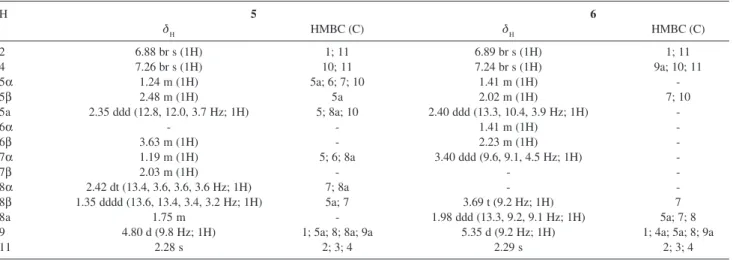
Table 2. 1H NMR and HMBC (H→C) data for dendryol E (5) and dendryol F (6) (500 MHz, CD 3OD)
H 5 6
δH HMBC (C) δH HMBC (C)
2 6.88 br s (1H) 1; 11 6.89 br s (1H) 1; 11
4 7.26 br s (1H) 10; 11 7.24 br s (1H) 9a; 10; 11
5α 1.24 m (1H) 5a; 6; 7; 10 1.41 m (1H)
-5β 2.48 m (1H) 5a 2.02 m (1H) 7; 10
5a 2.35 ddd (12.8, 12.0, 3.7 Hz; 1H) 5; 8a; 10 2.40 ddd (13.3, 10.4, 3.9 Hz; 1H)
-6α - - 1.41 m (1H)
-6β 3.63 m (1H) - 2.23 m (1H)
-7α 1.19 m (1H) 5; 6; 8a 3.40 ddd (9.6, 9.1, 4.5 Hz; 1H)
-7β 2.03 m (1H) - -
-8α 2.42 dt (13.4, 3.6, 3.6, 3.6 Hz; 1H) 7; 8a -
-8β 1.35 dddd (13.6, 13.4, 3.4, 3.2 Hz; 1H) 5a; 7 3.69 t (9.2 Hz; 1H) 7
8a 1.75 m - 1.98 ddd (13.3, 9.2, 9.1 Hz; 1H) 5a; 7; 8
9 4.80 d (9.8 Hz; 1H) 1; 5a; 8; 8a; 9a 5.35 d (9.2 Hz; 1H) 1; 4a; 5a; 8; 9a
11 2.28 s 2; 3; 4 2.29 s 2; 3; 4
All assignments were based on HMQC and HMBC experiments.
Table 1. 1H NMR and HMBC (H→C) data for anthraquinones 1-4
H 1a 2a 3b 4a
δH HMBC (C) δH HMBC (C) δH HMBC (C) δH HMBC (C)
2 7.05 d (1.4 Hz; 1H) 1; 4; 9a; 11 7.01 d (1.1 Hz; 1H) 1; 4; 11 7.67 dd (1.5, 0.5 Hz; 1H) 1; 4 ;9a; 11 7.21 d (0.6 Hz; 1H) 4; 9a; 11 4 7.53 d (1.4 Hz; 1H) 2; 9a; 10; 11 7.49 d (1.1 Hz; 1H) 2; 9a; 10; 11 7.12 dd (1.5, 0.8 Hz; 1H) 2; 9a; 10; 11 7.69 d (0.6 Hz; 1H) 2; 9a; 10; 11 5 8.05 d (8.5 Hz; 1H) 7; 8a; 10 7.39 d (2.4 Hz; 1H) 8a; 10 8.30 m (2H) - 8.02 d (8.6 Hz; 1H) 7; 8a; 10
6 7.19 dd (8.5, 1.6 Hz; 1H) 5a; 8 - - 7.81 m (2H) - 6.90 dd (8.6, 2.5 Hz; 1H) 5
7 - - 7.03 dd (8.5, 2.4 Hz; 1H) 5; 8a 7.81 m (2H) - -
-8 7.58 d(1.6 Hz; 1H) 5a; 9 8.05 d (8.5 Hz; 1H) 5a; 6; 9 8.30 m (2H) - 7.43 d (2.5 Hz; 1H) 5a; 6; 9
11 2.40 s (3H) 2; 3; 4 2.40 s (3H) 2; 3; 4 2.47 br s (3H) 2; 3; 4 4.68 s (2H) 2; 3; 4
1OH - - 8.55 s (1H) - 12.58 s (1H) 1; 2; 9a 8.54 s (1H)
-All assignments were based on the HMQC and HMBC experiments; aCD
3OD (500 MHz); bCDCl
3 (400 MHz).
Table 3.13C NMR data for compounds 1-6 (125 MHz)
C 1a 2a 3b 4a 5a 6a
1 163.2 163.7 163.0 - 158.1 163.6
2 123.4 124.9 124.3 121.1 123.1 123.1
3 149.4 148.7 148.8 153.3 140.5 140.3
4 120.7 121.1 121.0 117.9 119.5 119.0
4a 135.2 135.0 - - 133.6 132.5
5 131.0 115.4 127.5 131.6 35.9 31.6
5a 124.3 137.3 - 123.3 48.4 47.5
6 123.8 169.9 134.5 124.9 70.7 24.2
7 168.8 123.8 134.5 172.1 34.7 74.0
8 114.3 130.9 127.5 115.7 28.9 81.7
8a 136.4 124.4 - 137.1 47.3 50.5
9 190.2 188.4 - 190.6 73.8 74.1
9a 115.3 115.4 114.4 116.0 128.5 127.1
10 181.2 184.8 183.4 182.6 200.0 199.0
11 22.2 22.2 22.6 64.4 21.0 20.7
All assignments were based on the HMQC and HMBC experiments;
aCD 3OD;
bCDCl
Results and Discussion
The MeOH extract obtained from the cultivation of
Phoma sorghina, after chromatographic procedures, afforded three orange (1,2,4), one yellow (3) and two compounds (5-6) (Figure 1).
Compounds 1-3 exhibited typical UV, IR, NMR and MS data of hydroxyanthraquinones. Their physical data are in agreement with those previously reported for 1,7-dihydroxy-3-methyl-9,10-anthraquinone (1),19
1,6-dihydroxy-3-methyl-9,10-anthraquinone (phomarin,
2),20,21 and 1-hydroxy-3-methyl-9,10-anthraquinone
(pachybasin, 3).22 Although these anthraquinones have
already been isolated, only 3 has its 13C NMR data
published. For compounds 1 and 2 there are no previous
13C NMR data reported. HMQC and HMBC experiments
allowed us to assign the hydrogens and carbons for both compounds (Tables 1 and 3).
The molecular formula of compound 4 was established as C15H10O5 by HRESIMS, as well as 1H and 13C NMR
data. The IR spectrum of the compound 4 showed characteristic absorption bands from OH (broad, 3442 cm-1)
and α,β-unsaturated ketone (1638 cm-1). The 13C NMR
spectrum of 4 (Table 2) showed 13 carbon signals (two carbons were not observed): 2 carbonyls (δ 190.6 and δ
182.6), five quaternary sp2 carbons, five methine aromatic
carbons, and one sp3 methylene group. The 1H NMR
spectrum (Table 1) showed a singlet at δ 8.54, assigned to a hydroxyl group H-bonded to a carbonyl, and two meta -coupled aromatic hydrogens at δ 7.21 (d, J 0.6 Hz, H-2) and δ 7.69 (d, J 0.6 Hz, H-4), suggesting a 1,2,3,5-tetrasubstituted aromatic ring. The presence of a 1,2,4-trisubstituted aromatic ring was evident from the signals at δ 6.90 (dd, J 8.6 and 2.5 Hz, H-6), δ 7.43 (d, J 2.5 Hz, H-8) and δ 8.02 (d, J 8.6 Hz, H-5). The position of hydroxyl group at C-7 was unequivocally ascribed by HMBC correlations and splitting patterns of the 1H NMR
signals. Both hydrogens at δ 7.69 (H-4) and δ 8.02 (H-5) showed long range correlations with the carbon at δ 182.6,
establishing this ketone at C-10. H-5 was found to be orto
coupled to H-6, suggesting a substitution at C-7. The remaining ketone group was attributed to C-9, which presented long range correlation in the HMBC experiment with the hydrogen at δ 7.43 (d, J 2.5 Hz, H-8). The meta
coupling observed for H-8 is also only possible if the hydroxyl group is located at C-7. The typical signal for the methyl group attached to C-3 in anthraquinones was not observed. However, the cross peak in the HMQC between the hydrogens at δ 4.68 (s, 2H) and the carbon at
δ 64.4 suggested a hydroxymethyl group at C-3. The HMBC and HMQC data confirmed the location of the hydroxymethyl group at C-3 through the observed correlations amongst H-11 and C-2, C-3 and C-4.
The 1H and 13CNMR spectra of compounds 5 and 6
also showed the signals related to the aromatic ring bearing hydroxyl and methyl groups, typical of the anthraquinones (Tables 2 and 3). However, they did reveal only one signal of ketone carbonyl, and no signals of the additional aromatic ring. The 1H NMR spectra revealed signals at δ
1.19-2.48 and δ 3.40-5.35, suggesting the presence of hexahydroanthraquinone frameworks. Similar compounds were previously isolated from the pathogenic fungus
Dendryphiella sp., and were named dendryols.23
Compound 5 was obtained as a pale yellow amorphous solid. From its HRESIMS, as well as by the observed signals in the NMR spectra, it was possible to deduce its molecular formula as C15H18O4. The 1H
NMR, DEPT, HMBC and HMQC spectra revealed the presence of one methyl, three methylene, six methine, five quaternary carbons and three hydroxyl groups. COSY, HMBC and NOE experiments, and also J values, allowed us to unequivocally ascribe all the protons and carbons of the molecule, as well as the relative stereochemistries of the stereogenic carbons. Hydroxyl methine protons were observed at δ 3.63 (m) and δ 4.80 (d, J 9.8 Hz). In contrast with 9,10-anthraquinones
1-4, only one ketone carbonyl signal (δ 200.0) was observed in the HMBC experiment. The cross peak between the ketone carbon and the proton at δ 7.26 (br s, H-4), observed in the HMBC experiment, led us to locate the ketone at C-10. So, it was suggested that one hydroxyl group should be placed at C-9. Moreover, both deshielded chemical shifts for H-9 (δ 4.80) and C-9 (δ 73.8) suggested a benzylic position. Hydrogens H-9 (δ 4.80) and H-8a (δ 1.75) are coupled in a trans
relationship, as suggested by the J value of 9.8 Hz. The stereochemistry of an analogue structure, dendryol A, has been established through X-ray diffraction analysis.23 So, we assumed the same relative
con-figuration at C-9 for compound 5. In addition,
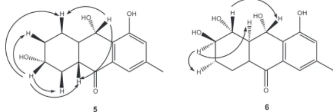
Figure 1. Anthraquinone derivatives produced by the endophytic fungus
irradiation of 9 resulted in NOE with 5a and H-8β, confirming an α-oriented hydroxyl group at C-9. The remaining hydroxyl methine hydrogen at δ 3.63 (H-6) was coupled to both methylene hydrogens at δ
1.19 (m) and δ 2.03 (m), as well as to additional methylene hydrogens at δ 1.24 (m) and δ 2.48 (m), as observed in the COSY experiment. COSY also showed the couplings of the methylene hydrogens at δ 1.35 (dddd) and δ 2.42 (dt) with both methylene hydrogens at δ 1.19 (m) and δ 2.03 (m) and with the methine at δ
1.75 (m). HMBC experiment revealed long range correlations between H-5α (δ 1.24) and the carbons at
δ 34.7 (C-7), 70.7 (C-6), 48.4 (C-5a) and 200.0 (C-10). These data allowed to locate the hydroxyl at C-6. Irradiation of H-6 (δ 3.63, m) showed NOE with four hydrogen signals at δ 1.35 (H-8β), 2.03 (H-7β), 2.35 (H-5a), and 2.48 (H-5β), which are only possible with H-6 in the axial configuration (Figure 2). Therefore, the hydroxyl group at C-6 should be α-oriented.
Compound 6 presented similar spectroscopic data compared to 5. The HRESIMS and NMR data suggested C15H18O5 as the molecular formula of compound 6. The additional hydroxyl group was evident from the MS data and also from the three hydroxyl methine protons at δ
5.35, 3.69 and 3.40, respectively attached to the carbons at δ 74.1, 81.7 and 74.0, as observed by the cross peaks in the HMQC experiment. These preliminary data also indicated a hexahydroanthraquinone framework for compound 6. The long range correlations of H-4 (δ 7.24, br s) with carbons at δ 20.7 and δ 199.0 led us to place the ketone group at C-10. Therefore, one hydroxyl group was also α-oriented at C-9. Proton H-9 (δ 5.35, d) was coupled to H-8a in trans stereochemistry, as suggested by the J value of 9.2 Hz. The HMBC experiment revealed a long range correlation between H-9 and the carbon at
δ 81.7, allowing us to locate another hydroxyl group at C-8. The splitting pattern and J values of H-8 signal (t,
J 9.2 Hz) suggested it should be axially oriented. Therefore, the hydroxyl group at C-8 had to be α -oriented. COSY experiment showed cross peaks of H-8 signal with protons at δ 1.98 (ddd, H-8a) and δ 3.40 (ddd), so the remaining hydroxyl group should be β
-oriented at C-7. In addition, NOE experiments are in agreement with the proposed stereochemistries. Irradiation of H-8 (δ 3.69) showed NOE with H-9 (δ
5.35), and irradiation of H-7 (δ 3.40) led to NOE on both H-8a (δ 1.98) and H-6α (δ 1.41) signals. Dendryol C was previously isolated from Dendryphiella sp.20 and
has the same structure proposed for compound 6. However, in dendryol C the 7-OH and 8-OH groups are
α- and β-oriented, respectively, as evidenced by the splitting patterns and J values of H-7 (δ 3.92, dt, J 3.2 and 2.8 Hz) and H-8 (δ 4.23, dd, J 2.8 and 3.2 Hz). Therefore, compound 6 and dendryol C were assumed as diastereomers.
Compounds 5 and 6 were named dendryol E and dendryol F, respectively, in analogy to the analogue structures of dendryols A-D, previously isolated from the phytopatogenic fungus Dendryphiella sp., and reported as phytotoxic against barnyardgras.20
Novel anthraquinone derivatives have recently been reported from the endophytic fungi Penicillium janthinellum11 and Pleospora sp.24 P. janthinellum
produced known antimicrobial anthraquinones and also a new modified anthraquinone, janthinone, containing a lactone between C-10 and C-4a.11 In addition,
deoxy-bostrycin, altersolanol B and dactylariol (1,2,3,4-tetrahydro-9,10-anthraquinones) and a new 1,2,3,4,4a,9a-hexahydro-9,10-anthraquinone (pleospdione) were isolated from Pleospora sp. Deoxybostrycin, altersolanol B and dactylariol exhibited significant cytotoxic activity against colon cancer and leukemia cell lines.24
The access to new biological diversity has often afforded new natural products.1 Few chemical
investigations were previously carried out only with pathogenic strains of Phoma sorghina, leading to the isolation of phytotoxins.25,26 In this work we described
the identification of compounds 4, 5 and 6 as novel anthraquinone derivatives. In addition, this is the first report of Phoma sorghina as an endophyte and its production of anthraquinones, although the production of anthraquinone derivatives by other Phoma species has already been reported.20,27
Acknowledgments
The authors are grateful to Fundação de Amparo à Pesquisa do Estado de São Paulo (FAPESP) for the financial support by the grants 03/07535-5 and 04/07935-6 (Bioprospecta-Biota-FAPESP). W.S.B. also thanks CAPES for the scholarship during his doctoral work. We also thank Dr. Norberto P. Lopes (FCFRP-USP) for the obtainment of high and low resolution mass spectra.
Supplementary Information
Spectra of compounds 1-6 are available free of charge at http://jbcs.sbq.org.br, as PDF file.
References
1. Clardy, J.; Walsh, C.; Nature2004,432, 829.
2. Strobel, G.; Daisy, B.; Castillo, U.; Harper, J.; J. Nat. Prod.
2004, 67, 257.
3. Schulz, B.; Boyle, C.; Mycol. Res.2005, 109, 661.
4. Arnold, A. E.; Maynard, Z.; Gilbert, G. S.; Coley, P. D.; Kursar, T. A.; Ecol. Lett.2000, 3, 267.
5. Schulz, B.; Boyle, C.; Draeger, S.; Rommert, A. K.; Krohn, K.;
Mycol. Res.2002, 106, 996.
6. Tan, R. X.; Zou, W. X.; Nat. Prod. Rep.2001, 18, 448. 7. Silva, G. H.; Teles, H. L.; Trevisan, H. C.; Bolzani, V. S.; Young,
M. C. M.; Pfenning, L. H.; Eberlin, M. N.; Haddad, R.; Costa-Neto, C. M.; Araujo, A. R.; J. Braz. Chem. Soc.2005, 16, 1463. 8. Teles, H. L.; Silva, G. H.; Castro-Gamboa, I.; Bolzani, V. S.; Pereira, J. O.; Costa-Neto, C. M.; Haddad, R.; Eberlin, M. N.; Young, M. C. M.; Araujo, A. R.; Phytochemistry2005, 66, 2363. 9. Cafeu, M. C.; Silva, G. H.; Teles, H. L.; Bolzani, V. S.; Araujo, A. R.; Young, M. C. M.; Pfenning, L. H.; Quim. Nova2005,
28, 991.
10. Santos, R. M. G.; Rodrigues-Fo, E.; J. Braz. Chem. Soc. 2003,
14, 722.
11. Marinho, A. M. R.; Rodrigues-Fo, E.; Moitinho, M. L. R.; Santos, L. S.; J. Braz. Chem. Soc.2005, 16, 280.
12. Barros, F. A. P.; Rodrigues-Fo, E.; Biochem. Syst. Ecol. 2005,
33,257.
13. Santos, R. M. G.; Rodrigues-Fo, E.; Phytochemistry 2002, 61, 907.
14. Gu, J. Q.; Gills, J. J.; Park, E. J.; Greenwood, E. M.; Hawthorne, M. E.; Axelrod, F.; Chavez, P. I.; Fong, H. H. S.; Mehta, R. G.; Pezzuto, J. M.; Kinghorn, A. D.; J. Nat. Prod. 2002, 65, 532. 15. Goffin, E.; Ziemons, E.; De Mol, P.; Madureira, M. C.; Martins,
A. P.; Cunha, A. P.; Philippe, G.; Tits, M.; Angenot, L.; Frederich, M.; Planta Med.2002, 68, 543.
16. Cos, P.; Hermans, N.; De Bruyne, T.; Apers, S.; Sindambiwe, J. B.; Vanden Berghe, D.; Pieters, L.; Vlietinck, A. J.; J. Ethnopharmacol. 2002, 79, 155.
17. Kongsaeree, P.; Prabpai, S.; Sriubolmas, N.; Vongvein, C.; Wiyakrutta, S.; J. Nat. Prod.2003, 66, 709.
18. Nam, J.; Kim, H.; Kwon, J.; Han, M. Y.; Son, K.; Lee, U. C.; Choi, J.; Kwon, B.; J. Nat. Prod. 2000, 63, 1303.
19. Imre, S.; Phytochemistry1969, 8, 315.
20. Bick, I. R. C.; Rhee, C.; Biochem. J.1966, 98, 112.
21. Arrebola, M. T.; Ringbom, T.; Verpoorte, R.; Phytochemistry
1999, 52, 1283.
22. Semmelhack, M. F.; Ho, S.; Cohen, D.; Steigerwald, M.; Lee, M. C.; Lee, G.; Gilbert, A. M.; Wulff, W. D.; Ball, R. G.; J. Am. Chem. Soc.1994, 116, 7108.
23. Tanaka, M.; Ohra, J.; Tsujino, Y.; Fujimori, T.; Ago, H.; Tsuge, H.; Z. Naturforsch. (C) 1995, 50, 751.
24. Ge, H. M.; Song, Y. C.; Shan, C. Y.; Ye, Y. H.; Tan, R. X.; Planta Med. 2005, 71, 1063.
25. Steyn, P. S.; Rabie, C. J.; Phytochemistry1976, 15, 1977. 26. Venkatasubbaiah, P.; Vandyke, C. G.; Chilton, W. S.; Mycologia
1992, 84, 715.
27. Parisi, A.; Piatelli, M.; Tringali, C.; Lio, G. M. D. S.;
Phytochemistry1993, 32, 865.
Received: February 3, 2006
Published on the web: June 29, 2006