Centric relation registration: intra- and interexaminer agreement
after a calibration program
Avaliação da reprodutibilidade intra e interexaminador no registro
da relação cêntrica, após um programa de calibração
Evelyn Mikaela Kogawa* Luis Fernando Risso Lopes** Melissa Thiemi Kato** Fernando Tsuyoshi Ueno** Carlos Neanes Santos*** José Roberto Pereira Lauris**** Paulo César Rodrigues Conti*****
ABSTRACT:Centric relation (CR) has been considered a maxillomandibular position of choice for some dental and prosthetic procedures. Although regarded as a fully reproducible relation, there is great controversy about its clinical use and recording technique, especially in patients with signs and symptoms of temporomandibular disorders (TMD). This study aimed at evaluating the effectiveness of a training program on intra- and interexaminer agreement when determining the clinical record of the CR position. Forty individuals constituted the sample, divided into symptomatic (TMD) and asymptomatic groups. Three previously calibrated examiners performed the initial assessment and the sec-ond evaluation after 30 days in a blind design, using Dawson’s bilateral manipulation technique with and without an anterior stop. The amount of frontal and sagittal deviations in relation to intercuspal position, the presence of pain and discomfort during manipulation, and the first occlusal contact in CR were analyzed. Kendall and Kappa tests with a 5% level of significance were used to determine agreement. Values for both intra- and interexaminer agreement were deemed good. The best results were obtained for frontal deviations and assessment of pain (or absence of it) during ma-nipulation. Sagittal deviations showed the lowest agreement in both examinations. The authors concluded that a cali-bration program could be effective for intra- and interexaminer agreement when recording centric relation. However, caution is recommended when analyzing some isolated items.
DESCRIPTORS:Centric relation; Temporomandibular joint disorders.
RESUMO:Na tentativa de estabelecer uma posição maxilomandibular reprodutível em pacientes sem dentes suportes posteriores ou portadores de oclusão instável, o conceito de relação cêntrica (RC) foi introduzido. Apesar de ser utiliza-da como uma posição de referência, existe uma considerável divergência de opiniões sobre a sua reprodutibiliutiliza-dade. Este estudo procurou avaliar se os métodos de treinamento profissional são efetivos na análise e obtenção de RC. Para isso utilizou-se uma amostra de 40 indivíduos, divididos em 20 assintomáticos e 20 portadores de disfunções da ATM. Os indivíduos foram avaliados por 3 examinadores, previamente calibrados. Foram realizados 2 exames: inicial e final (30 dias após o inicial), possibilitando dessa forma, análise da concordância interexaminadores, assim como intra-examinadores. Posteriormente, os resultados foram submetidos à análise estatística, utilizando-se os testes de con-cordância de Kendall e de Kappa. Os resultados foram considerados bons, sendo os melhores obtidos para a análise do desvio frontal e relato de dor (ou ausência) durante a manipulação. Os autores concluíram que os programas de cali-bração podem ser efetivos para análise da RC. No entanto, cautela é recomendada quando da análise de alguns itens isolados.
DESCRITORES:Relação central; Transtornos da articulação temporomandibular.
INTRODUCTION
The concept of centric relation (CR) was intro-duced in dentistry with a view to reproduce the mandibular position during the fabrication of den-tures, therefore providing conditions for complete
*Graduate Student, MSc level, Oral Rehabilitation Area; **DDS; ***Graduate Student, PhD level, Oral Rehabilitation Area; ****PhD, Professor, Department of Public Health; *****PhD, Professor, Department of Prosthodontics – School of Dentistry of Ba-uru, University of São Paulo.
dentures to develop all their functions in harmony with the other components of the stomatognathic system.
One of the first reports on CR was made by Gysi8, in 1910, who presented the gothic arch
initia-ting a new phase in dentistry. After this study, dif-ferent concepts have appeared about this occlusal relationship, which were altered according to opi-nions from that time period. The current concept of CR most accepted by the scientific community was provided by the 7th
edition of the “Glossary of Prosthetic Terms” (1999), as follows: “the maxillo-mandibular relationship in which the condyles ar-ticulate with the medial portion of their respective disks, being this complex (disk-condyle) in an an-tero-superior position against the surface of the articular eminence”. When employed for prosthetic reconstructions, this is a suitable position because of its reproducibility4
. One review of the reproduc-tion techniques reveals that most of them (inclu-ding the bilateral manipulation, the chin point gui-dance, the Lucia jig or the laminated calibrator) are able to achieve a consistent condylar position in nearly all patients. Yet, some studies5,10
have de-monstrated that the precision of most of these techniques is severely limited in the presence of temporomandibular disorders.
Several studies addressing the reproducibility of CR have related small variations in the condylar position between several CR recordings15,16,21,22
. Despite of its reproducibility, there is a millimetric variation, i.e. an accurate achievement of CR re-cording is not always feasible12,13,23.
Some evidence suggests that the CR position may vary with time and with the different record-ing methods. Moreover, several factors may influ-ence its registration, such as emotional stress, TMJ and facial muscle pain, neuromuscular con-ditioning, manipulation technique or operator’s guidance2. On this basis, the need to evaluate the
efficacy of a calibration procedure for CR analysis is highlighted.
The present study aimed at evaluating the in-tra- and interexaminer agreement in the analysis of CR position, comparing these findings in asymptomatic individuals and patients with TMJ internal derangements.
MATERIAL AND METHODS
The present study comprised a sample of 40 in-dividuals, divided into 2 groups (asymptomatic and symptomatic patients). Group I (asymptoma-tic) included 20 individuals equally distributed between genders, which were randomly selected from the dental clinics, School of Dentistry of
Bau-ru (FOB/USP). These patients presented no signs and symptoms of TMD.
Group II (symptomatic) consisted of 20 indivi-duals equally distributed between genders presen-ting with signs and symptoms of arthrogenic TMD. All these patients were randomly selected from the individuals attending the TMD and Orofacial Pain Center, Department of Prosthodontics (FOB/USP). Inclusion criteria for this group was accomplished after anamnesis and detailed physical examinati-on, comprising muscle and TMJ palpatiexaminati-on, evalua-tion of the mandibular movement and inspecevalua-tion of joint sounds.
The individuals received information on the ob-jectives of the research, and, after all procedures had been fully explained, they signed an informed consent term, in agreement with Regulation #196/96 of the Brazilian National Health Council.
The examiners were trained to perform the ma-nipulation technique and the CR recording met-hod. For that purpose, the research coordinator demonstrated the technique and subsequently the three examiners carried out the same procedure in four dental students, simulating the study evalua-tion.
The bilateral manipulation technique suggested by Dawson was selected, with or without an anteri-or stop.
Manipulation of the patients was initially per-formed with no anterior stop. During manipula-tion, the examiners identified the first centric den-tal contact, by means of an articulating paper (AccuFilm II, USA). Vertical and sagittal deviations were also recorded in a standardized form, as well as the report of pain or discomfort upon manipula-tion. Afterwards, a cotton roll was placed between the incisors for 5 minutes to act as a stop, in order to eliminate occlusal contact and mechanorecepti-on of the periodmechanorecepti-ontal ligament. After this period, the same procedures were repeated in an attempt to verify the influence of the stop on the CR recor-ding.
The groups of 10 patients evaluated in each ses-sion always comprised 5 asymptomatic and 5 symptomatic individuals, who were randomly eva-luated. Yet, examiners were blinded to group dis-tribution.
both. The Kappa (K) and Kendall (W) values are in-terpreted as follows: from 0 to 0.20 - poor agree-ment; from 0.21 to 0.40 - regular; from 0.41 to 0.60 - moderate; from 0.61 to 0.80 - satisfactory; and from 0.81 to 1.00 - excellent.
RESULTS
Tables 1 and 2 demonstrate the results of inte-rexaminer agreement of the 1st
and 2nd
evaluations for the different study variables, according to the Kendall test.
DISCUSSION
The literature on interexaminer agreement for the clinical evaluation of CR reproducibility is qui-te large. Previous studies3,6,7,24have focused on the
interexaminer agreement for evaluation of caries, signs and symptoms of periodontal disease and ra-diographic examinations. All these studies have employed relatively objective data, such as pocket probing depth, bone loss and presence or absence of caries, whereas standardization of CR manipu-lation is based on less objective data.
TABLE 1 -Agreement value (W) for the study variables between the different examiners (interexaminer analysis), with no distinction of group (symptomatic or asymptomatic) – 1stand 2ndevaluation (Kendall test).
Evaluated item 1
stevaluation 2ndevaluation
W p W p
Orthodontic class 0.69 < 0.001 0.60 = 0.001
Frontal deviation (WOAS) 0.73 < 0.001 0.70 < 0.001
Sagittal deviation (WOAS) 0.50 = 0.027 0.62 = 0.001
Report of pain during manipulation (WOAS) 0.87 < 0.001 0.78 < 0.001
1st
contact (WOAS) 0.72 < 0.001 0.74 < 0.001
Frontal deviation (WAS) 0.63 = 0.001 0.73 < 0.001
Sagittal deviation (WAS) 0.53 = 0.013 0.66 < 0.001
Report of pain during manipulation (WAS) 0.88 < 0.001 0.90 < 0.001
1stcontact (WAS) 0.66 < 0.001 0.83 < 0.001
WOAS: without anterior stop; WAS: with anterior stop.
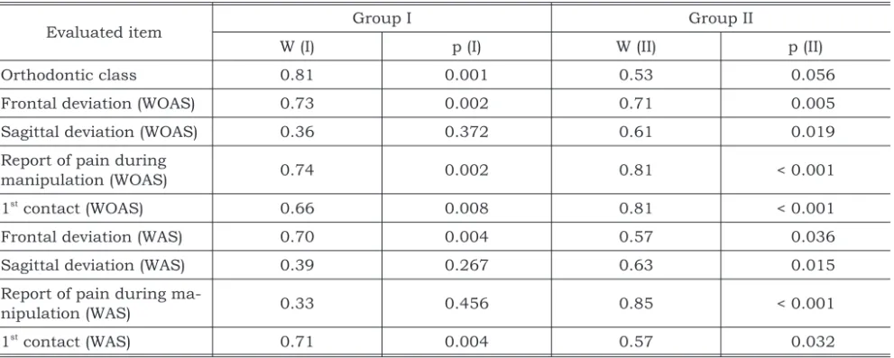
TABLE 2 -Agreement value (W) for the study variables between the different examiners (interexaminer analysis), with distinction between symptomatic (I) and asymptomatic (II) groups.
Evaluated item Group I Group II
W (I) p (I) W (II) p (II)
Orthodontic class 0.81 0.001 0.53 0.056
Frontal deviation (WOAS) 0.73 0.002 0.71 0.005
Sagittal deviation (WOAS) 0.36 0.372 0.61 0.019
Report of pain during
manipulation (WOAS) 0.74 0.002 0.81 < 0.001
1st
contact (WOAS) 0.66 0.008 0.81 < 0.001
Frontal deviation (WAS) 0.70 0.004 0.57 0.036
Sagittal deviation (WAS) 0.39 0.267 0.63 0.015
Report of pain during
ma-nipulation (WAS) 0.33 0.456 0.85 < 0.001
1st
contact (WAS) 0.71 0.004 0.57 0.032
The literature unanimously states that stan-dardizing the examination is paramount to assure reliability of the information obtained.
The interexaminer evaluations performed in the present study demonstrated a satisfactory agree-ment for most study variables: 0.645 (orthodontic class), 0.715 (frontal deviation without anterior stop - FDWOAS), 0.730 (contact WOAS), 0.68 (frontal deviation with anterior stop - FDWAS), 0.745 (contact WAS). Results were excellent for the items report of pain WOAS (0.825) and report of pain WAS (0.890), therefore demonstrating the im-portance of calibration procedures before examin-ers initiate the evaluations.
The statistical outcomes in Table 1 (1st and 2nd
interexaminer evaluations) revealed that the sagittal deviations without anterior stop (SDWOAS) and sagittal deviation with anterior stop (SDWAS) demonstrated the lowest agreement levels (0.50 and 0.53 at the 1stevaluation, and 0.62
and 0.66 at the 2nd evaluation, respectively). A
small percentage of individuals presented coinci-dent CR and intercuspal (IC) positions1,17
. A much higher percentage of individuals have a difference of 0.1 to 1.5 mm between CR and IC. The discrep-ancy between both positions, commonly named centric slide or centric discrepancy, may occur in all three planes of the space and is estimated to be 0.1 to 1.5 mm in the vertical direction, 0.1 to 1.0 mm in the horizontal direction and smaller than 1.0 mm in the transverse direction. This small variability of sagittal displacement observed in the literature, combined to the difficulty experi-enced by examiners upon measuring, has influ-enced the agreement levels. The difficulty to evalu-ate the discrepancy is transmitted to the judgment form itself, in which differences below 0.5 mm may yield disagreement between examiners (IC = CR; up to 1.0 mm; and higher than 1.0 mm)17
.
In the present study, the report of possible pain during manipulation in symptomatic patients demonstrated an optimal agreement, possibly due to the fact that most patients in this group had a complaint of pain during manipulation, thus sim-plifying analysis of this item by the examiners and therefore increasing the agreement level. Accord-ing to Harper, Schineidermen9(1996), the
determi-nation of the condylar hinge axis with the condyle in CR was more reproducible in patients with TMJ internal derangement than in asymptomatic pa-tients. This might be the outcome of the anatomic obstruction of a displaced disk without reduction
or the presence of adherences of TMJ, limiting the condylar position during the translation phase. The present study, however, did not evaluate disc displacement without reduction patients.
Regarding the manipulation technique sug-gested by Dawson5(1996), employed in the present
study, there are three possible reasons for the oc-currence of pain in patients when firm pressure is applied: bad positioning of the condyle, improper alignment between the condyle and its disk, and joint pathology.
Many other factors influencing the CR record-ing are also observed in the literature2
, including physical or emotional stress, pain affecting the TMJ and other components of the masticatory sys-tem, neuromuscular conditioning, manipulation or guidance of the operator, soft tissue alterations, different examiners and different recording meth-ods.
Some evidence suggests that the CR position may vary with time and the different periods of the day. Latta11
(1992) reported that recordings in edentulous patients demonstrated differences in the condylar position throughout the day as high as 2.63 mm. Shafagh et al.20
(1975) reported that different outcomes were observed when the CR re-cordings were performed in dentate patients at day and at night, probably due to the daily variation of shape and synovial fluid.
During CR recording in this study, after place-ment of the anterior stop, the examiners reported an easier manipulation when compared to the re-cording without anterior stop, although no signifi-cant difference was detected. According to Mezzomo, Frasca14
(1996), depending on the inten-sity of pain and the degree of muscle hyperactivity, manipulation of the mandible aiming at reaching CR is difficult at first. Thus, allowing the patient to rest for 10 to 15 minutes with no dental contact may be helpful for neuromuscular deprogrammi-ng. As previously mentioned, this procedure may increase agreement, yet it did not yield any signifi-cant differences in the present study.
It is important to distinguish between the two types of agreement evaluation: one refers to the re-liability of each examiner when performing the same task different times (intraexaminer), while the other indicates whether this same reliability also exists between examiners when observing the same variable (interexaminer).
agreement levels were not very similar. Yet, this study did not aim to discuss the validity of the clin-ical employment of CR as a therapy for TMJ pathosis. It is known that TMJ internal derange-ments may cause the joint structure to become more sensitive to alterations in condylar position. The items sagittal deviation (with or without an-terior stop) presented the same agreement levels, maybe because of the more difficult observation, since this analysis was performed through lateral visualization of the posteroanterior slide of the mandible when assuming the intercuspation posi-tion. This smaller agreement points toward the need to be careful when this item is regarded alone as the parameter for comparison between groups. Except for the items sagittal deviation with and without anterior stop, especially in asymptomatic patients, and for the possible report of pain during manipulation, no detectable statistical differences were found between groups I (asymptomatic) and II (symptomatic).
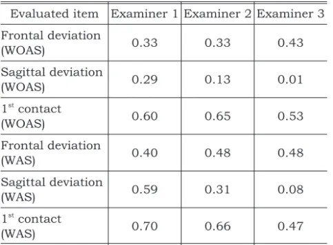
As regards the intraexaminer evaluation (Ta-ble 3), the agreement levels were generally smaller than the interexaminer values, suggesting the pos-sibility that the time period of one month between the first and second evaluations may have affected the accuracy of the manipulation technique and observation of the study items for all three
examin-ers, which is in agreement with previous stud-ies18,19
.
In spite of the relatively poor agreement, the frontal deviation revealed an even intraexaminer agreement for the three examiners. This noticeably lower level of agreement for the item sagittal devia-tion for both intra- and interexaminer evaluadevia-tions possibly demonstrates the more difficult observa-tion of such item by the examiners. Thus, it may be stated that the calibration program was effective for the achievement of agreement between examin-ers. Yet, after one month, these values were re-duced, even though still maintaining acceptable levels. This difference may probably have occurred due to natural alterations affecting the joint struc-tures, related to the synovial fluid, disk shape and muscular condition. This also leads us to question the adoption of CR as a rigid position, absolutely required for stomatognathic health. The difficulty to judge some important items and the report of pain in patients with TMD may suggest that this position might just be an initial guide for extensive prosthetic and occlusal procedures, yet being highly susceptible to individual variation.
CONCLUSIONS
Considering the results obtained in the present study, it can be concluded that:
1. The training and calibration programs demon-strated to be efficient for the achievement of interexaminer agreement in CR recording. 2. The main difficulty experienced by the
examin-ers was related to sagittal deviation, which con-sequently demonstrated the lowest agreement values.
3. Time and the physiological variation of the stomatognathic system led to a lower level of intraexaminer agreement.
4. Patients presenting with temporomandibular dysfunctions do not present differences in the reproducibility of CR position when compared to normal patients.
ACKNOWLEDGEMENTS
To the Fundação de Amparo à Pesquisa do Estado de São Paulo (FAPESP), for the financial support provided for the development of this study (grant #00/14881-9).
TABLE 3 -Agreement value (W) for the study variables within the same examiner during the 1stand 2nd
evalua-tions (intraexaminer evaluation).
Evaluated item Examiner 1 Examiner 2 Examiner 3
Frontal deviation
(WOAS) 0.33 0.33 0.43
Sagittal deviation
(WOAS) 0.29 0.13 0.01
1st
contact
(WOAS) 0.60 0.65 0.53
Frontal deviation
(WAS) 0.40 0.48 0.48
Sagittal deviation
(WAS) 0.59 0.31 0.08
1st
contact
(WAS) 0.70 0.66 0.47
REFERENCES
1. Beyron H. Optimal occlusion. Dent Clin North Am 1969; 13:537-54.
2. Calagna LJ, Silverman SI, Garfinkel L. Influence of neuromuscular conditioning of centric relation registra-tions. J Prosthet Dent 1973;30:598-606.
3. Carlsson GE, Egermark-Eriksson I, Magnusson T. Intra and inter-observer variation in functional examination of the masticatory system. Swed Dent J 1980;4:187-94. 4. Celenza FV. The theory and clinical management of centric
positions: II centric relation and centric relation occlusion. Int J Periodontics Restorative Dent 1984;4:63-86. 5. Dawson PE. A classification system for occlusion that
re-lates maximal intercuspation to the position and condition of the temporomandibular joints. J Prosthet Dent 1996; 75:60-6.
6. Fleiss JL, Chilton NW. The measurement of interexaminer agreement in periodontal disease. J Periodontal Res 1983; 18:601-6.
7. Fleiss JL, Slakter MJ, Fischman SL, Park MH, Chilton NW. Interexaminer reliability in caries trials. J Dent Res 1979;58(2):604-9.
8. Gysi A. The problem of articulation. Dental Cosmos 1910; 52:1-19.
9. Harper RP, Schneiderman E. Condylar movement and centric relation in patients with derangement of the temporomandibular joint. J Prosthet Dent 1996;75:67-71. 10. Kantor ME, Silverman SI, Garfinkel L. Centric relation
re-cording techniques - a comparative investigation. J Prosthet Dent 1972;28(6):593-600.
11. Latta GH Jr. Influence of circadian periodicity on repro-ducibility of centric relation records for edentulous pa-tients. J Prosthet Dent 1992;68:780-3.
12. McKee JR. Comparing condylar position repeatibility for standardized versus nonstandardized methods of achiev-ing centric relation. J Prosthet Dent 1997;77:280-4.
13. Mcneill C. Ciência e Prática da Oclusão. Estados Unidos: Quintessence; 2000. p. 407-42.
14. Mezzomo E, Frasca LCF. Dor na ATM – o que fazer? In: Todescan FF, Bottino MA Atualização na clínica odon-tológica: a prática da clínica geral. São Paulo: APCD, Artes Médicas; 1996. Cap.14. p. 357-82.
15. Piehslinger E, Celar A, Celar R, Jäger W, Slavicek R. Reproducibility of the condylar reference position. J Orofac Pain 1993;7:68-75.
16. Posselt V. Studies on the mobility of the human mandi-bule. Acta Odontol Scand 1952;10:1-160.
17. Rieder CE. The prevalence and magnitude of mandibular displacement in a survey population. J Prosthet Dent 1978;39:324-9.
18. Schubert R. Zur Frage der Reproduzierbarkeit der terminalen Scharnierachsenposition. Dtsch Zahnärztl Z 1985;40:96-9.
19. Seiler F, Hupfauf L. Untersuchungen über die Repro-duzierbarkeit der terminalen Scharnierachsenpunkte. Dtsch Zahnärztl Z 1973;28:775.
20. Shafagh I, Yoder JL, Thayer KE. Diurnal variance of centric relation position. J Prosthet Dent 1975;34:574-82. 21. Suvinen TI, Reade PC. Temporomandibular disorders: a
critical review of the nature of pain and its assessment. J Orofac Pain 1995;9:317-39.
22. Tripodakis AP, Smulow JB, Mehta NR, Clark RE. Clinical study of location and reproducibility of three mandibular positions in relation to body posture and muscle function. J Prosthet Dent 1995;73:190-8.
23. Tuppy F, Celar RM, Celar AG, Piehslinger E, Jäger W. The reproducibility of condylar hinge axios positons in pa-tients, by differents operators, using the electronic man-dibular position indicator. J Orofac Pain 1994;8:315-9. 24. Valachovic RW, Douglass CW, Berkey CS, McNeil BJ,
Chauncey HH. Examiner reliability in dental radiography. J Dent Res 1986;65:432-6.