Universidade do Minho
Escola de Ciências
Andreia Dóris Pedras Rodrigues
Functional characterization of purified
vacuoles and evaluation of their role in
yeast apoptosis induced by acetic acid.
Universidade do Minho
Escola de Ciências
Andreia Dóris Pedras Rodrigues
Functional characterization of purified
vacuoles and evaluation of their role in
yeast apoptosis induced by acetic acid.
Dissertação de Mestrado
Mestrado em Genética Molecular
Trabalho efectuado sob a orientação do
Professor Doutor Hernêni Varanda Gerós
e da
Acknowledgments
Foram muitas as pessoas que me ajudaram e apoiaram de uma maneira ou de outra durante estes dois anos em que frequentei o Mestrado em Genética Molecular. A todas quero deixar os meus agradecimentos e dedicar este trabalho que muito trabalho me deu...
Começo por agradecer aos meus orientadores, Professor Hernâni Gerós e Professora Manuela Côrte-Real, pelo imenso apoio, dedicação e confiança que depositaram em mim. Aprendi imenso com eles, e posso até dizer que foram como uns “pais” para mim no Departamento de Biologia. Quero agradecer também à Professora Ana Cunha pelo interesse no meu trabalho, pelas sugestões, e pela simpatia e boa disposição durante as nossas longas reuniões das sextas-feiras. Agradeço também a todos os técnicos e funcionários que me ajudaram sempre, nomeadamente Cristina Ribeiro, sempre uma querida, Amaro Rodrigues, Adelino Carneiro, Magda Graça, Manuela Rodrigues, Manuela Teixeira, e todos os outros que a minha memória não reteve os nomes.
Aos meus colegas de laboratório, à Natacha por me ter “guiado” pelo laboratório nos primeiros tempos, ao Shark pela ajuda prestada e pelos conselhos científicos sempre prontos e perspicazes, apesar das nossas “pequenas discussões” de laboratório, ao Noronha e ao Richard que foram uns queridos, por todo o apoio e ajuda prestada, ao Noronha por me ter ensinado a trabalhar com a ultra, ao Richard por me ter deixado “usar” azoto líquido, à Lisa, apesar do pouco tempo que passou na nossa companhia, à Viviana, grande Vi! Por tudo, mesmo tudo! Espero que a tua estadia na Tunísia esteja a ser melhor que óptima e que te tornes uma ainda maior investigadora do que já és, se é que isso é possível. Obrigada linda, foste (e és) a colega com quem todo o investigador deseja trabalhar! A todos os outros, Rute, Rambo, Rómulo, Sara, Juliana, Daniel, Humberto e Vítor pelas pequenas ajudas aqui e ali e pela convivência durante a minha passagem pelo laboratório.
Ao pessoal de Micro I, Andreia, António, Dário, Sara, Susana, Rui, por me terem aturado todas as vezes (e não foram poucas) que lá fui pedir zimoliase ou β-mercapto
Ao pessoal de Biotecnologia por me terem deixado usar e abusar da incubadora e do espectrofotómetro.
Aos meus “padrinhos”, Tiago e Carla, pela força que me deram. Adoro-vos! Obrigada por acreditarem sempre em mim. Às minhas mulheres, Judite e Carlinha, friends forever! Judite, nunca nos iremos esquecer uma da outra mesmo se um dia o sr. Alzheimer bater à nossa porta, tenho a certeza! Às minhas outras mulheres, Vera aka Tóxica II, ¡te quiero, cariño! Tenho muitas saudades! Dri, amo-te minha “vadia”, tu sabes! Rita, apesar de tudo, a nossa amizade é mais dura do que qualquer calhau, daqueles a que eras alérgica em Materiais! À Rosarinho... 14 anos de amizade é muito ano!... Estás no meu coração para sempre.
Ao César, o único. Pelos nossos momentos, pela nossa ligação, pela nossa amizade... Por tudo. Adoro-te.
Aos meus pais, simplesmente por me terem dado vida. Vida essa fascinante e cheia de segredos por descobrir... Por isso estou em Biologia, para tentar descobrir alguns desses segredos.
Obrigada a todos!
This work was supported by the following projects: (PTDC/BIA-BCM/69448/2006) and by PTDC/AGR-ALI/100636/2008.
Abstract
The vacuole is the largest organelle of yeast cells and is functionally equivalent to animal lysosome and plant vacuole. Vacuoles are the most acidic organelles of the cell, and play major roles in protein degradation, ion and metabolite storage, as well as in ion homeostasis, response to nutrient deprivation, osmotic and ionic stress, autophagy and even in apoptosis. Lysosome membrane permeabilization and the consequent release of lysosomal proteases, namely cathepsins, are now widely accepted to trigger apoptosis. Of all lysosomal cathepsins, the aspartic cathepsin D was the first identified protein with apoptogenic properties. Acetic acid was shown to induce apoptosis in yeast, associated with changes in mitochondria and release of pro-apoptotic proteins. Pep4p, a vacuolar yeast protease orthologue of the lysosomal human cathepsin D was also shown to be involved in acetic acid-induced apoptosis.
The ultimate objective of the present study was to contribute to the understanding of the crosstalk between the vacuole and mitochondria in yeast programmed cell death induced by acetic acid. For these purposes the effect of acetic acid in isolated vacuoles and whole cells with different genetic backgrounds, namely on vacuole membrane permeabilization as well on the release of Pep4p and their relation with alterations in vacuole function, was investigated. The functional characterization of isolated vacuoles as well the effect of acetic acid on vacuolar function was monitored with fluorescent probes combined with flow cytometry and fluorescence microscopy. Functional characterization of isolated vacuoles was also monitored through the activity of V-H+-ATPase by spectrofluorimetry.
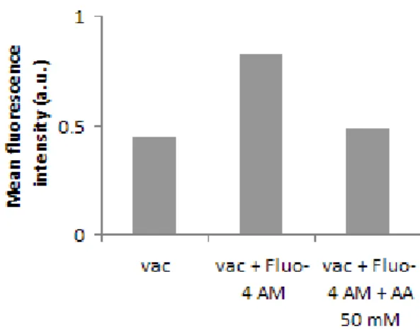
Epifluorescence microscopy imaging showed that the vacuolar membrane stains strongly with the styryl dye FM1-43 and that most of the vacuoles accumulate Ca2+, as assessed with
Fluo-4 AM. Flow cytometry analysis of vacuole samples incubated with these fluorescent probes confirmed a well-defined population of intact and functional vacuoles. Consistently spectrofluorimetric assays with the pH-sensitive probe ACMA suggested that the isolated vacuoles were intact and functional, the vacuolar membrane being able to generate and maintain a pH gradient through a concanamycin A-sensitive V-H+-ATPase. The addition of acetic acid
induced the release from the vacuole lumen of an EGFP-Pep4p fusion protein. Changes in fluorescence of vacuoles stained with acridine orange and Fluo-4 AM suggest that the acid induces a transient perturbation of vacuolar pH and Ca2+ release. It has been described that
release of Ca2+ is an event involved in cell death, as well as the release of H+ and consequent
cytosol acidification. The main novelty of the present study with isolated vacuoles is the finding that acetic acid is able to directly induce a partial permeabilization of the vacuolar membrane, similar to LMP in mammalian cells, without the involvement of other organelles or triggering upstream pathways.
Sumário
O vacúolo é o maior organelo da levedura e é funcionalmente equivalente ao lisossoma das células animais e ao vacúolo das células vegetais. Este organelo, cujo lúmen tem o pH mais ácido, participa em processos celulares importantes tais como degradação proteica e armazenamento de iões e metabolitos, bem como na homeostase iónica, resposta à privação de nutrientes, stress osmótico e iónico, autofagia e mesmo na apoptose. Sabe-se que a permeabilização da membrana lisossomal (LMP) e a consequente libertação de proteases lisossomais, nomeadamente de catepsinas, induz apoptose. De todas as proteases lisossomais, a catepsina D foi a primeira proteína a ser identificada com propriedades apoptóticas. Foi demonstrado que o ácido acético induz apoptose em leveduras, associada a alterações mitocondriais e à libertação de proteínas pró-apoptóticas. Foi igualmente demonstrado que a protease vacuolar Pep4p, ortóloga da catepsina D humana, está envolvida na apoptose induzida por ácido acético.
O presente estudo teve como principal objectivo contribuir para a compreensão da interligação entre o vacúolo e a mitocôndria na morte celular programada induzida por ácido acético na levedura Saccharomyces cerevisiae. Para tal foi investigado o efeito do ácido acético em vacúolos isolados e em células inteiras de leveduras com diferentes backgrounds genéticos, nomeadamente no que respeita à permeabilização da membrana vacuolar bem como à libertação de Pep4p e sua relação com alterações nas funções do vacúolos. A caracterização funcional de vacúolos isolados bem como o efeito da adição de ácido acético na função vacuolar foram realizadas por citometria de fluxo e microscopia de fluorescência associada à utilização de diferentes sondas fluorescentes. A caracterização funcional dos vacúolos isolados foi complementada pela determinação da actividade da V-H+-ATPase por espectrofluorimetria.
Os estudos de microscopia de fluorescência mostraram uma marcação forte da membrana vacuolar com a sonda lipofílica FM1-43 e que a maioria dos vacúolos acumulavam Ca2+ com
base na marcação pela sonda Fluo-4 AM. A análise por citometria de fluxo de amostras de vacúolos incubadas com estas sondas fluorescentes evidenciou uma população bem definida de vacúolos intactos e funcionais. Consistentemente os ensaios de espectrofluorimetria com a sonda ACMA, sensível ao pH, sugeriram que os vacúolos isolados se encontravam intactos e funcionais, dado que a membrana vacuolar era capaz de gerar e manter um gradiente de pH através da actividade da bomba V-H+-ATPase, sensível à concanamicina A. A adição de ácido
acético promoveu a libertação da proteína de fusão EGFP-Pep4p em vacúolos isolados e induziu variações de fluorescência de vacúolos marcados com laranja de acridina e com Fluo-4 AM indicativas de uma perturbação transitória do pH vacuolar e de uma libertação de Ca2+. Está
o ácido acético é capaz de induzir directamente uma permeabilização parcial da membrana vacuolar, semelhante à LMP em mamíferos, sem o envolvimento de outros organelos ou sem a activação de vias a montante.
Index
Abbreviations... 1. Introduction... 1.1. The yeast vacuole... 1.1.1. The role of V-H+-ATPase in the acidification of the vacuolar lumen...
1.1.2. Regulation of vacuolar acidification... 1.1.3. The vacuolar morphology... 1.1.4. Vacuolar functions... 1.1.4.1. Degradation... 1.1.4.2. Metabolite and ion storage and homeostasis... 1.1.4.3. Detoxification... 1.1.4.4. Response to nutrient deprivation... 1.1.4.5. Response to ionic and osmotic shock... 1.2. Programmed cell death...
1.2.1. Apoptosis and its importance... 1.2.2. Apoptotic morphological features... 1.2.3. Key components of the apoptotic machinery... 1.2.4. Apoptotic signalling pathways... 1.2.4.1. The extrinsic pathway... 1.2.4.2. The intrinsic pathway... 1.2.4.3. Type I and type II cells... 1.2.5. The crucial role of mitochondria in apoptosis... 1.2.5.1. Permeability transition pore... 1.2.6. Regulatory mechanisms... 1.2.7. Apoptosis in yeast... 1.2.8. Apoptosis induced by acetic acid... 1.3. Role of vacuole/lysosome in apoptosis...
1.3.1. Mechanisms underlying lysosome membrane permeabilization... 1.3.2. The crosstalk between lysosomes and mitochondria during cell death... 1.3.3. Lysosomal cathepsins...
1.3.3.1. The role of cathepsin D... 1.3.3.2. The role of Pep4p in yeast... 2. Aims... 3. Experimental procedures... 3.1. Yeast strains and growth conditions... 3.2. Reagents... 1 3 3 4 5 6 7 7 9 10 10 11 11 12 13 13 14 14 15 15 16 16 17 19 20 22 22 23 24 26 27 29 30 30 30
3.3. Vacuole isolation and purification... 3.4. Fluorochrome solutions and staining protocols... 3.5. Analysis by epifluorescence microscopy... 3.6. Analysis by flow cytometry... 3.7. Determination of the proton pumping activity of V-H+-ATPase... 4. Results...
4.1. Characterization of isolated vacuoles from S. cerevisiae... 4.2. Flow cytometric analysis of vacuole populations... 4.3. Study of the effect of acetic acid on vacuole functionality and membrane permeabilization by flow cytometry... 4.3.1. Effect of acetic acid on vacuolar pH... 4.3.2. Effect of acetic acid in vacuolar Ca2+ homeostasis...
4.3.3. Effect of acetic acid and H2O2 on Pep4p release from the vacuole...
4.4. Effect of ATP on vacuole dynamics... 5. Discussion...
5.1. Isolated vacuoles from S. cerevisiae are intact and functional... 5.2. V-H+-ATPase regulates vacuole dynamics...
5.3. Acetic acid causes partial vacuole membrane permeabilization... 5.4. Future prospects... 6. References... 31 32 33 33 34 35 35 37 40 40 41 41 43 44 44 45 46 48 49
Abbreviations
Δψm ACMA AIF ANT Apaf-1 ATP a.u. Bcl-2 BH3 Caspase dATP DED DD Diablo DISC DMSO DNA EGFP EGTA ER FM1-43 FADD Fas FasL H2O2 HtrA2 IAP IMM LMP MES MgCl2 MOMPTransmembrane mitochondrial potential 9-amino-6-chloro-2-methoxyacridine Apoptosis-inducing factor
Adenine nucleotide translocator Apoptotic protease-activating factor-1 Adenosine-5’-triphosphate
Arbitrary units B-cell lymphoma-2 Bcl-2 homology 3
Cysteine-dependent aspartate-specific protease Deoxyadenosine triphosphate
Death-effector domain Death domain
Direct IAP binding protein with low pI Death inducing signaling complex Dimethyl sulfoxide
Deoxyribonucleic acid
Enhanced green fluorescent protein
Ethylene glycol-bis[β-aminoethylether]-N, N, N’, N’-tetraacetic acid Endoplasmatic reticulum
N-(3-triethylammoniumpropyl)-4-(4-(dibutylamino)styryl)pyridinium dibromide
Fas-associated death domain Apoptosis stimulating fragment Fas ligand
Hydrogen peroxide
High temperature requirement A2 Inhibitor of apoptosis protein Inner mitochondrial membrane
Lysosomal membrane permeabilization 2-(Nmorpholino)ethanesulfonic acid Magnesium chloride
MOPS OMM PCD PLA2 PT PTP ROS rpm SC siRNA Smac SNARE tBid TNF TNFR-1 TRAIL TRADD Tris UV V-H+-ATPase VDAC XIAP YEPD 3-(N-morpholino)propanesulfonic acid Outer mitochondrial membrane Programmed cell death
Phospholipase A2 Permeability transition Permeability transition pore Reactive oxygen species Rotations per minute Synthetic complete medium Small-interfering RNA
Second mitochondria-derived activator of caspases Soluble NSF attachment receptors
Truncated Bid
Tumor necrosis factor
Tumor necrosis factor receptor-1 TNF-related apoptosis inducing ligand TNFR1-associated death domain protein Tris(hydroxymethyl)aminomethane Ultraviolet
Vacuolar-type proton-translocating ATPase Voltage-dependent anion channel
X-chromosome linked inhibitor of apoptosis protein Yeast extract/peptone/dextrose medium
1. Introduction
The yeast vacuole is the most acidic organelle and is equivalent to the lysosome of animal cells and to the vacuole of plant cells. Besides its well known degradative and storage functions, this organelle is also involved in cell death processes, namely in apoptosis. Apoptosis is a type of programmed cell death (PCD) that eliminates excess or damaged cells in a controlled way. This cell death process is vital for embryonic development, immune system and tissue homeostasis in metazoans. However, it is currently recognised that apoptosis also occurs in single-celled organisms, for instance in the budding yeast Saccharomyces cerevisiae. Thus this simple and genetic tractable unicellular eukaryotic organism has been used as a valuable model system for cell death research.
In this section the yeast vacuole, its characteristics and various functions as well its responses to stress stimuli will be focused. Next, the mammalian apoptotic pathways will be briefly described as well as the apoptotic components and regulators in yeast so far identified. Particular focus will be given to apoptosis induced by acetic acid since it was the apoptotic agent used in this study. Finally, an overview of the role of the vacuole and lysosome in apoptosis will be presented, as well as the release of Pep4p, the orthologue of the human lysosomal cathepsin D, which was addressed in this study.
1.1. The yeast vacuole
The vacuole is the largest and most acidic organelle of yeast cells playing major roles in protein degradation, ion and metabolite storage as well in detoxification. It also has an important function in ion homeostasis, response to nutrient deprivation, osmotic and ionic stress, autophagy and even in apoptosis (Klionsky et al., 1990; Guicciardi et
al., 2004; Li and Kane, 2009). These features support the recent view of vacuoles as
dynamic, highly sensitive and responsive organelles to environmental changes, and not only as storage compartments.
1.1.1. The role of V-H+-ATPase in the acidification of the vacuolar lumen
Almost all vacuolar and lysosomal functions are dependent either on the acidic pH of vacuolar lumen or on the pH gradient across vacuolar membrane. It has been shown that yeast vacuolar pH varies from <5-6.5 depending mostly on growth conditions (Plant et al., 1999; Brett et al., 2005; Padilla-Lopez et al., 2006; Martinez-Munoz and Kane, 2008). In both vacuoles and lysosomes, acidification is achieved through the action of a proton pump V-H+-ATPase. This proton pump is responsible for the coupling the free energy of ATP hydrolysis to proton transport from the cytosol to the organelle lumen. V-H+-ATPases are highly conserved enzymes, consisting of a complex V1 that contains the sites for ATP hydrolysis in the cytosolic side, bound to a complex
V0 that is integrated into the vacuolar membrane (Figure 1).
The yeast V-H+-ATPase consists of 14 subunits all encoded by a single gene, except for the largest membrane subunit a (Manolson et al., 1994) which exists in two isoforms. Mammalian V-H+-ATPases have a similar structure overall, but the number of isoforms is much higher (reviewed by Li and Kane, 2009), which complicates the biochemical analysis of lysosomal V-H+-ATPases. This is one of the main reasons why
yeast is an appropriate model system to study the V-H+-ATPase.
In contrast to other organisms, genetic deletions of ubiquitously expressed V-H+
-ATPase subunits in fungi are not lethal. Thus, yeast mutants lacking V-H+-ATPase
subunits (vma mutants) offer important insights into the functional importance of vacuolar acidification (Kane, 2006). Genetic deletion of any of V-H+-ATPase subunits appears to abolish organelle acidification, resulting in a phenotype characterized by sensitivity to high extracellular pH and high extracellular calcium concentrations as well to heavy metals and oxidants. Moreover these mutants are unable to grow on non-fermentative carbon sources, and are multidrug sensitive (Kane, 2006).
Calcium sensitivity of vma mutants results from the dependence of the vacuolar Ca2+/H+ exchanger (Vcx1p) on the proton gradient established by V-H+-ATPase (Forster and Kane, 2000). Vacuolar sequestration of heavy metals is mediated by some transporters and by binding to polyphosphate vacuolar stores and is compromised in mutants lacking V-H+-ATPase activity (Eide et al., 1993; MacDiarmid et al., 2002; Freimoser et al., 2006). Vma mutants are also unable to grow on a medium containing low iron and calcium concentrations (Davis-Kaplan et al., 2004; Yadav et al., 2007) and
at high pH (Serrano et al., 2004), both reflecting the important role of the vacuole as a storage compartment and in pH homeostasis, respectively.
1.1.2. Regulation of vacuolar acidification
Vacuolar acidification is regulated by distinct mechanisms. In yeast, V-H+ -ATPase is regulated at the level of assembly of its V1 and V0 complexes (Forgac, 2007;
Kane, 2000, 2006). Disassembly is rapid and regulated post-transcriptionally (Forgac, 2007; Kane, 2006) and leads to inactivation of both ATP hydrolysis and proton translocation. Disassembly of V-H+-ATPase is reversible and occurs in order to adjust the level of assembled enzyme to the availability of ATP and/or the need for proton pump activity. Glucose metabolism also regulates V-H+-ATPase assembly. Fructose-6-phosphate is necessary to maintain V-H+-ATPase assembly (Parra and Kane, 1998), as well as some glycolytic enzymes that also support its assembly by signalling glucose deficiency (Lu et al., 2001, 2003, 2007). Additionally, there are mechanisms to regulate vacuolar acidification without acting on the V-H+-ATPase itself (Paroutis et al., 2004).
Due to the electrochemical gradient produced by V-H+-ATPase, counterion transport
plays a critical role for continued pumping (Arai et al., 1989). Hence, a number of exchangers such as Nhx1p (a Na+/H+ exchanger), transport H+ against the pH gradient
created by the V-H+-ATPase, and Nhx1p has been shown to limit acidification in the compartment where it resides, the late endosome/ multivesicular body, and indirectly, to control vacuolar pH (Ali et al., 2004).
Buffering systems also influence the pH of the vacuole. In yeast, polyphosphate is present at very high concentrations inside the vacuole (Beauvoit et al., 1991; Thomas and O'Shea, 2005; Freimoser et al., 2006), and is the major vacuolar buffering system. Recently it has been suggested that vacuolar proteins may also act as buffers (Brett et
al., 2006). Together, these systems operate to determine the final vacuolar pH and each
1.1.3. The vacuolar morphology
Vacuolar morphology depends on growth phase and undergoes alterations in response to different stress conditions. During exponential phase, active yeast cells contain medium-sized vacuoles, which fuse into one large vacuole in stationary phase or under glucose deprivation. Under conditions of osmotic stress, vacuoles also adjust their shape and number, to facilitate the release or uptake of water and ions. The exposure of vacuoles to hyper-osmotic conditions leads to fragmentation into numerous small vesicles, whereas under hypo-osmotic conditions they fuse into one large vacuole (Li and Kane, 2009). These changes in vacuole morphology rely in fusion/fission machinery and on the equilibrium between these two processes.
Vacuolar morphology is dictated by the balance between fusion and fission (Baars
et al., 2007). Vacuolar fusion and fission are necessary for the merging of small
vacuolar vesicles and fragmentation of a large vacuole, for delivering cargo into and out of the vacuole as well for regulation of vacuolar volume in response to stress conditions. Fusion includes four major steps namely, priming, tethering, docking and fusion. It also involves coordinated and regulated interactions between many proteins, such as soluble NSF attachment receptors (SNAREs), SNARE chaperones, GTPases and specific lipids like phosphoinositides which are widely present in the vacuolar membrane (reviewed by Li and Kane, 2009). On the other hand, fission-mediated vesicle formation during endocytosis requires coat proteins like COPI, COPII and clathrin (Peplowska and Ungermann, 2005). The molecular mechanisms underlying fission of vacuolar vesicles are hard to define comparatively to the knowledge of those associated to vesicle fission during endocytosis.
In yeast, vacuolar fission is necessary for vacuolar inheritance during cell division and in response to osmotic shock. Interestingly, V-H+-ATPase is required for both fusion and fission processes (Baars et al., 2007). A recent study has shown that fusion requires the presence of the V0 membrane complex of V-H+-ATPase but not its
pumping activity, while proton translocation activity is required for fission (Baars et al., 2007). It was observed that cells with no V-H+-ATPase activity due either to the deletion of V-H+-ATPase or its inhibition by concanamycin A (a specific inhibitor drug of V-H+-ATPase) were defective for fission and possessed a large vacuole. In contrast, the deletion of a V0 subunit (vph1Δ) complex that still maintained a partial acidification
led to a defective fusion and cells had fragmented vacuoles. Because cells treated with concanamycin A contained big vacuoles, defective fission caused by the lack of proton pumping activity likely dominates over defective fusion by physical absence of the total or of a subunit of the V0 sector (Baars et al., 2007). These observations support the
interpretation that the equilibrium between vacuolar fusion and fission determines vacuolar morphology.
1.1.4. Vacuolar functions
As referred to above vacuoles are highly dynamic organelles, playing constitutive roles in different processes such as degradation, storage, homeostasis and detoxification (Figure 1), as well as in response to stress conditions like nutrient deprivation and osmotic shock. In the following sections the most important functions of the vacuole are detailed.
1.1.4.1. Degradation
Similarly to the lysosomes, vacuoles are the major degradation systems of the cell. Both soluble (e.g. proteinase A, Pep4p; carboxypeptidade Y, CPY; proteinase B, Prb1p; and carboxypeptidade S, CPS) and membrane-bound (e.g. dipeptidyl aminopeptidase B, DPAP-B) have been described. Usually, vacuolar proteases are relatively non-specific and are engaged in the degradation of a large number of different substrates. Vacuolar proteases are transported to the vacuole in a zymogen form and are activated in a complex cascade at low pH (Van Den Hazel et al., 1996), but since vacuolar protease activity occurs in the absence of V-H+-ATPase activity, acidic vacuolar pH does not appear to be critical for zymogen activation. For instance Pep4p activation inside the acidic vacuole can occur by two different pathways, a one-step process to release mature proteinase A, involving the intervention of proteinase B, or a step-wise pathway via the autoactivation product known as pseudo-proteinase A. Once active, S. cerevisiae proteinase A is essential to the activities of other yeast vacuolar hydrolases, including proteinase B and carboxypeptidase Y (Parr et al., 2007).
Fig. 1. The yeast vacuole is involved in many cellular processes. a) Degradation: the vacuole is highly
enriched in hydrolases responsible for the breaking down of molecules and/or organelles delivered to the vacuole via different pathways. b) Storage: the vacuolar membrane contains multiple transporters that import amino acids, ions, and metals. Activity of V-H+-ATPase is
necessary for hydrolases maturation and for the generation of the proton gradient that drives many transporters. c) Buffering: the vacuolar membrane contains transporters that export amino acids, ions, and metals, which combined to the activity of importers contribute to ion homeostasis and amino acid recycling. The vacuole also accumulates polyphosphate that buffers cations. d) Detoxification: the vacuole sequesters toxic metals and potentially harmful metabolic side-products via ABC transporters (adapted from Li and Kane, 2009).
Vacuolar proteins and protein substrates are directed to the vacuole by different pathways (Bowers and Stevens, 2005). While resident vacuolar proteins are sent to the vacuole by biosynthetic pathways (from Golgi or cytosol), proteins targeted for degradation are sent via endocytosis (plasma membrane proteins) or autophagy (cytosolic and organellar proteins).
1.1.4.2. Metabolite and ion storage and homeostasis
Vacuole is the major storage compartment for amino acids, phosphate, Ca2+ and
metal ions (Klionsky et al., 1990; Van Ho et al., 2002). Basic and neutral amino acids accumulate in yeast vacuole at high levels, but there is little vacuolar storage of acidic amino acids (Klionsky et al., 1990), which mostly require the proton gradient for their accumulation. There are many specific amino acid transporters identified in yeast responsible for the bidirectional transport across the vacuolar membrane. Influx and efflux is highly balanced to allow storage or amino acid recycling, depending on cellular conditions and needs.
Vacuole contains very high concentrations of phosphate, stored as polyphosphate. It acts as both a macromolecular anion that facilitates the uptake and retention of positive charged ions and amino acids, and as a phosphate store, serving as a cytosolic phosphate buffer during conditions of phosphate depletion (Thomas and O'Shea, 2005).
Vacuole also serves as the main intracellular Ca2+ store. Yeast vacuoles membrane contains an H+/Ca2+ antiporter (Vcx1p) which drives Ca2+ uptake at the expense of the proton gradient, and a P-type Ca2+-ATPase which operates independently of the proton gradient (Miseta et al., 1999). Vcx1p generates a high uptake of Ca2+ but with low
affinity, while Pmc1p is important for Ca2+ uptake in absence of a functional V-H+
-ATPase or under high concentrations of extracelular Ca2+ (Forster and Kane, 2000). In
addition, there is a transient receptor potential calcium channel, Yvc1p that allows the release of Ca2+ from the vacuole (Denis and Cyert, 2002; Zhou et al., 2003).
The storage and release of other metal ions, like Zn2+ and Fe3+ follow a similar pattern. Yeast vacuolar membrane contains two zinc transporters, Zrc1p and Cot1p that direct zinc uptake into the vacuole when Zn2+ is in excess, while deficiency in zinc leads to transcription of a Zn2+ exporter, Zrt3p (MacDiarmid et al., 2000, 2002). Regulation of iron is particularly important because it is a redox-active metal as well as a scarce and essential nutrient. Ccc1p is responsible for iron import (Li et al., 2001), while Fet5p/Fth1p complex and Smf3p transporter are responsible for iron mobilization when Fe3+ supply is limited (Singh et al., 2007).
As a storage compartment for a wide variety of ions, the vacuole is the major organelle responsible for intracellular ion homeostasis and response to ion shock. Vacuolar transporters mediate ion homeostasis by regulating ion transport between the vacuole and the cytosol, along with plasma membrane ion transporters that regulate ion
influx/efflux between the cytosol and the extracellular environment. The activity of many vacuolar transporters is dependent on the pH gradient across vacuolar membranes generated by the V-H+-ATPase. For instance, the late endosomal transporter Nhx1p confers osmotolerance following acute hypertonic shock (Nass and Rao, 1999).
1.1.4.3. Detoxification
Besides its storage functions, vacuole also plays an important role in detoxification. Different toxic molecules are sequestered into the vacuole away from cytosol and other organelles. Some of these molecules are redox-active heavy metals or even nutrients and metabolites that become toxic when in excess. There are two different ATP binding cassette (ABC) transporters present in the vacuolar membrane responsible for detoxification: Ycf1p and Bpt1p (Ghosh et al., 1999; Sharma et al., 2002). These two pumps have analogous mechanisms and specificities, but different regulatory mechanisms. The toxic metals arsenite and cadmium, and accumulated side-products of adenine metabolism are some of their substrates.
1.1.4.4. Response to nutrient deprivation
Autophagy is the vesicular sequestration of cytosolic cargo for vacuolar degradation. In starvation conditions, autophagy can be triggered as a stress response, in order to provide supplementary reserves for the cell (Kim and Klionsky, 2000). There are two main types of autophagy: macroautophagy and microautophagy. Macroautophagy involves the enclosing of cytosolic components by the autophagosome, a double membrane vesicle, with subsequent fusion with the vacuole. Both the inner membrane and autophagosome luminal components are digested and the resulting macromolecules are released back into the cytosol. In microautophagy, the vacuole membrane forms invaginations and cytosolic materials are directly engulfed by the vacuole (Mizushima et al., 2008).
Under starvation conditions, vacuolar hydrolases are produced at a higher rate induced by the influx of material into the vacuole through autophagy, which results in an increase of vacuolar hydrolytic activity (Teichert et al., 1989; Scott et al., 1996; Kim and Klionsky, 2000). The influx of material to the vacuole also leads to the accumulation of membranes. Microautophagy has been observed to occur after
macroautophagy, suggesting that this may be a mechanism for vacuolar membrane recycling (Mijaljica et al., 2007).
1.1.4.5. Response to ionic and osmotic shock
Yeast cells have two different responses to osmotic shock. A long-term response that involves the HOG (high osmolarity glycerol) pathway, leading to production of high levels of glycerol, a cellular osmoprotectant (Hohmann, 2002, 2007), and in contrast, an immediate response that leads to vacuolar fragmentation and release of calcium.
Upon salt shock, vacuoles release calcium via Yvc1p, resulting in a transient increase in the cytosolic Ca2+ concentration (Denis and Cyert, 2002). The exact role of transient Ca2+ release remains unclear. Ca2+ is a key signalling molecule and its release probably activates other cellular responses that alleviate osmotic shock.
Vacuolar fragmentation may be a result of the release of water from vacuoles to maintain proper osmotic pressure in the cytosol. Interestingly, high cytosolic levels of Ca2+ also induce vacuolar fragmentation (Kellermayer et al., 2003). Possibly fragmentation is a normal response to Ca2+ stress by increasing the surface/volume ratio
of the vacuole, to enable maximal sequestration of calcium at the vacuole (Kellermayer
et al., 2003).
1.2. Programmed cell death
Since the mid-nineteenth century, many observations have already indicated that cell death plays considerable roles in multicellular organisms, particularly during embryogenesis and metamorphosis (Gluecksmann, 1951; Lockshin and Williams, 1964). The term programmed cell death (PCD) was introduced in 1964, proposing that cell death during development is not of accidental nature but follows a sequence of controlled steps leading to locally and temporally defined self-destruction (Lockshin and Williams, 1964). The term PCD also includes the concept that cell death has been genetically programmed, as this is the case during development and aging. The development and maintenance of multicellular biological systems depends on a complex interaction between the cells of the organism, sometimes involving an
altruistic behaviour of individual cells that trigger suicide in the benefit of the organism as a whole. For instance, during the development, several cells are produced in excess which then undergo PCD, thus contributing to the final shape of the many organs and tissues (Meier et al., 2000). Balance between cell division and cell death is of utmost importance for the development and maintenance of multicellular organisms. Disorders of either process have pathological consequences and can lead for instance to disturbed embryogenesis, neurodegenerative diseases, or the development of cancer.
1.2.1. Apoptosis and its importance
The most frequent and well-defined form of PCD is apoptosis. Apoptosis is of Greek origin, meaning "falling off” or “dropping off", in analogy to leaves falling off trees. This analogy emphasizes that the death of living matter is an integral and necessary part of the life cycle of organisms. The term apoptosis has been coined in order to describe the morphological processes leading to controlled cellular self-destruction and was first introduced in a publication by Kerr and colleagues (Kerr et al., 1972), but other, non-apoptotic types of cell death also might be of biological significance (Leist and Jäättelä, 2001). Therefore, it is worth noting that “programmed cell death” and “apoptosis” are not synonyms because cell death, as it occurs during physiological development, can manifest non-apoptotic features (Barkla and Gibson, 1999; Roach and Clarke, 2000; Baehrecke, 2002).
Apoptotic processes are of a widespread biological importance, being involved in the development, differentiation, proliferation, homeostasis, regulation and function of immune system and in the removal of defective cells. Therefore, defects in apoptotic processes are implicated in several pathological conditions, which may result in cancer, autoimmune diseases and spreading of viral infections, while neurodegenerative diseases, AIDS and isquemias are caused or enhanced by excessive apoptosis (Fadeel et
al., 1999a). Due to its importance in such biological processes, apoptosis is a very
common phenomenon, occurring in all kinds of metazoans (Tittel and Steller, 2000). Moreover, it plays an important role in plant biology (Solomon et al., 1999), and even unicellular organisms such as yeast exhibit apoptotic-like mechanisms, hence being
used as a simpler eukaryotic model system for apoptosis studies (Fröhlich and Madeo, 2000; Skulachev, 2002).
1.2.2. Apoptotic morphological features
Apoptotic cells can be distinguished by their typical morphological changes: cell shrinks, becomes deformed and loses contact with neighbour cells. Chromatin condenses, plasma membrane buds and finally cell fragments in membranar structures, the apoptotic bodies, which contain cytosol, cell organelles and fragments of condensed chromatin. Apoptotic bodies are then engulfed by phagocytosis by surrounding cells and removed from the tissue (in multicellular organisms), without triggering an inflammatory response (unlike during a necrotic process). All these morphological changes are a result of many biochemical and molecular events that take place in an apoptotic cell, essentially the activation of proteolytic enzymes, which eventually carry out DNA cleavage in fragments, as well as cleavage of multiple proteins involved in cytoplasm integrity and organelle’s shape (Saraste and Pulkki, 2000).
1.2.3 Key components of the apoptotic machinery
During apoptosis, the cell is mainly degraded by proteases and endonucleases. The main class of proteases involved in the apoptotic process are caspases (cysteine-dependent aspartate-specific proteases), which play an important role in the triggering and execution of apoptosis. These proteases, when activated, acquire the capacity of cleaving intracellular key-substrates, resulting in morphological and biochemical modifications characteristic of apoptosis (Earnshaw et al., 1999).
Another set of proteins that play a central role in apoptosis is the Bcl-2 family. Bcl-2 family can be defined by the presence of conserved sequence motifs known as Bcl-2 homology domains (BH1 to BH4). In mammals, up to 30 relatives have been described of which some belong to pro-apoptotic members and others to a group of anti-apoptotic members (Borner, 2003). Bcl-2, Bcl-XL, Bcl-w, A1, and Mcl-1 are examples
The pro-apoptotic group of Bcl-2 members can be divided into two subgroups: the Bax-subfamily consisting of Bax, Bak, and Bok, all possessing the domains BH1, BH2, and BH3, and the BH3-only proteins (Bid, Bim, Bik, Bad, Bmf, Hrk, Noxa, Puma, Blk, BNIP3, and Spike) which possess only the short BH3 motif, an interaction domain that is both necessary and sufficient for their killing action (Cory and Adams, 2002; Mund et
al., 2003).
Inhibitor apoptosis proteins (IAPs) are another class of apoptosis regulators which neutralise pro-apoptotic components and that can in turn be antagonised by other proteins.
1.2.4 Apoptotic signalling pathways
Apoptosis can be triggered by different extra- or intracellular stimuli, such as attachment of death ligands to membrane death receptors, DNA damage caused by faults in DNA repair mechanisms, treatment with cytotoxic compounds or irradiation, lack of survival signals, or development of death signals. Cell death through apoptosis is tightly controlled by changes in protein expression, protein-protein interactions, and various post-translational modifications, including proteolytic cleavage and phosphorylation. For some of these events to occur, proteins must re-localize from one sub-cellular compartment to another to gain access to their substrates or interacting partners. Thus, compartmentalization is yet an important regulatory mechanism, preventing accidental triggering of cell death signalling.
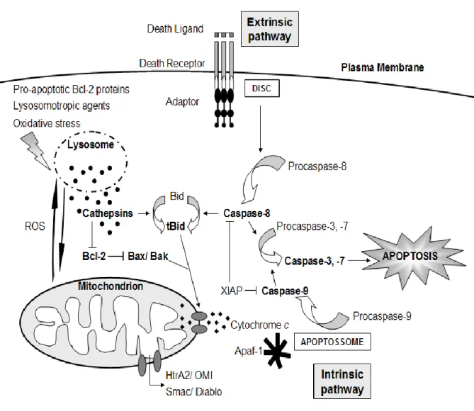
Two main signalling pathways lead to apoptosis: the extrinsic or death receptor pathway and the intrinsic or mitochondrial pathway (Figure 2). These two pathways differ in the initiator caspases that transmit the signal but that may later converge at the level of activation of executioner caspases (caspases-3, -6, and -7) (Hengartner, 2000).
1.2.4.1. The extrinsic pathway
The extrinsic pathway is triggered upon binding of specific ligands (such as TNF-α, FasL or TRAIL) to their membrane death receptors (such as TNFR-1 or Fas), which allows the exposure of their death domain (DD) in the cytosol side. Through the DD, adaptor molecules such as FADD or TRADD are recruited to form the death inducing
signalling complex (DISC). The procaspase-8 is then recruited to the DISC by its death-effector domain (DED) and activated (Sartorius et al., 2001; Denault and Salvesen, 2002). Active caspase-8 initiates a caspase cascade by processing the executioner caspases which in turn cleave different protein substrates.
1.2.4.2. The intrinsic pathway
Mitochondrial apoptotic signalling includes the release of pro-apoptotic factors from the mitochondrial intermembrane space to the cytosol such as cytochrome c (Salvesen and Renatus, 2002b), apoptosis inducing factor AIF (Susin et al., 1999), the endonuclease endoG (Li et al., 2001), Smac/Diablo (Verhagen et al., 2000) and HtrA2/Omi (Verhagen et al., 2002). In the cytosol, cytochrome c contributes to the formation of the apoptosome, a complex that consists of cytochrome c, Apaf-1 (an adaptor molecule) and dATP (Acehan et al., 2002), which activates another initiator caspase, caspase-9 (Denault and Salvesen, 2002), hence mediating a caspase cascade by activating caspase-3 (Slee et al., 1999).
1.2.4.3. Type I and type II cells
In some cells (type I cells), the signal coming from the DISC generates a caspase signalling cascade strong enough for execution of cell death on its own. Cells that have the capacity to induce such direct and mainly caspase-dependent apoptotic pathway belongs to the so-called type I cells (Scaffidi et al., 1998).
In type II cells, the amount of active caspase-8 generated is limited and activation of caspase-3 can be blocked by overexpression of Bcl-2 (Scaffidi et al., 1998). Thus, signal coming from the activated receptor must be amplified by the proteolytic activation of the pro-apoptotic protein Bid by caspase-8. In its truncated form (tBid), is translocated to mitochondria with subsequent induction of apoptotic events (Luo et al., 1998). However, it has been recently suggested that the level of XIAP, an inhibitor of apoptosis protein, is the critical discriminator between type I and type II apoptosis signalling (Jost et al., 2009).
1.2.5. The crucial role of mitochondria in apoptosis
Besides amplifying and mediating extrinsic apoptotic pathways, mitochondria also play a central role in the integration and propagation of death signals originating from inside the cell. Most of the events inducing apoptosis involve disruption of trans-membrane mitochondrial potential (Δψm), as well as the so-called permeability
transition (PT), a sudden increase of outer mitochondrial membrane (OMM) permeability to low molecular mass solutes. Simultaneously, an increase of mitochondrial volume takes place caused by water influx to the matrix, eventually originating the rupture of OMM and resulting in a non specific release of pro-apoptotic factors from the mitochondrial intermembrane space to the cytosol (Bernardi et al., 1999; Loeffler and Kroemer, 2000).
1.2.5.1. Permeability transition pore
Several possible mechanisms for PT have been proposed. Although, there is some consensus regarding the formation of the so-called permeability transition pore (PTP), the molecular components involved have not yet been completely identified. The adenine nucleotide translocator (ANT) and the voltage-dependent anion channel (VDAC) have been proposed as their major components, since ANT is the most abundant protein in inner mitochondrial membrane (IMM), and VDAC the most common protein in OMM. Indicated by direct protein-protein interactions, VDAC-ANT complexes presumably could be able to connect IMM and OMM to the so-called “contact sites” corresponding to a close association of the two membranes and thus possibly constituting the PT pore (Beutner et al., 1998). However, subsequent genetic ablation studies have unequivocally shown that neither the ANT nor any VDAC isoform are essential for the PT to occur (Kokoszka et al., 2004; Baines et al., 2007).
Since PT, loss of Δψm, and release of mitochondrial proteins are of central
importance in mediating and enhancing apoptotic pathways, those mitochondrial events must be kept under strict control of regulatory mechanisms which are in many ways dependent on members of the Bcl-2 family. How cytochrome c can cross the OMM is not exactly known, but many different mechanisms have been proposed (Muchmore et
al., 1966; Reed, 1997; Adams and Cory, 1998; Gross et al., 1999; Antonsson and
2002), with none of them having been proven definitively: formation of a non specific pore, through which cytochrome c and other intermembrane space proteins can escape; heterodimerization between pro- and anti-apoptotic family members, since Bax and Bak display structural similarities with bacterial pore-forming toxins, leading to the hypothesis that these proteins themselves might directly form pores in the OMM; interaction with other mitochondrial proteins, either to generate a pore for cytochrome c exit, or to modulate mitochondrial homeostasis (for example, opening of the PTP); oligomerization to form a weakly selective ion channel; and interaction of activated Bax and Bak with OMM lipids leading to membrane bending and, ultimately, formation of transient lipid pores or inverted micelles. Additionally, it was recently suggested that protein release from mitochondria can occur through ceramide channels. The hypothesis of such channels are involved early in apoptosis is strongly supported by their regulation by the Bcl-2 family proteins. The regulation observed is in the direction concurrent with their role in apoptosis: anti-apoptotic proteins inhibit ceramide channels while the pro-apoptotic proteins enhance these channels (Ganesan and Colombini, 2010).
In addition to mitochondrial factors release, dissipation of Δψm and PT also cause
changes in cell homeostasis: synthesis of ATP is interrupted, molecules involved in redox reactions oxidize, and reactive oxygen species (ROS) are generated (Kroemer and Reed, 2000; Kroemer et al., 1997). High levels of ROS directly cause oxidation of macromolecules, thus contributing to Δψm disruption and acting as a positive feedback
(Marchetti et al., 1997).
1.2.6. Regulatory mechanisms
The pro-apoptotic factor Smac/Diablo, which is released from mitochondria, acts by inhibiting the inhibitors of apoptosis proteins (IAPs; Du et al., 2000), a family of proteins with anti-apoptotic activity by directly inhibiting caspases (Salvesen and Duckett, 2002). IAP expression is upregulated in response to survival signals, hence providing a means to suppress apoptosis signalling (Raff et al., 1993; Ameisen, 2002).
Members of the BH3-only subfamily are required for the activation of pro-apoptotic Bax/Bak (Bouillet and Strasser, 2002). Bax is a cytosolic monomer in viable
cells that during apoptosis changes its conformation, integrates into the OMM and oligomerizes (Nechushtan et al., 2001). Although the mechanism is still controversial, Bax and Bak oligomers are believed to contribute to the mitochondrial PT, either by forming channels by themselves (Antonsson et al., 2000) or by interacting with components of the PT pore such as VDAC (Tsujimoto and Shimizu, 2000).
BH3-only proteins interfere with the fine-tuned balance of homo- or hetero-oligomerization between pro-apoptotic multidomain members Bax/Bak and anti-apoptotic members Bcl-2/Bcl-XL. BH3 domains of Bid and Bim can directly mediate
Bax/Bak oligomerization, whereas Bad and Bik BH3 domains preferentially interact with anti-apoptotic Bcl-2/Bcl-XL. As a consequence, activated Bad/Bik might be able to
displace Bid/Bim from the binding pocket of anti-apoptotic Bcl-2/Bcl-XL, and thus
released Bid/Bim induce Bax/Bak oligomerization and cytochrome c release, even at low levels (Letai et al., 2002).
The anti-apoptotic Bcl-2 family members play a central role by guarding mitochondrial integrity and by controlling the release of mitochondrial proteins into the cytoplasm, thus counteracting the action of pro-apoptotic proteins and inhibiting mitochondrial pro-apoptotic events (Cory and Adams, 2002). Anti-apoptotic Bcl-2 members sequester proapoptotic Bcl-2 members by binding to their BH3 domains and hence preventing Bax or Bak activation/ oligomerization and consequently inhibiting mitochondrial proapoptotic events. Overexpression of Bcl-2 or Bcl-XL potently inhibits
apoptosis in response to many cytotoxic insults, e.g. by suppressing the generation of ROS, stabilizing Δψm, preventing PT and consequently blocking the release of
cytochrome c (Reed, 1999).
BH3-only proteins are regulated either by post-translational modifications or by transcription. For instance, Bid is activated by cleavage as referred before, Bim and Bad are activated by dephosphorylation, and Puma and Noxa are transcriptionally regulated by the p53 transcription factor. Similar events occur with anti-apoptotic Bcl-2 proteins (Youle and Strasser, 2008). The fate of the cell is therefore determined by the balance between the intracellular levels of anti-apoptotic and pro-apoptotic proteins (Brenner and Mak, 2009).
Apoptotic signals coming from the inside of the cell frequently have their origin within the nucleus. DNA damage usually results in the activation of p53, which promotes expression of pro-apoptotic Bcl-2 members and suppresses anti-apoptotic
members Bcl-2 and Bcl-XL. Other organelles besides mitochondria and the nucleus,
such as the endoplasmatic reticulum (ER) and lysosomes also have been implicated in apoptotic signalling pathways, and it should be kept in mind that probably hundreds of proteins are part of an extremely fine-tuned regulatory network consisting of pro- and anti-apoptotic factors (Hengartner, 2000).
1.2.7. Apoptosis in yeast
The occurrence of an apoptotic process in yeast was first described in cells with a mutation in the AAA-ATPase CDC48 gene that plays a role in cell division and vesicle trafficking (Madeo et al., 1997). These cells displayed apoptotic markers such as chromatin condensation, DNA fragmentation, externalization of phosphatidylserine to the outer leaflet of plasma membrane and ROS generation. Moreover, apoptotic scenarios have been observed in yeast undergoing cell death such as mitochondrial fragmentation, cytochrome c release, perturbations of the cytoskeleton and modifica-tions in the chromatin (Carmona-Gutierrez et al., 2010). In addition, if not engulfed by neighboring cells or in cell culture, where phagocytosis does not usually happen, dead cells in the late stages of apoptosis may present necrotic features due to the loss of cellular energy and plasma membrane integrity. This process is called “apoptotic necrosis” or “secondary necrosis” (Majno and Joris, 1995) and constitutes the ultimate fate of the yeast apoptotic cell. Since the first description of apoptosis in yeast, a growing number of proteins homologues of apoptotic regulators in higher eukaryotes have been identified in yeast, such a caspase Yca1p/Mca1p (Madeo et al., 2002), Nma111p the orthologue of HtrA2/Omi (Fahrenkrog et al., 2004), Aif1p the orthologue of AIF (Wissing et al., 2004), Bir1p, an yeast IAP (Walter et al., 2006), and the yeast endonuclease G (Büttner et al., 2007). Additionally, it has been suggested that Rad9 protein from Schizosaccharomyces pombe could belong to Bcl-2 family, being the first protein from this family identified in yeast (Komatsu et al., 2000). These results have suggested that apoptosis occurring in yeast has many similarities to apoptosis in metazoans. A large number of external stimuli and stress conditions have also been described as apoptosis inducers in yeast, including H2O2 (Madeo et al., 1999), acetic
ageing (Herker et al., 2004), UV radiation (Del Carratore et al., 2002), exposure to high concentrations of mating pheromone (Severin and Hyman, 2002), salt stress (Huh et al., 2002), hyper-osmotic stress (Silva et al., 2005), and exposure to yeast killer toxins (Reiter et al., 2005). Moreover yeast cells undergoing replicative ageing (Laun et al., 2001) or chronological ageing (Herker et al., 2004) also die by apoptosis.
An explanation for such a process in which a unicellular organism commits to cellular suicide, has been provided by demonstrating its role in several physiological scenarios, for example, during meiosis, ageing and mating (Büttner et al., 2006; Laun et
al., 2001; Fabrizio et al., 2004; Henker et al., 2004). Budding yeast grows as
multicellular colonies that are capable of simple differentiation. Each colony arises from a single cell that has undergone many divisions. Thus sacrifice of an aged cell containing high levels of ROS and potentially damage DNA acts as a selective advantage for the yeast population as a whole, protecting the integrity of the original cell’s genome and allowing the spreading of the clone. Hence, in the case of S.
cerevisiae one should consider it as a multicellular entity in which apoptosis acts as a
self-preservation mechanism for the colony as a whole (Gourlay et al., 2006; Carmona-Gutierrez et al., 2010).
1.2.8. Apoptosis induced by acetic acid
Acetic acid is a final product of alcoholic fermentation produced by S. cerevisiae. Glucose-repressed yeast cannot metabolize this compound that enters the cell in its non-dissociated form by simple diffusion. Within the cell, acetic acid dissociates and promotes internal acidification, ion accumulation and inhibition of cell metabolic activity, namely fermentation/respiration (Leão and van Uden, 1986; Cássio et al., 1987; Pampulha and Loureiro, 1989). Moreover, it was demonstrated that, in S.
cerevisiae, under certain conditions acetic acid compromises cell viability, resulting in
cell death (Pinto et al., 1989). Nevertheless, the process by which yeast lose viability when damaged by acetic acid is not yet completely understood.
Cell death in stress conditions induced by acetic acid has been characterized in our Laboratory (Ludovico et al., 2001, 2002 2003). After treatment with acetic acid, apoptotic markers such as chromatin condensation, phosphatidylserine exposure and
DNA fragmentation were observed, indicating that acetic acid, like H2O2 (Madeo et al.,
1999), is able to activate an apoptotic process in S. cerevisiae (Ludovico et al., 2001). Moreover, high doses of acetic acid (120-200 mM) lead to a necrotic phenotype in exponential phase cells of S. cerevisiae whereas low doses (20-80 mM) trigger a PCD exhibiting characteristics of mammalian apoptosis (Ludovico et al., 2001).
Yeast mitochondria play a key role in the apoptotic death process activated by acetic acid (Ludovico et al., 2002; 2003). S. cerevisiae cells committed to apoptosis in response to acetic acid display mitochondrial ROS accumulation, and mitochondria become permeabilized, promoting the release of lethal factors like cytochrome c (Ludovico et al., 2002) and Aif1p (Wissing et al., 2004), hence contributing to the death process.
Yeast genetic approaches revealed that while deletion of POR1 (yeast VDAC) enhances apoptosis triggered by acetic acid, absence of ADP/ATP carrier (AAC) proteins (yeast orthologues of ANT) protects cells exposed to acetic acid (Pereira et al., 2007). Absence of AAC proteins does not completely prevent acetic acid-induced apoptosis, suggesting that alternative redundant pathways are involved. One such pathway may be the translocation of Aif1p from the mitochondria to the nucleus in response to acetic acid (Wissing et al., 2004). Other mitochondrial proteins have been implicated in the execution of the yeast apoptotic program induced by acetic acid, including those involved in fission/fusion, namely Fis1p, Dnm1p, Mdv1p (Fannjiang et
al., 2004) and Nuc1p, the yeast ortholog of the mammalian endonuclease G (Büttner et al., 2007). Ysp2p is another mitochondrial protein with a direct function in
mitochondria-mediated PCD, since its absence confers resistance to acetic acid-induced PCD (Sokolov et al., 2006). The metacaspase Yca1p is activated in cells undergoing acetic acid-induced apoptosis (Pereira et al., 2007), although cells lacking Yca1p also undergo apoptosis in response to acetic acid, which suggests the existence of a caspase-independent pathway (Guaragnella et al., 2006).
The Kex1p protease, involved in PCD caused by defective N-glycosylation, also contributes to the active cell death program induced by acetic acid stress (Hauptmann et
al., 2006). Transient proteasome activation is also necessary for protein degradation
1.3. Role of vacuole/lysosome in apoptosis
Over the last decade, the lysosome has appeared as an important component of the cellular death machinery. In the classic concept of cell death, lysosomes were exclusively considered involved in necrotic and autophagic cell death. The concept of lysosomal constituents participating in cellular pathology and degeneration was originally proposed by De Duve (De Duve, 1966). After the proposal that lysosomal components could be involved in programmed cellular degeneration, several studies have attempted to evidence the role of lysosomes in triggering apoptosis.
There are several lines of evidence that sustain the participation of lysosomes in apoptosis. Leakage of lysosomal proteases into the cytosol is involved in the activation of caspases (Ishisaka et al., 1998). Active participation of lysosomal proteases, cathepsins, has also been observed in cell death induced by several stimuli, including TNF-α (Guicciardi et al., 2000; Foghsgaard et al., 2001; Guicciardi et al., 2001; Werneburg et al., 2002), bile salt-induced apoptosis (Roberts et al., 1997; Canbay et al., 2003), and chemotherapeutic drugs (Johansson et al., 2003; Bröker et al., 2004). Moreover, lysosomal apoptotic pathway, either cathepsin-dependent or independent, is believed to proceed through mitochondria (Boya et al., 2003a; Droga-Mazovec et al., 2008), suggesting a crosstalk between lysosomes and mitochondria.
1.3.1. Mechanisms underlying lysosome membrane permeabilization
Lysosome membrane permeabilization (LMP) can be triggered by an extensive range of apoptotic stimuli (reviewed by Johansson et al., 2010). These include pro-apoptotic Bcl-2 family proteins, ROS, cathepsins, calpains, caspases, changes in lysosomal membrane lipid composition, proteins from pathogenic bacteria and virus, and p53.
Several studies have suggested that the magnitude of LMP and consequently the amount of proteolytic enzymes released into the cytosol are key factors in determining the type of cell death (necrosis versus apoptosis) mediated by lysosomal enzymes (Li et
al., 2000). Massive collapse of lysosomes causes necrosis, while moderate
some contexts, LMP is an early event essential for apoptosis signalling to proceed, while under other circumstances it occurs late in the apoptotic process, contributing to amplification of the death signal.
The mechanism underlying LMP is not yet completely understood. Besides cathepsins, other hydrolases (Nylandsted et al., 2004; Kågedal et al., 2005; Blomgran et
al., 2007; Miao et al., 2008), H+ (causing acidification of the cytosol) (Nilsson et al., 2006), Ca2+ (Mirnikjoo et al., 2009), and many lysosomotropic dyes (Brunk et al., 1997; Kågedal et al., 2001b; Werneburg et al., 2002) can escape into the cytosol at the onset of apoptosis. This may suggest a nonspecific release mechanism due to limited membrane damage or through a pore, rather than selective transport across the membrane. However, the release of H+, Ca2+, and lysosomotropic dyes does not necessarily reflect a change in membrane integrity, but could merely be due to a change in ion pump activity (Johansson et al., 2010).
1.3.2. The crosstalk between lysosomes and mitochondria during cell death
Increasing evidence indicates that lysosomes communicate with mitochondria during cell death. This may occur through the selective leakage of proteins that activate some forms of apoptotic cell death, or through the massive lysosomal permeabilization that leads to an uncontrolled cell dismantling characteristic of necrosis. Although cells manifest large-scale autophagy shortly before or during cell death, autophagy is a pro-survival process. In response to metabolic stress, autophagy can indeed delay apoptotic cell death, and in apoptosis-defective cells it was demonstrated that inactivation of autophagy promoted cell death by necrosis (White, 2008).
Similarly to other cellular organelles, damaged mitochondria are removed by autophagy (mitophagy; Chu et al., 2007). In apoptosis, this mechanism becomes particularly significant. For instance, degradation of mitochondria that undergo PT are frequently removed by mitophagy (Rodriguez-Enriquez et al., 2006), therefore preventing the release of pro-apoptotic factors (Geisler et al., 2010).
Lysosomes are the end point of autophagic pathway. So that if lysosomes are destabilized prior to mitochondria, the cell loses the capacity of degrade autophago-cytosed mitochondria. Thus, induction of apoptosis and LMP would therefore not only
amplify the pathway leading to MOMP but also prevent cells from rescue by autophagy (Groth-Pedersen et al., 2007; Hoyer-Hansen and Jäättelä, 2008; Turk and Turk, 2009). In conclusion, LMP amplify cell death in two ways: i) amplifying MOMP by cleaving Bid and degrading other anti-apoptotic factors following the release of cathepsins, and ii) inhibiting autophagy downstream of autophagosome formation.
1.3.3. Lysosomal cathepsins
Lysosomes are equipped with more than 50 acid hydrolases, including phosphatases, nucleases, glycosidases, proteases, peptidases, sulphatases, and lipases (De Duve, 1983), thus representing the main reservoir of nonspecific proteases in mammalian cells. The family of cathepsins, which belongs to lysosomal hydrolases, is the best described. Cathepsins are subdivided, according to their active site amino acids, into cysteine (cathepsins B, C, F, H, K, L, O, S, V, W, and X), serine (cathepsins A and G), and aspartic cathepsins (cathepsins D and E; Turk and Stoka, 2007).
Cathepsins have been thought to be involved only in protein degradation within lysosomes, and their function outside lysosomes has been ignored because of their instability at neutral pH (Turk et al., 2000, 2002). However, it has become evident that cathepsins can remain active at neutral pH from several minutes to several hours (Turk
et al., 1993), allowing temporary activity in the cytosol. Intralysosomal concentration of
cathepsins can reach 1 mM (Mason, 1996), so they represent a huge destructive potential if released from lysosomes. The localization of cathepsins into the lysosome prevents the cell from their harmful action (Turk and Stoka, 2007), and allows the regulation of the release of cathepsins to the cytosol. Thus, compartmentalization is essential to prevent accidental leakage of components and triggering cell death signalling pathways (Figure 2) (Boya and Kroemer, 2008).
It is now commonly accepted that apoptosis is frequently associated with release of cathepsins into the cytosol. Accordingly, it has been reported that microinjection of cathepsins into the cytosol is enough to induce apoptosis (Roberg et al., 2002; Bivik et
Fig. 2. The link between lysosome, mitochondria and apoptosis. Pro-apoptotic Bcl-2 proteins,
lysosomotropic agents or oxidative stress induce LMP and the release of lysosomal content into the cytosol. Cathepsins cleave Bid and degrade anti-apoptotic Bcl-2 proteins. Bax and Bak are activated and OMM is permeabilized, triggering the intrinsic apoptotic pathway, with the release of cytochrome c and the formation of the apoptosome. Extrinsic apoptotic pathway is triggered by the binding of death ligands to the death receptors, with the recruitment of adaptor proteins and the formation of the DISC. In both pathways, executioner caspases are activated, with Bid cleavage being the linker between both. Also in both pathways, ROS and other factors are involved in the feedback between lysosomes and mitochondria, which amplifies apoptotic cell death (adapted from Repnik and Turk, 2010).