Current methodological designs of fMRI studies of panic
disorder: Can data be compared?
Gisele Pereira Dias
1, Marcele Regine de Carvalho
1, Anna Claudia Domingos Silveira
1, Valfrido
Leão de Melo Neto
1,2, Mário Cesar do Nascimento Bevilaqua
1, Patricia Franca Gardino
1and
Antonio Egidio Nardi
11 - Universidade Federal do Rio de Janeiro, Rio de Janeiro, RJ, Brazil
2 - Universidade Federal de Alagoas, Maceió, AL, Brazil
Abstract
Panic disorder (PD) is a pluridimensional condition that leads to psychological suffering. Due to advances in neuroimaging techniques, important contributions have been made in the understanding of the neurobiological basis of PD. However, because of diverging research designs and protocols, more conclusive data concerning the neurocircuitry of PD remain dificult to achieve. To address this issue, a bibliographical search was performed using the Institute for Scientiic Information Web of Science and Medline/PubMed databases. Fifteen articles were found, and their research methodology including sample, comorbidity, gender, and pharmacological criteria were explored. Although current functional magnetic resonance imaging studies of PD constitute fundamental tools for health sciences, more uniform research protocols must be implemented to provide more consistent and conclusive data concerning the neural substrates of PD. Keywords: functional magnetic resonance imaging, panic disorder, methodology, research design criteria.
Received 5 July 2011; received in revised form 4 September 2011; accepted 5 September 2011. Available online 29 December 2011
Gisele, Marcele, Anna Claudia, Valfrido and Antonio Egidio Laboratory of Panic & Respiration, Institute of Psychiatry, Universidade Federal do Rio de Janeiro, Rio de Janeiro, Brazil. INCT Translational Medicine (CNPq), Brazil. Gisele, Mario Cesar, Patricia Gardino, Laboratory of Neurobiology of the Retina, Program of Neurobiology, Institute of Biophysics, Universidade Federal do Rio de Janeiro, Rio de Janeiro, Brazil. INNT Translational Neuroscience (CNPq), Brazil. Valfrido Núcleo de Ensino e Pesquisa em Psiquiatria de Alagoas (NEPPAL), Universidade Federal de Alagoas, Maceió, Brazil. Correspondence regarding this article should be directed to: Gisele Pereira Dias, Av. Venceslau Bras, 71. CEP: 22290-140 Fundos, Rio de Janeiro, RJ, Brazil. Phone: +55-21-22294445. Cell: +55-21-86653430. Fax: +55-21-2562-6594. E-mail: [email protected]
Introduction
Panic disorder (PD) is an incapacitating psychiatric condition characterized by recurrent and unexpected panic attacks (PAs), fear of new attacks and their consequences, and important behavioral alterations in an attempt to avoid new PAs (American Psychiatric Association, 2000). Once initiated, the PA is characterized by a growing series of autonomic symptoms such as palpitations, dyspnea, sweating, trembling, suffocation, increased heart rate, and dizziness, often lasting from 10 to 15 min. A strong fear of dying or losing control is often present. Panic disorder has a chronic course and represents a debilitating condition for the patient,
leading to severe social costs as a consequence of absenteeism and medical expenses (Ballenger, 1989).
The biological basis of PD has yet to be totally elucidated, but it continues to be studied and improved. Indeed, the neurobiology of PD has assumed a central position in neuroscience studies and in recent years has been the subject of exhaustive research (Bourin,
Baker, & Bradwejn, 1998). The ield of neuroscience
has received important contributions in the form of new technologies including functional magnetic resonance imaging (fMRI) in the pursuit of elucidating diverse issues related to neural circuits, especially those concerning fear and anxiety (De Carvalho
et al., 2010). Functional MRI presents signiicant
2003b), midbrain, pons, and left insula (Gorman et al., 2000; Uchida et al., 2008). Although human cortical complexity has been proposed to be the seat of anxiety disorders (Berkowitz, Coplan, Reddy, & Gorman, 2007), noncortical regions also appear to play major roles in the etiology and development of PD, making fMRI an appropriate neuroimaging approach because it provides anatomical resolution that distinguishes between small and deep structures. Additionally, functional studies allow brain functioning assessment in response to different sensorial stimuli and during cognitive and affective tasks (Amaro & Yamashita, 2001), reasons why this technique has been widely used in recent studies seeking to unravel how the brain works and what characterizes anxious responses in terms of neurobiology.
Although functional neuroimaging studies represent outstanding contributions to the understanding of mental disorders such as PD, they use a wide range
of research criteria making data comparisons dificult
for researchers and may be one of the reasons why no consensus has been reached regarding more conclusive
models of the neurobiological substrates of speciic
disorders. To further investigate the current research criteria used in fMRI studies of PD, a bibliographic
search was performed using the Institute for Scientiic Information Web of Science and Medline/PubMed
databases. Fifteen articles were selected, and their research methodology components such as laterality, sample, comorbidity, gender, and pharmacological criteria were described.
Methods
A bibliographic search was performed using the
Institute for Scientiic Information Web of Science and
Medline/PubMed databases to collect studies of PD and the functional investigation of its neurocircuitry using fMRI.
We used keywords “fMRI” and “panic disorder.” Only
original articles written in English and published during the last 10 years were selected. Articles about regional lesions, epilepsy, and panic attack challenges in healthy subjects were excluded. Fifteen articles were selected. Two of these articles refer to fMRI genetic studies of PD.
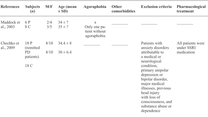
Results are organized in Table 1 (subjects [n], gender, mean age, presence of agoraphobia, other comorbidities, exclusion criteria, pharmacological treatment) and Table 2 (clinical design, type of stimulus, psychological assessment [scales used], objective measures of anxiety, scanner type, image processing, statistical analysis). The following items are described and discussed: sample, gender, laterality criteria, agoraphobia, other comorbidities, stimulus modalities, and psychological evaluation. Considerations of imaging genomics are included at the end of the Results section.
Results
Study samples
Most fMRI studies used a small number of patients in their investigation. Sample sizes of the collected studies varied from 1 (case studies) to 20. Although pioneering and highly relevant, the current studies’ samples remain to be considered representative of the population of PD. Because increasingly more studies of PD are being conducted using fMRI, increased sample sizes are expected in future investigations. Notably, sample sizes need to not only be increased but also need to be homogeneous with regard to both demographics (e.g., economic status, ethnicity) and clinical factors (e.g., past and current medical history).
Controlling these variables may be an important step toward more direct comparisons among studies in the pursuit of identifying the neurocircuitry of PD. Conducting cross-cultural fMRI studies of PD might also lead to a deeper understanding of the cultural aspects that contribute to the etiology and development of this disorder.
Gender
As discussed in a recent article, gender may play an important role in the neural activation of
emotional processing (Ohrmann et al., 2010). Women
display stronger activation of the insula, pre-central gyrus, middle cingulate cortex, left middle temporal gyrus, occipital gyri, and caudate in response to fearful facial stimuli compared with men. In women,
signiicant activation of the bilateral amygdala was
observed in response to fearful faces but, in men, no
signiicant activation of the amygdala was observed
(Ohrmann et al., 2010). Not all studies selected in the present systematic review balanced the sample with regard to gender.
Interestingly, a recent study demonstrated that women with Social Anxiety Disorder required less emotional intensity to recognize faces that expressed fear, sadness, and happiness on a computer screen, suggesting gender differences with regard to the sensitivity of evaluating threat- and approval-related social cues (Arrais et al., 2010). Because PD is also an anxiety disorder, men and women with PD may also respond differently to anxiogenic stimuli, an issue that must be addressed by future studies.
Laterality criteria
left-handed compared with right-left-handed individuals (Cuzzocreo et al., 2009). Some studies have indicated that these mental functions are not necessarily related to handedness and that hemispheric lateralization
could vary with material- or task-speciic paradigms. Hemispheric specialization can be speciic for particular
tasks, and a wide variety of mental functions could be successfully processed by both hemispheres depending
on their differences in processing eficacy (Gazzaniga,
Ivry, & Mangun, 2002).
Considering language as an example, left hemisphere dominance is not strongly related to handedness dominance. Approximately 50% of all left-handed subjects have left hemisphere dominance for language, although they are only 7% to 8% of the general population. Therefore, more than 96% of humans have left hemisphere dominance for language (Gazzaniga et al., 2002).
The decision to choose right-handed subjects to compose the sample for fMRI studies is based on minimizing possible confounds with regard to lateralization of some brain functions and the resulting brain activation, although this is not always true.
Speciically, in right-handers, the correlation between
cerebral dominance and handedness is much higher. Importantly, fMRI studies should use objective measures that help determine handedness. Although most studies suggest that a sample inclusion criterion must be that only right-handed subjects are selected, only two articles reported having used an objective measure to assess handedness. The authors used the Edinburgh Handedness Inventory (EHI) to select right-handed individuals for their study.
Agoraphobia
Agoraphobia is a common PD comorbidity. From 25% to 50% of individuals diagnosed with PD in community samples also have agoraphobia, although a much higher rate of agoraphobia is encountered in
clinical samples (Weissman et al., 1997). However,
a dearth of studies have used agoraphobia groups as experimental samples. Only six of the present studies included agoraphobic patients, but only one selected these subjects to form an experimental group. Future research should consider this issue. As discussed in more detail below, the severity of PD and agoraphobia—and not only the presence or absence of these illnesses— must be considered when interpreting fMRI data because a patient with mild agoraphobia, for example, although meeting clinical criteria, may perceive and interpret stimuli differently from a patient with severe agoraphobia.
Comorbidity
Most of the studies did not provide descriptions of the comorbidities presented by the PD patients. Seven
of the studies described comorbidities of PD but did not use this information to evaluate their results. The results were attributed to the characteristics of brain activation associated with PD. Thus, it is unclear whether other
disorders possibly interfered with their indings. Comorbidities relect clinical complexity and are the rule
rather than the exception. For example, in four panic-related subgroups, one or more comorbid conditions were found in 71.9% of the isolated panic-without-agoraphobia subgroup, 83.1% of the PD-without-agoraphobiasubgroup, and 100% of the isolated panic-with-agoraphobia and PD-panic-with-agoraphobia subgroups
(Kessler, Chiu, Jin, Ruscio, Shear, & Walters, 2006). In
this context, studying PD without addressing current and past comorbidities may yield no consistency.
Experimental protocols: stimulus modality
Comparing results of the 15 articles may be misleading because of several methodological differences including different experimental protocols. Divergent neuronal activation patterns can be extracted from different task performances (Davidson, 2002). For example, emotional induction using visual stimuli activated the amygdala in healthy subjects, whereas imagery stimulated the anterior cingulate cortex and
insula (Phan, Wager, Taylor, & Liberzon, 2002). As
presented in Table 2, PD studies used different modes of stimulus presentation. Eleven articles used visual stimuli in their fMRI paradigms. Three studies used auditory stimuli. One study used motor activation stimuli, and one visual study used electrodermal
stimulation to determine “threat” and “safe” conditions.
This may impact the brain areas that are activated. The three studies that applied auditory stimuli were very different in their methodological designs. The case report registered a PA during scanning. Despite their differences, two studies showed hyperactivation of the parahippocampal gyrus. Boshuisen, Ter Horst, Paans, Reinders, & den Boer (2002) found increased activity of the parahippocampal gyrus during anticipatory anxiety. This structure is related to memory and context and participates in the visceromotor network (Hasler, Nugent, Carlson, Carson, Geraci, & Drevets, 2008).
This activation may be explained by a compensatory effect attributable to parahippocampal gray matter
deicits (Massana et al., 2003b). Lai, Hsu, & Wu
(2010) demonstrated a gray matter volume decrease in the parahippocampal gyrus in a drug-naive sample
of PD patients with a irst episode of major depressive
disorder, which may be associated with abnormalities of interoceptive awareness and emotion modulation.
Important features of the third article that used auditory stimuli were increased activity of the right middle temporal gyrus, left anterior insula, and inferior parietal lobe during the third block of auditory
References Subjects (n)
M/F Age (mean ± SD)
Agoraphobia Other comorbidities
Exclusion criteria Pharmacological treatment
Marchand et al., 2008
12 P
18 C
F 27.5 ± 4.9
26.4 ± 3.9
________ GAD (1), GAD and DD (1), SP (1), SP and PTSD (1)
History of head injury, neurological disorder, medical disorder that could impact the central nervous system, any contraindications to fMRI and any psychiatric comorbidity other than another coexistent anxiety disorder or dysthymic disorder
BZD (2; needed basis/wash out 24 h), SSRI (1), SSRI and BZD (3; needed basis)
Pleiderer et al., 2007
1 F 26 x ________ Cardiologic,
neurological, comorbid psychiatric disorders
SSRI for 3 months prior to the MRI investigation
Pillay et al., 2007
8 P 8 C
4/4 4/4
36 ± 8.3 25.8 ± 3.5
________ ________ History of head injury with loss of consciousness or other medical illness that might affect cognitive function; history of any form of substance abuse or dependence; any other current Axis I disorder
SSRI and BZD (5); SSRI, BZD, and gabapentin (1); BZD (2)
Pillay et al., 2006
8P 8C
4/4 4/4
36 ± 8.3 31.6 ± 8.8
________ ________ History of head injury with loss of consciousness or other medical illness that might affect cognitive function; history of any form of substance abuse or dependence; any other current Axis I disorder.
SSRI and BZD (5); SSRI, BZD, and gabapentin (1); BZD (2)
van der Heuvel et al., 2005
15P (PD) 18P (OCD) 14P (H)
8/7 6/12 10/9
33.7 ± 2.5 33.4 ± 2.4 40.6 ± 3.2
________ ________ Major internal or neurological illness, other psychiatric disorders, and the use of psychotropic medication
Wash out for at least 4 weeks
analyzed the association between serotonin transporter gene variations and auditory processing and found that altered auditory habituation was associated with the less-active 5-HTTLPR S allele and the less-active 5-HTTLPR/rs25531 haplotype.
Among visual stimuli paradigm studies, paradoxical features can be found such as increased or diminished cingulate cortex and amygdala activity. As discussed by Davidson (2002), some results may
relect different levels of paradigm dificulty or may be inluenced by therapy modalities. Most studies did
not use physiological measures to determine whether the affective response corresponded to the intended emotion in the paradigm.
Psychological evaluation: the use of scales in fMRI studies of PD
All papers reviewed here used the DSM-IV criteria for diagnosing PD, with the exception of one study that used DSM-III criteria. The fMRI studies selected in this review used the following instruments: Structured Clinical Interview-Axis I, DSM-IV-R (SCID-I), Mini-Mental State Examination (MMSE), Edinburgh Handedness Inventory (EHI), Panic-Associated Symptom Scale (PASS), Hamilton Anxiety Scale (HAM-A), Hamilton Depression Rating Scale (HAM-D), Anxiety Sensitivity Index (ASI), State-Trait Anxiety Inventory – Trait scale (STAI-T), State-Trait Anxiety Inventory-State scale (STAI-S), Anxiety Control Questionnaire (ACQ), Body Sensation Questionnaire (BSQ), Beck Depression Inventory (BDI), Brief Symptom Inventory (BSI),
Clinical Global Impressions (CGI) scale, Differential Emotions Scale (DES), Mobility Inventory (MI), Positive and Negative Affect Schedule (PANAS), State-Trait Anger Expression Inventory (STAXI), and Panic and Agoraphobia Scale (PAS). Different types of scales were used among the studies, and only the following scales were used with more frequency: HAM-A, HAM-D, STAI-T, and EHI. For this reason,
these psychometric tools will be briely reviewed.
Hamilton Anxiety Scale (HAM-A)
The HAM-A (Hamilton, 1959) was one of the
irst rating scales developed to measure the severity
of anxiety symptoms and is still widely used today in both clinical and research settings. The scale consists
of 14 items, each deined by a series of symptoms, and
measures both psychic anxiety (i.e., mental agitation and psychological distress) and somatic anxiety (i.e., physical complaints related to anxiety). Although the HAM-A remains widely used as an outcome measure in clinical trials, it has been criticized for its sometimes poor ability to discriminate between anxiolytic and antidepressant effects and between somatic anxiety and somatic side effects (Maier, Buller, Philipp, & Heuser, 1988). The HAM-A does not provide any standardized probe questions. Despite this, the reported levels of inter-rater reliability for the scale appear to be acceptable. Each item is scored on a scale from 0 (not present) to 4 (severe), with a total score of 0 to 56 in which <17 indicates mild severity, 18-24 indicates mild to moderate, and 25-30 indicates moderate to severe. Six studies used the HAM-A.
Table 1. Continued
Maddock et al., 2003
6 P 8 C
2/4 3/5
34 ± ? 35 ± ?
x Only one pa-tient without agoraphobia
________ ________ ________
Chechko et al., 2009
18 P (remitted PD patients)
18 C
8/10
8/10
34.4 ± 8
30 ± 6.4
________ ________ Patients with anxiety disorders attributable to a medical or neurological condition, primary unipolar depression or bipolar disorder, major medical illnesses, previous head injury with loss of consciousness, and substance abuse or dependence
All patients were under SSRI medication References Subjects
(n)
M/F Age (mean ± SD)
Agoraphobia Other comorbidities
Hamilton Depression Rating Scale (HAM-D)
One year after publishing the HAM-A, the depression version of the scale was presented (Hamilton, 1960). The HAM-D has proven useful for many years as a method of determining a patient’s level of depression before, during, and after treatment. It should be administered by a clinician experienced in working with psychiatric patients. Although the HAM-D form
consists of 21 items, the scoring is based on the irst
17 items. It generally takes 15-20 min to complete the interview and score the results. Eight items are scored on a 5-point scale ranging from 0 (not present) to 4 (severe). Nine items are scored from 0 to 2. Since its development, the scale has been widely used in clinical practice and became a standard in pharmaceutical trials. The HAM-D score is the result of summing the scores
from the irst 17 items, with 0-7 indicating Normal, 8-13 indicating Mild Depression, 14-18 indicating Moderate Depression, 19-22 indicating Severe
Depression, and ≥23 indicating Very Severe Depression (Hamilton, 1960; Williams, 2001). Several studies
evaluated this scale and concluded that the HAM-D exhibited a relatively stable factorial structure based on a large sample of outpatients with unipolar depressive disorders (O’Brien & Glaudin, 1988), but its total score may be a weak index of depressive syndrome severity (Gibbons, Clark, & Kupfer, 1993). Only three studies used the HAM-D; the other two studies used the BDI to assess depressive states.
State-Trait Anxiety Inventory-Trait (STAI-T) scale
The STAI scale, which is appropriate for those who have at least a sixth grade reading level, consists of a four-point Likert scale to assess anxiety. The STAI scale assesses state anxiety (i.e., a transient condition characterized by tension, apprehension, and hyperactivity of the autonomic nervous system) and trait anxiety (i.e., a general tendency that an individual has to respond, with anxiety in response to environmental
stimuli). The STAI scale was irst published in 1970
(Spielberger, Gorsuch, & Lushene, 1970) with the purpose of measuring anxiety in adults, although currently a version for children is available. It has been widely used by clinicians and researchers, although some suggest that the trait scale may assess both depression and anxiety (Bieling, Antony, & Swinson, 1998; Bados, Gómez-Benito, & Balaguer, 2010). The instrument is divided into two sections, each with 20 questions. The STAI scale has three forms. The current variation is the STAI Form Y, which differentiates between temporary or emotional state anxiety and long-standing personality
trait anxiety in adults. The STAI Form X is the irst
version of the STAI, which is still available. The third form is the STAI for children.
The STAI Form Y serves as an indicator of two types of anxiety: state and trait anxiety. It measures
the severity of the overall level of anxiety and is an administered analysis of reported anxiety symptoms.
The irst subscale measures state anxiety, and the
second subscale measures trait anxiety. The scores range from 20 to 80, with higher scores indicating greater anxiety (Spielberger et al., 1970). Some of the questions are related to the absence of anxiety and are reverse-scored. Results of the STAI scale can be used in the formulation of clinical diagnoses, for psychological and health research, and for the assessment of clinical anxiety in patients in medical, surgical, and psychiatric settings. Four studies from those selected in the present review used this scale to assess anxiety levels among participants.
Edinburgh Handedness Inventory (EHI)
As mentioned above, establishing laterality criteria is an important component of fMRI research designs. The EHI is probably the most widely used scale for
assessing this feature (Oldield, 1971). Briely, the
inventory is composed of 10 items representing daily activities such as drawing, writing, using a spoon, and
throwing objects. By relecting on the way these tasks
are performed, the participant is encouraged to check his/her preference in using his/her left or right hand. The inventory includes the possibility that the preference is so strong that the participant would never use the other hand unless absolutely forced to, a situation where the participant may place two checks, resulting in different scoring. Only one study from the selected articles of this systematic review assessed this parameter.
Psychological evaluation and fMRI studies of PD: considerations of the application of scales
An important aspect to be considered concerning the use of scales in fMRI studies is the time during which the scale is applied. Only two studies applied the scales at the time of scanning to quantify the severity of the panic symptoms present at the exact time of the exam, although three other studies reported anxiety symptoms during the fMRI exam. As a measure of state anxiety, one study applied the DES immediately after the fMRI exam to assess the intensity of anxiety during fMRI scanning.
Only 7/15 selected studies reported patients’ scores on the scales. This represents important information for correlating the severity of PD and the imaging activation found and should be considered in future studies.
Lack of consensus concerning the choice of scales used, time at which they are applied, and absence of a correlation between severity of the diagnosis and
imaging results make it dificult to analyze the data as a
whole and to compare them with other studies.
of agoraphobia. These parameters reveal discrete but important aspects that may show the level of gravity
and idiosyncratic speciicities of the study’s sample. In
this sense, these scores should not be neglected when interpreting fMRI data in PD studies.
Imaging genomics
The knowledge about genetics in the pathogenesis
of PD has experienced signiicant advances during the
last several years. However, the relationship between PD and genetics is complex. A large number of small-effect genes may contribute to vulnerability to the disease (Rothe et al., 2006).
Only three of the reviewed articles discussed this
emerging ield of science. All of these studies had small
samples. They included both male and female patients, medicated and non-medicated patients, and comorbid psychiatric conditions. Two of the three studies did not utilize a healthy volunteer control group. These may be confounding factors in the discussion of the results. Two of the three studies used neuronal activation in response to visual stimuli as an endophenotype of PD to be associated with polymorphisms. One study analyzed the association between neuronal activation
in response to auditory stimulation and the inluence
of the 5-HTTLPR polymorphism. Similar studies with
reined and larger samples are necessary in order to
make comparisons.
One of the studies discussed the gene that codes for the enzyme catechol-O-methyltransferase (COMT). COMT is an enzyme that metabolizes monoaminergic neurotransmitters including adrenaline, noradrenaline,
and dopamine (Weinshilboum, Otterness, & Szumlanski,
1999). The COMT gene is located on chromosome 22q11.2 (Rothe et al., 2006). A nucleotide substitution polymorphism (guanine to adenosine) in codon 158 of the COMT gene results in an amino acid change from valine to methionine, with the valine allele relating to higher COMT activity. The COMT genetic variation
may inluence limbic and prefrontal brain activation in
response to unpleasant stimuli (Domschke et al., 2008).
Information processing deicits appear to play a crucial
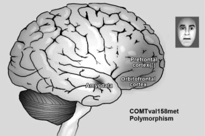
role in the pathogenesis of PD. Domschke et al. (2008) used neuronal activation elicited by emotional stimuli as an endophenotype to study the association with PD and found that patients who carried at least one 158val allele showed increased activation of the amygdala in response to fearful faces and increased activation of the left orbitofrontal cortex (Fiigure 1). Other alterations were also observed, indicating that activation of the amygdala and prefrontal cortex in response to emotional
faces may be inluenced by the genetic variation. The
COMT variation may alter the neuronal processing of anxiety-related emotional cues.
In a previous study, Domschke et al. (2006) studied serotonergic polymorphisms 1019C/G 5-HT1A
and 5-HTTLPR in PD patients. The serotonergic system appeared to play an important role in the etiology of PD. The G allele of the –1019C/G promoter polymorphism in the gene that codes for the 5-HT1A receptor located on chromosome 5q12.3 was proposed to depress 5-HT1A autoreceptor expression, reducing serotonergic neurotransmission (Domschke
Figure 1. Differential activation of brain areas in PD patients with the COMT polymorphism.
et al., 2006). Another serotonergic polymorphism was the variant site 5HTTLPR ( serotonin-transporter-linked promoter region). The short “s” allele of the
serotonin transporter (5-HTT) is associated with reduced transcription and functional capacity of the serotonin transporter. Perna, Favaron, Di Bella, Bussi, & Bellodi (2005) showed that monozygotes for the short allele (s/s) of 5HTTLPR had a worse response to
paroxetine. Domschke et al. (2006) found a signiicant
decrease in the activation of the right ventromedial and orbitofrontal cortex and right anterior cingulate cortex in patients homozygous for the high-risk G allele of the 5-HT1A–1019 C/G promoter polymorphism. They also concluded that both the 5-HT1A–1019 C/G and 5HTTLPR polymorphisms play a role in amygdala activation in response to positive emotional stimuli. This study also had a small sample and no control group with healthy volunteers; additionally, some patients were under treatment and others had psychiatric comorbidities. Despite these limitations, its contribution to the understanding of gene-environment interactions for the PD phenotype is notable.
Discussion
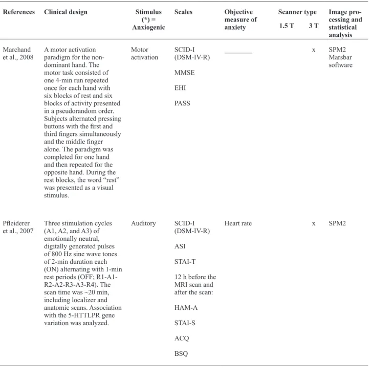
Table 2. Parameters compared among studies with regard to experimental designs and protocols
References Clinical design Stimulus (*) = Anxiogenic
Scales Objective measure of anxiety
Scanner type Image pro-cessing and statistical analysis 1.5 T 3 T
Marchand et al., 2008
A motor activation paradigm for the non-dominant hand. The motor task consisted of one 4-min run repeated once for each hand with six blocks of rest and six blocks of activity presented in a pseudorandom order. Subjects alternated pressing buttons with the irst and third ingers simultaneously and the middle inger alone. The paradigm was completed for one hand and then repeated for the opposite hand. During the rest blocks, the word “rest” was presented as a visual stimulus.
Motor activation
SCID-I (DSM-IV-R)
MMSE
EHI
PASS
________ x SPM2
Marsbar software
Pleiderer et al., 2007
Three stimulation cycles (A1, A2, and A3) of emotionally neutral, digitally generated pulses of 800 Hz sine wave tones of 2-min duration each (ON) alternating with 1-min rest periods (OFF; R1-A1-R2-A2-R3-A3-R4). The scan time was ~20 min, including localizer and anatomic scans. Association with the 5-HTTLPR gene variation was analyzed.
Auditory SCID-I (DSM-IV-R)
ASI
STAI-T
12 h before the MRI scan and after the scan:
HAM-A
STAI-S
ACQ
BSQ
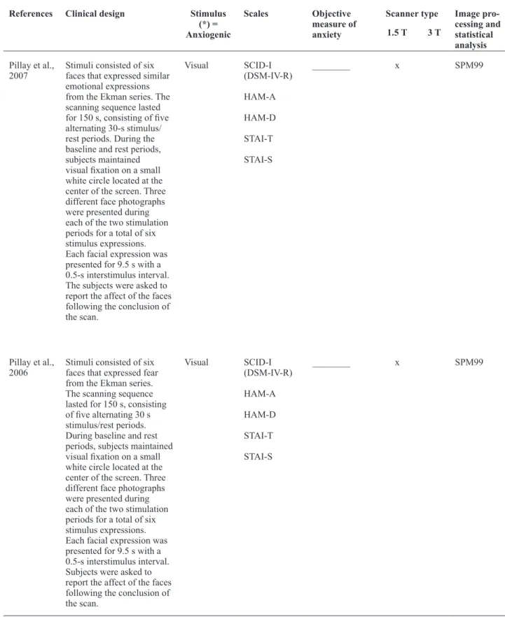
Table 2.Continued
Pillay et al., 2007
Stimuli consisted of six faces that expressed similar emotional expressions from the Ekman series. The scanning sequence lasted for 150 s, consisting of ive alternating 30-s stimulus/ rest periods. During the baseline and rest periods, subjects maintained visual ixation on a small white circle located at the center of the screen. Three different face photographs were presented during each of the two stimulation periods for a total of six stimulus expressions. Each facial expression was presented for 9.5 s with a 0.5-s interstimulus interval. The subjects were asked to report the affect of the faces following the conclusion of the scan.
Visual SCID-I (DSM-IV-R)
HAM-A
HAM-D
STAI-T
STAI-S
________ x SPM99
Pillay et al., 2006
Stimuli consisted of six faces that expressed fear from the Ekman series. The scanning sequence lasted for 150 s, consisting of ive alternating 30 s stimulus/rest periods. During baseline and rest periods, subjects maintained visual ixation on a small white circle located at the center of the screen. Three different face photographs were presented during each of the two stimulation periods for a total of six stimulus expressions. Each facial expression was presented for 9.5 s with a 0.5-s interstimulus interval. Subjects were asked to report the affect of the faces following the conclusion of the scan.
Visual SCID-I (DSM-IV-R)
HAM-A
HAM-D
STAI-T
STAI-S
________ x SPM99
References Clinical design Stimulus (*) = Anxiogenic
Scales Objective measure of anxiety
van der Heuvel et al., 2005
A cognitive and emotional Stroop task consisting of congruent and incongruent color words, obsessive compulsive disorder-related and panic-related negative words, and neutral words. Stimuli were presented in a block design, consisting of 18 randomized blocks (three blocks of each condition), each containing 16 words. Each word was presented for 2 s followed by a 200-ms blank screen. Subjects were asked to respond as fast as possible by pressing the button that corresponded to the color of the ink, regardless of the meaning of the word. After performing the task, subjects were asked to rate subjective distress using a 100-point analog scale.
Visual SCID-I (DSM-IV-R)
BSQ (panic patients)
________ x SPM99
Maddock et al., 2003
Stimuli consisted of 10 threat words and 10 emotionally neutral words matched for word length and frequency of usage. Each word was presented once in pseudorandom order in each 16-s block of 10 words of the same type. Sixteen alternating blocks of threat-related and neutral words were presented for 256 s following a 32-s baseline. Subjects were instructed to form a silent judgment of the valence of each word. After scanning, the subjects were questioned about stimulus audibility, task performance, and their emotional state during the scan.
Auditory* SCD-I (DSM-III-R)
HAM-A
Panic diaries
________ x Medx
software
BrainMRI software Table 2.Continued
References Clinical design Stimulus (*) = Anxiogenic
Scales Objective measure of anxiety
Table 2.Continued
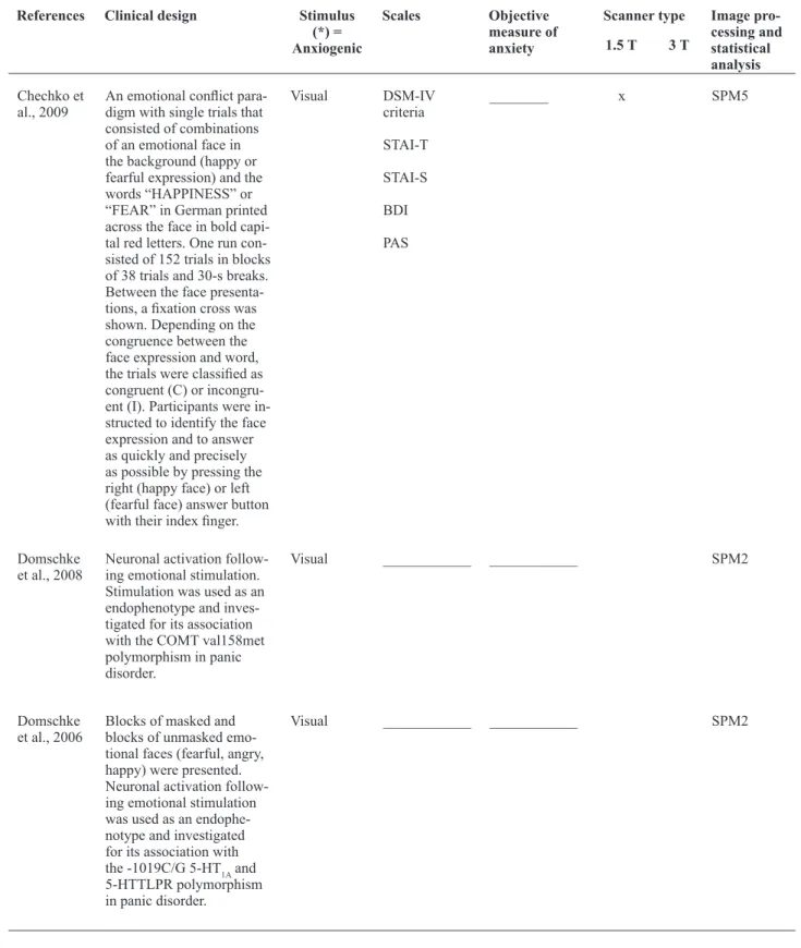
Chechko et al., 2009
An emotional conlict para-digm with single trials that consisted of combinations of an emotional face in the background (happy or fearful expression) and the words “HAPPINESS” or “FEAR” in German printed across the face in bold capi-tal red letters. One run con-sisted of 152 trials in blocks of 38 trials and 30-s breaks. Between the face presenta-tions, a ixation cross was shown. Depending on the congruence between the face expression and word, the trials were classiied as congruent (C) or incongru-ent (I). Participants were in-structed to identify the face expression and to answer as quickly and precisely as possible by pressing the right (happy face) or left (fearful face) answer button with their index inger.
Visual DSM-IV
criteria
STAI-T
STAI-S
BDI
PAS
________ x SPM5
Domschke et al., 2008
Neuronal activation follow-ing emotional stimulation. Stimulation was used as an endophenotype and inves-tigated for its association with the COMT val158met polymorphism in panic disorder.
Visual ____________ ____________ SPM2
Domschke et al., 2006
Blocks of masked and blocks of unmasked emo-tional faces (fearful, angry, happy) were presented. Neuronal activation follow-ing emotional stimulation was used as an endophe-notype and investigated for its association with the -1019C/G 5-HT1A and 5-HTTLPR polymorphism in panic disorder.
Visual ____________ ____________ SPM2
References Clinical design Stimulus (*) = Anxiogenic
Scales Objective measure of anxiety
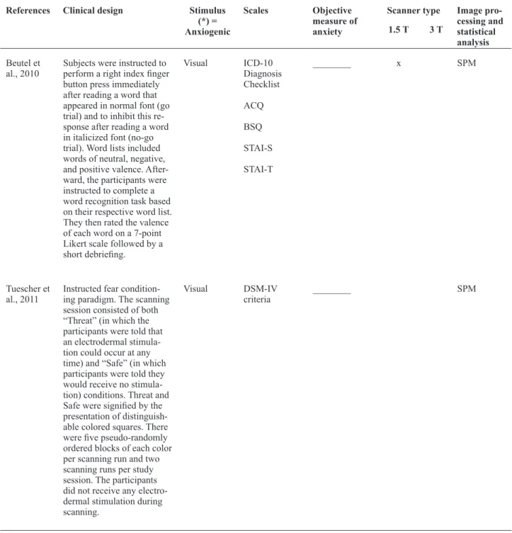
Beutel et al., 2010
Subjects were instructed to perform a right index inger button press immediately after reading a word that appeared in normal font (go trial) and to inhibit this re-sponse after reading a word in italicized font (no-go trial). Word lists included words of neutral, negative, and positive valence. After-ward, the participants were instructed to complete a word recognition task based on their respective word list. They then rated the valence of each word on a 7-point Likert scale followed by a short debrieing.
Visual ICD-10
Diagnosis Checklist
ACQ
BSQ
STAI-S
STAI-T
________ x SPM
Tuescher et al., 2011
Instructed fear condition-ing paradigm. The scanncondition-ing session consisted of both “Threat” (in which the participants were told that an electrodermal stimula-tion could occur at any time) and “Safe” (in which participants were told they would receive no stimula-tion) conditions. Threat and Safe were signiied by the presentation of distinguish-able colored squares. There were ive pseudo-randomly ordered blocks of each color per scanning run and two scanning runs per study session. The participants did not receive any electro-dermal stimulation during scanning.
Visual DSM-IV
criteria
________ SPM
Table 2.Continued
References Clinical design Stimulus (*) = Anxiogenic
Scales Objective measure of anxiety
Table 2.Continued
Ohrmann et al., 2010
Facial stimuli consisted of gray-scale-normalized im-ages that depicted fearful, angry, happy, and neutral expressions of 10 male and female individuals. The subjects were presented with alternating 30-s blocks of one of the four facial expressions or a no-face control stimulus (a gray rectangle). The order of the blocks was counterbalanced across subjects. Each face epoch was preceded by a no-face control epoch and was presented twice, result-ing in an overall presenta-tion time of 8 min.
Visual DSM-IV
criteria
DES
EHI
STAI-S
STAI-T
________ SPM2
Wittmann et al., 2010
Twenty-four neutral pic-tures and 24 agoraphobia pictures were presented in a cued manner. The other remaining 48 neutral and agoraphobia pictures were presented uncued by a ran-dom combination of charac-ters. The picture sequence was randomized for each subject. Each picture was presented for 2,000 ms, and cues were presented for 250 ms, separated by the pre-sentation of a ixation cross. The complete presentation duration was ~15 min. The participants were instructed to pay attention to the con-tent of the pictures, experi-ence the presented situation, and pay attention to the cue and its predictive content before picture presentation. They were requested to push a button each time a picture was presented.
Visual
CAPI-WHO-CIDI
HAM-A
CGI
BSI
BDI
MI
EHI
________ SPM5
References Clinical design Stimulus (*) = Anxiogenic
Scales Objective measure of anxiety
Pleiderer et al., 2010
Three stimulation cycles (A1-A3) of digitally gener-ated pulsed (5 Hz frequen-cy) 800-Hz sine wave tones of 2 min duration, which alternated with rest periods (R1-R3) of 1 min duration. Auditory stimulation was presented binaurally via pneumatic headphones. The hearing threshold was deter-mined individually within the magnet. All subjects had comparable hearing thresholds, and each subject was stimulated with a sound pressure level of 85 dB above their individual hear-ing threshold.
Auditory SCD-I (DSM-IV)
HAM-A
HAM-D
________ SPM5
Dresler et al., 2011
Participants had to passively view emotional faces. Four different emotions (anxious, happy, sad, and neutral) were used in a block para-digm. Each block contained eight faces (four female, four male) and was present-ed four times in a random order. Each face was shown for 2 s, directly followed by the next one. Face blocks were alternated with blocks of the same length that showed a white ixation cross. The patients were in-structed to attentively look at the faces and empathize with the expression.
Visual ICD-10
criteria
STAI-S
STAI-T
STAXI
PANAS
VAS
BDI
________ x SPM5
Table 2.Continued
and new psychotherapeutic techniques that address specific components of the fear neurocircuitry that underlies PD.
Neuroimaging studies play a crucial role in this scenario because they allow researchers to achieve a deeper understanding of the phenomenology associated with the brain circuits that underlie anxiety disorders (Davidson, 2002). Such knowledge emerges from research designs that allow a
better analysis of speciic affective and cognitive
processing. Additionally, these studies can reveal the heterogeneity of these disorders, which may contribute to descriptions of more accurate subtypes of psychopathologies. Functional neuroimaging
techniques can verify the impact of certain therapeutic interventions on brain function.
Although many aspects of this anxiety disorder have been investigated from different perspectives, much remains to be discovered. The purpose of this review was to highlight the importance of fMRI studies of PD by acknowledging the value of using different available methodological resources in current science and encouraging researchers to address some important points concerning research criteria. In this way, data published by different groups worldwide can be more easily integrated and compared. This may represent an important step towards establishing more conclusive conceptual models of PD.
References Clinical design Stimulus (*) = Anxiogenic
Scales Objective measure of anxiety
When considering the use of fMRI in the study of a
certain disorder, it would be interesting for researchers to address some of the highlighted points; otherwise, consistency of the data may be compromised or the
scientiic literature in regard to the issue may be
composed of data that cannot be generalized in terms of building consensus and a more uniform delineation of the neurocircuitry of PD. Davidson (2002) suggested that when evaluating treatment-related differences,
researchers commonly associate the neural indings with vulnerability to a speciic disorder; however,
the possibility that the differences are attributable to nonclinical features as a consequence of the pathology should not be ruled out. Additionally, in studies where patients are scanned on two or more occasions, test-retest reliability should be considered, highlighting the importance of associating psychometrics to psychophysiological measures and neuroimaging
indings. One of the chosen parameters analyzed
among fMRI studies of PD was use of psychological assessment, and consideration should be made of the data obtained from scales and inventories when interpreting neuroimages.
The research criteria that diverged among the selected papers of this present systematic review or that need to be reconsidered in future investigations include the following: (1) samples were small and cannot be considered representative of the PD population and we strongly encourage designing cross-cultural studies in order that a more universal model of the neurocircuitry of PD can be developed; (2) samples must be balanced with regard to gender because men and women display different brain activation patterns; (3) fMRI studies need to use objective measures that help assess handedness, such as the EHI, because handedness can provide evidence of laterality dominance, accounting for differential brain activation; (4) high levels of comorbidity between PD and agoraphobia indicate the need to include agoraphobia in future studies so that the samples are as similar as possible to what is found in the population; (5) comorbidities presented by PD patients should be described and considered in the interpretation of the results because differential activation may be attributable to PD, comorbidities, or their interaction; (6) stimulus modalities varied, which is important with regard to understanding how PD patients process different sensory information; (7) future studies that investigate a given sensory modality should consider applying similar methodologies so that the data can be compared and analyzed across studies rather than just described; (8) the studies used a wide range of scales and inventories to perform psychological evaluations, but these instruments varied considerably among studies both in terms of the phenomena assessed and the psychometric instruments chosen. However, what was most evident was the lack
of consideration of the obtained scores in the analysis
of fMRI indings.
Additionally, an important parameter to be assessed in PD patients is agoraphobic avoidance behavior and frequency of panic attacks, which can be consistently addressed with the Mobility Inventory for Agoraphobia
(MI; Chambless, Caputo, Jasin, Gracixy, & Williams,
1985). Interestingly, none of the studies mentioned in this present review used this scale as part of the psychological assessment.
Conclusion
Panic disorder leads patients to severe psychological suffering, and fMRI studies constitute important tools for revealing the fear neurocircuitry that underlies this
condition. However, the mental health ield may beneit
from these studies, both in terms of establishing more
precise diagnostic criteria for PD based on speciic neural
activation and designing more accurate pharmacological and psychotherapeutic treatments that may remodulate this circuitry if research designs attempt to follow the complexity of PD and address its multifactorial features. The present systematic review was intended to provide helpful guidelines for future fMRI studies of PD. These include increasing sample sizes, using homogeneous samples, addressing severity and comorbidities issues, and analyzing the psychological evaluations performed because these scores may provide important features of the studied sample and the possibility of correlating them with the neural networks activated by anxiogenic and panicogenic stimuli.
Acknowledgements
The authors received inancial support for this article from the Brazilian Council for Scientiic
and Technological Development (CNPq), FAPERJ/ PRONEX, INCT/CNPq/National Institute of Translational Medicine, and INCT/CNPq/National Institute of Translational Neuroscience. The authors have
no other relevant afiliations or inancial involvement with any organization or entity with a inancial interest in, or inancial conlict with, the subject matter or
materials discussed in the manuscript, apart from those disclosed. No writing assistance was utilized in the production of this manuscript.
References
Amaro, E., Jr., & Yamashita, H. (2001). Aspectos básicos de
tomograia computadorizada e ressonância magnética. Revista Brasileira de Psiquiatria, 23, 2-3.
American Psychiatric Association. (2000). Diagnostic and statistical manual of mental disorders, 4th edition, text revision. Washington,
D.C.: American Psychiatric Association.
anxiety disorder women easily recognize fearful, sad and happy
faces: The inluence of gender. Journal of Psychiatric Research,
44, 535-540.
Bados, A., Gómez-Benito, J., & Balaguer, G. (2010). The State-Trait Anxiety Inventory, trait version: Does it really measure anxiety?
Journal of Personality Assessment, 92, 560-567.
Ballenger, J. C. (1989). Toward an integrated model of panic disorder.
American Journal of Orthopsychiatry, 59, 284-293.
Bieling, P. J., Antony, M. M., & Swinson, R. P. (1998). The State-Trait Anxiety Inventory, trait version: structure and content re-examined.
Behaviour Research and Therapy, 36, 777-788.
Berkowitz, R. L., Coplan, J. D., Reddy, D. P., & Gorman, J. M. (2007). The human dimension: How the prefrontal cortex modulates the subcortical fear response. Reviews in the Neurosciences, 18, 191-207. Boshuisen, M. L., Ter Horst, G. J., Paans, A. M., Reinders, A.
A., & den Boer, J. A. (2002). rCBF differences between panic disorder patients and control subjects during anticipatory anxiety and rest. Biological Psychiatry,52, 126-135.
Bourin, M., Baker, G. B., & Bradwejn, J. (1998). Neurobiology of panic disorder. Journal of Psychosomatic Research, 44, 163-180. Chambless, D. L., Caputo, G. C., Jasin, S. E., Gracely, E. J., &
Williams, C. (1985). The Mobility Inventory for Agoraphobia. Behaviour Research and Therapy,23, 35-44.
Chechko, N., Czisch, M., Erhardt, A., Hoehn, D., Wehrle, R., &
Sämann, P. (2007). Control of the anterior cingulate/mPFC over the amygdala: a longitudinal fMRI study in patients with panic disorder. Pharmacopsychiatry, 40(5), 230.
Cuzzocreo, J. L., Yassa, M. A., Verduzco, G., Honeycutt, N. A., Scott, D. J., & Bassett, S.S. (2009). Effect of handedness on fMRI activation in the medial temporal lobe during an auditory verbal memory task. Human Brain Mapping,30, 1271-1278.
Davidson, R. J. (2002). Activation paradigms in affective and cognitive neuroscience: Probing the neuronal circuitry underlying mood and anxiety disorders. In K. L. Davis, D. Charney, J. T. Coyle, & C. Nemeroff (Eds.), Neuropsychopharmacology: The
ifth generation of progress (pp. 373-381). New York: Lippincott
Williams and Wilkins.
De Carvalho, M. R., Dias, G. P., Cosci, F., de-Melo-Neto, V. L., Bevilaqua, M. C. N., Gardino, P. F., & Nardi, A. E. (2010).
Current indings of fMRI in panic disorder: contributions for
the fear neurocircuitry and CBT effects. Expert Reviews of Neurotherapeutics, 10, 291-303.
Domschke, K., Braun, M., Ohrmann, P., Suslow, T., Kugel, H., Bauer, J., … Deckert, J. (2006). Association of the functional -1019C/ G 5-HT1A polymorphism with prefrontal cortex and amygdala activation measured with 3 T fMRI in panic disorder. International Journal of Neuropsychopharmacology, 9(3), 349-355.
Domschke, K., Ohrmann, P., Braun, M., Suslow, T., Bauer, J.,
Hohoff, C., … Kugel, H. (2008). Inluence of the catechol-O -methyltransferase val158met genotype on amygdala and prefrontal cortex emotional processing in panic disorder. Psychiatry Research,
163(1), 13-20.
Dresler, T., Hahn, T., Plichta, M. M., Ernst, L. H., Tupak, S. V., Ehlis, A. C., … Fallgatter, A. J. (2011). Neural correlates of spontaneous panic attacks. Journal of Neural Transmission, 118(2), 263-9. Gazzaniga, M. S., Ivry, R. B., & Mangun, G. R. (2002). Cognitive
neuroscience: The biology of the mind. New York: W. W. Norton.
Gibbons, R. D., Clark, D. C., & Kupfer, D. J. (1993). Exactly what does the Hamilton Depression Rating Scale measure? Journal of Psychiatric Research, 27, 259-273.
Gorman, J. M., Kent, J. M., Sullivan, G. M., & Coplan, J. D. (2000). Neuroanatomical hypothesis of panic disorder, revised. American Journal of Psychiatry, 157(4), 493-505.
Hamilton, M. (1959). The assessment of anxiety states by rating.
British Journal of Medical Psychology, 32, 50-55.
Hamilton, M. (1960). A rating scale for depression. Journal of Neurology, Neurosurgery, and Psychiatry, 23, 56-62.
Hasler, G., Nugent, A. C., Carlson, P. J., Carson, R. E., Geraci, M., &
Drevets, W. C. (2008). Altered cerebral γ-aminobutyric acid type
A-benzodiazepine receptor binding in panic disorder determined by [11C]lumazenil positron emission tomography. Archives of
General Psychiatry, 65(10), 1166-1175.
Kessler, R. C., Chiu, W. T., Jin, R., Ruscio, A. M., Shear, K., & Walters, E. E. (2006). The epidemiology of panic attacks, panic
disorder, and agoraphobia in the National Comorbidity Survey Replication. Archives of General Psychiatry, 63, 415-424.
Lai, C. H., Hsu, Y. Y., & Wu, Y. T. (2010). First episode drug-naïve
major depressive disorder with panic disorder: Gray matter
deicits in limbic and default network structures. European Neuropsychopharmacology, 20(10), 676-682.
Maddock, R. J., Buonocore, M. H., Kile, S. J., & Garrett, A. S. (2003). Brain regions showing increased activation by threat-related words in panic disorder. Neuroreport, 14(3), 325-328.
Maier, W., Buller, R., Philipp, M., & Heuser, I. (1988). The Hamilton
Anxiety Scale: Reliability, validity and sensitivity to change in anxiety and depressive disorders. Journal of Affective Disorders, 14, 61-68.
Marchand, W. R., Lee, J. N., Healy, L., Thatcher, J. W., Rashkin,
E., Starr, J., Hsu, E. (2008). An fMRI motor activation paradigm demonstrates abnormalities of putamen activation in females with panic disorder. Journal of Affective Disorders, 116(1-2), 121-125. Massana, J., Serra-Grabulosa, J. M., Salgado-Pineda, P., Gaso, C.,
Junque, C., Massana, J., ... Salamero, M. (2003a). Amygdalar atrophy in panic disorder patients detected by volumetric magnetic resonance imaging. Neuroimage, 19, 80-90.
Massana, G., Serra-Grabulosa, J. M., Salgado-Pineda, P., Gastó,
C., Junqué, C., Massana, J., ... Mercader, J. M. (2003b).
Parahippocampal gray matter density in panic disorder: a voxel-based morphometric study. American Journal of Psychiatry, 160, 566-568.
O’Brien, K. P., & Glaudin, V. (1988). Factorial structure and factor reliability of the Hamilton Rating Scale for Depression. Acta Psychiatrica Scandinavica, 78, 113-120.
Ohrmann, P., Pedersen, A., Braun, M., Bauer, J., Kugel, H., Kersting, A., … Suslow, T. (2010). Effect of gender on processing threat-related stimuli in patients with panic disorder: Sex does matter.
Depression and Anxiety, 27, 1034-1043.
Oldield, R. C. (1971). The assessment and analysis of handedness:
The Edinburgh Inventory. Neuropsychologia, 9, 97-113.
Perna, G., Favaron, E., Di Bella, D., Bussi, R., & Bellodi, L.
(2005). Antipanic eficacy of paroxetine and polymorphism
within the promoter of the serotonin transporter gene.
Neuropsychopharmacology, 30, 2230-2235.
Pleiderer, B., Zinkirciran, S., Arolt, V., Heindel, W., Deckert, J.,
& Domschke, K. (2007). fMRI amygdala activation during a spontaneous panic attack in a patient with panic disorder. World Journal of Biological Psychiatry, 8(4):269-72.
Pleiderer, B., Zinkirciran, S., Michael, N., Hohoff, C., Kersting, A.,
Arolt, V., … Domschke, K. (2010). Altered auditory processing in patients with panic disorder: A pilot study. World Journal of Biological Psychiatry, 11(8), 945-955.
Pillay, S. S., Gruber, S. A., Rogowska, J., Simpson, N., & Yurgelun-Todd, D. A. (2006). fMRI of fearful facial affect recognition in panic disorder: The cingulate gyrus-amygdala connection. Journal of Affective Disorders, 94(1-3), 173-181.
Pillay, S. S., Rogowska, J., Gruber, S. A., Simpson, N., & Yurgelun-Todd, D. A. (2007). Recognition of happy facial affect in panic disorder: an fMRI study. Journal of Anxiety Disorders, 21(3): 381-393.
Phan, K. L., Wager, T., Taylor, S. F., & Liberzon, I. (2002). Functional
neuroanatomy of emotion: A meta-analysis of emotion activation studies in PET and fMRI. Neuroimage, 16(2), 331-348.
Rocha, E. T., Alves, T. C. T. F., Garridoc, G. E. J., Buchpiguel, R.,
Nitrinid, R., & Filho, G. B. (2001). Novas técnicas de neuroimagem
em psiquiatria: Qual o potencial de aplicações na prática clínica?
Revista Brasileira de Psiquiatria, 23, 58-60.
Rothe, C., Koszycki, D., Bradwejn, J., King, N., Deluca, V., Tharmalingam, S., ... Macciardi, F. (2006). Association of the Val158Met catechol O-methyltransferase genetic polymorphism with panic disorder. Neuropsychopharmacology, 31, 2237-2242. Schunck, T., Erb, G., Mathis, A., Gilles, C., Namer, I. J., Hode, Y.,
… Demaziere, A. (2006). Functional magnetic resonance imaging characterization of CCK-4-induced panic attack and subsequent anticipatory anxiety. Neuroimage, 31, 1197-1208.
Spielberger, C. D., Gorsuch, R. L., & Lushene, R. E. (1970). Manual for the State-Trait Anxiety Inventory. Palo Alto: Consulting Psychologists Press.
Tuescher, O., Protopopescu, X., Pan, H., Cloitre, M., Butler, T., Goldstein, M., … Stern, E. (2011). Differential activity of subgenual cingulate and brainstem in panic disorder and PTSD.
Journal of Anxiety Disorders, 25(2), 251-257.
Psychiatry Research, 63, 21-29.
van den Heuvel, O. A., Veltman, D. J., Groenewegen, H. J., Witter, M.
P., Merkelbach, J., Cath, D. C., … van Dyck, R. (2005).
Disorder-speciic neuroanatomical correlates of attentional bias in
obsessive-compulsive disorder, panic disorder, and hypochondriasis. Archives of General Psychiatry, 62(8), 922-933.
Weinshilboum, R. M., Otterness, D. M., & Szumlanski, C. L. (1999).
Methylation pharmacogenetics: Catechol O-methyltransferase, thiopurine methyltransferase, and histamine N-methyltransferase.
Annual Review of Pharmacology and Toxicology, 39, 19-52.
Weissman, M. M., Bland, R. C., Canino, G. J., Faravelli, C.,
Greenwald, S., Hwu, H. G., … Yeh, E. K. (1997). The cross-national epidemiology of panic disorder. Archives of General Psychiatry, 54, 305-309.
Williams, J. B., (2001). Standardizing the Hamilton Depression Rating
Scale: Past, present, and future. European Archives of Psychiatry and Clinical Neuroscience, 251(Suppl. 2), II6-II2. (Please verify II6-II2)
Wittmann, A., Schlagenhauf, F., John, T., Guhn, A., Rehbein, H., Siegmund, A., … Ströhle, A. (2010). A new paradigm