Case Report
Key Words
Aortic valve / surgery; acute coronary syndrome; coronary vessels; dissection.
Late aortic dissection can occur after aortic valve replacement surgery, but rarely in the first postoperative month. Coronary artery dissection is rare and usually occurs after coronary angiography. We report a rare case of coronary artery dissection followed by myocardial infarction in the immediate postoperative period of a successful aortic valve replacement with a good postoperative evolution.
Acute Coronary Artery Dissection after Aortic Valve Replacement
Fernando de Paula Machado, Roney Orismar Sampaio, Fernanda Lopez Mazzucato, Flávio Tarasoutchi, Guilherme
Sobreira Spina, Max Grinberg
Instituto do Coração do Hospital das Clínicas da Universidade de São Paulo, São Paulo, SP - Brazil
Mailing address: Roney Orismar Sampaio •
Divisão de Clínica - Unidade de Cardiopatias Valvares - Instituto do Coração - Av. Enéas de Carvalho Aguiar, 44, andar AB -05403-000 - São Paulo, SP - Brazil E-mail: [email protected], [email protected],
Manuscript received September 16, 2008; revised manuscript received February 05, 2009; accepted August 18, 2009.
Introduction
The prevalence of aortic dissection after aortic valve replacement surgery is estimated at 0.6%. Usually, it occurs one month to 16 years after the cardiac surgery. In addition, 13% of patients with aortic dissection had previously undergone aortic valve replacement1. Aortic dissection after
cardiac surgery with cardiopulmonary bypass and myocardial protection using cardioplegic solution is even rarer (0.16% of cases)2. Cannulation of the thoracic aorta associated
with clamping and anastomosis is a known cause of this phenomenon. Most cases are recognized during surgery and promptly corrected.
Coronary artery dissection can occur by itself or accompanied by aortic dissection, usually after catheter manipulation at the time of coronary angiography, or even spontaneously. The main complication is arterial occlusion, which used to occur in 11% of angioplasties in the pre-stent era. The incidence of this event has been reduced to less than 1% with the introduction of stents3.
Below we give the description of a case of acute myocardial infarction caused by coronary artery dissection after aortic valve replacement.
Case Report
A 73-year old woman, with a history of high blood pressure, hypothyroidism, asthma, and severe aortic stenosis associated with moderate aortic regurgitation had functional class III (New York Heart Association) at the time of surgery. The electrocardiogram showed preoperative left ventricular hypertrophy (Fig. 1A). The echocardiogram showed a mean gradient of 61 mmHg, and a peak gradient of 105 mmHg, with a left ventricular ejection fraction of 0.65 (Teicholz method). The coronary angiography showed an 80% obstruction of the diagonal artery (small caliber and short length), coupled with significant left ventricular hypertrophy, and an 80 mmHg pressure gradient between the left ventricle and the aorta.
During surgery, we found a calcified tricuspid aortic valve, which was replaced by a bioprosthesis number 23; the cardiopulmonary bypass time was 84 minutes. The surgery had no complications, except for the infusion of sodium nitroprusside for blood pressure control.
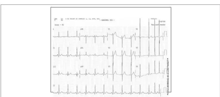
On the second day after surgery, an ST-segment elevation in the anteroseptal wall (Figure 1B) and increased creatinine kinase MB levels were observed. The patient had no symptoms until the third day after surgery, when she presented typical anginal pain at rest. Once the diagnosis of acute myocardial infarction was hypothesized, she was referred for emergency coronary angiography (Figure 2). The angiography showed a dissection extending from the left main coronary artery to its branches, i.e., the left anterior descending artery and the left circumflex artery. The dissection also affected the right coronary artery. The patient underwent coronary artery bypass grafting surgery: the left internal mammary artery was anastomosed to the left anterior descending artery, and saphenous vein grafts were placed from the aorta to the left marginal branch of the circumflex artery and to the right coronary artery. There was no dissection of the aorta.
After revascularization, the patient remained in good condition, with resolution of angina and ECG abnormalities.
Comments
We describe a case of coronary artery dissection after aortic valve replacement surgery, probably due to high blood pressure in the perioperative and postoperative periods. Acute myocardial infarction was diagnosed by electrocardiogram changes and myocardial necrosis markers, and angina appeared later on. There was no relief of angina after blood pressure control, leading to an urgent hemodynamic study
Case Report
Machado et al Coronary artery dissection after aortic valve replacementArq Bras Cardiol 2010;94(2): e23-e25
Figure 1A -Preoperative electrocardiogram showing sinus rhythm and left ventricular hypertrophy.
Figure 1B -Second postoperative day showing sinus rhythm with ST segment elevation in leads V1 to V6 and DI and aVL.
with subsequent surgical treatment.
Although not very common, there are case reports of aortic dissection after aortic valve replacement. However, the authors have found no descriptions of cases in which a coronary artery dissection occurred after aortic valve replacement1,2. Aortic
wall weakness or thinness, usually due to collagen deficiency, and aortic regurgitation are independent risk factors for the occurrence of proximal aortic dissection at the time of aortic valve replacement. Other risk factors include high blood pressure and a bicuspid valve4.
Aortic dissection during surgery is a rare but serious complication after cardiac surgery, and its incidence
varies from 0.16% to 0.25%5. Predisposing factors include
atherosclerosis, significantly high blood pressure, cystic medial necrosis, collagen vascular diseases, and a thin or dilated ascending aorta5,6. The dissection is often identified after aortic
declamping, but may also occur at the time of cannulation, clamping, or decannulation. In this situation, a transesophageal echocardiogram is a good and practical method to confirm the diagnosis. If the expansion of the dissection is limited, a small plication technique can be used without mobilization of the aortic cannula.
When major dissections or low output syndrome occur, the cannula should be removed to another arterial access, such as the femoral and axillary arteries7. In our case, there were
Case Report
Machado et alCoronary artery dissection after aortic valve replacement
Arq Bras Cardiol 2010;94(2): e23-e25
Figure 2 -Coronary angiography showing dissection of the trunk, left coronary artery, anterior descending artery and circumlex artery (arrows).
no complications during surgery that could be indicated as the cause of the coronary dissection.
Acute myocardial infarction can occur after cardiac surgery, especially after coronary revascularization. Modern methods of myocardial protection and surgical techniques have been developed. However, occasionally there are cases of atherosclerotic disease in the distal coronary artery, spasm, embolism or thrombus in native vessels or grafts7. Other causes
of myocardial ischemia are inadequate myocardial protection during surgery, increased oxygen demand, as seen in patients with left ventricular hypertrophy, and hemodynamic instability. The diagnosis of acute infarction in the postoperative period of a cardiac surgery can be difficult due to the absence of pain during sedation and analgesia, and the frequent elevation of myocardial necrosis markers by surgical manipulation. In this patient, there were a single prior obstruction of the coronary artery in a branch of little importance and left ventricular hypertrophy, but increased enzyme levels associated with angina indicated the need for coronary angiography.
Coronary artery dissection can cause myocardial infarction, particularly after percutaneous transluminal coronary angioplasty. Another rare form is a spontaneous dissection of the coronary arteries. The vast majority (80%) occurs in young
women in the postpartum period or in those who use oral contraceptives. The treatment varies, but follows the usual procedure for other causes of acute myocardial infarction8.
Our patient was a rare case of coronary artery dissection after aortic valve replacement, probably due to high blood pressure in the perioperative and postoperative periods. Acute myocardial infarction was diagnosed by ECG changes and laboratory markers of myocardial necrosis with late onset of angina. Its refractoriness indicated coronary angiography with subsequent emergency surgical treatment.
Potential Conflict of Interest
No potential conflict of interest relevant to this article was reported.
Sources of Funding
There were no external funding sources for this study.
Study Association
This study is not associated with any post-graduation program.
References
1. von Kodolitsch Y, Simic O, Schwartz A, Dresler C, Loose R, Staudt M, et al. Predictors of proximal aortic dissection at the time of aortic valve replacement. Circulation. 1999; 100: II287-II294.
2. Still RJ, Hilgenberg AD, Akins CW, Daggett WM, Buckley MJ. Intraoperative aortic dissection. Ann Thorac Surg. 1992; 53 (3): 374-9.
3. Rogers JH, Lasala JM. Coronary artery dissection and perforation complicating percutaneous coronary intervention. J Invasive Cardiol. 2004; 16 (9): 493-9.
4. Shen CH, Wu CC, Hung CM, Ho WM. Intraoperative aortic dissection--a case report. Acta Anaesthesiol Sin. 2002; 40 (2): 85-9.
5. Sakakibara Y, Matsuda K, Sato F, Matsuzaki K, Jikuya T, Mitsui T. Aortic
dissection complicating cardiac surgery in a patient with calcified ascending aorta. Jpn J Thorac Cardiovasc Surg. 1999; 47 (12): 625-8.
6. Shinichi M, Akihiko U, Toshiaki A, Yuichi U. Management of intraoperative aortic dissection with a direct cannulation on the intimal flap. Interactive Cardiovascular and Thoracic Surgery. 2003; 2: 636-8.
7. Obarski TP, Loop FD, Cosgrove DM, Lytle BW, Stewart WJ. Frequency of acute myocardial infarction in valve repairs versus valve replacement for pure mitral regurgitation. Am J Cardiol. 1990; 65 (13): 887-90.
8. Leone F, Macchiusi A, Ricci R, Cerquetani E, Reynaud M. Acute myocardial infarction from spontaneous coronary artery dissection a case report and review of the literature. Cardiol Rev. 2004; 12 (1): 3-9.