Arq Bras Oftalmol. 2006;69(6):919-22
Biomicroscopia ultra-sônica na comparação da morfometria do segmento anterior
antes e após vitrectomia “pars plana”
1Post-Doctorate and Research Fellow, The New York Medical College and The New York Eye and Ear Infir-mary. Assistant Professor of Clinical Ophthalmology, São Geraldo Eye Hospital, School of Medicine, Univer-sidade Federal de Minas Gerais - UFMG - Belo Horizon-te (MG) - Brazil.
2Post-Graduated in Ophthalmology (Doctorate), São Geraldo Eye Hospital, School of Medicine UFMG -Belo Horizonte (MG) - Brazil.
3Associate Professor of Clinical Ophthalmology, São Geraldo Eye Hospital, School of Medicine UFMG -Belo Horizonte (MG) - Brazil.
4Clinical Assistant Ophthalmologist, Department of Glaucoma, Institute of Vision - IOV- Belo Horizonte (MG) - Brazil.
Address to correspondence: Flávio A. Marigo. Rua dos Otoni, 881 13º andar Belo Horizonte (MG) Brazil -CEP 30150-270
E-mail: [email protected]
Recebido para publicação em 19.11.2005 Última versão recebida em 21.05.2006 Aprovação em 04.06.2006
No financial interest. Supported by a Research Grant from CAPES - The Ministry of Education -Brazil. Flavio de Andrade Marigo1
Márcio Zisman2 Márcio Bittar Nehemy3
Patrícia Vianna Brandão Marigo4
INTRODUCTION
Pars plana vitrectomy (PPV) is considered the standard surgery to approach a large number of vitreoretinal disorders. Ultrasound biomicros-copy (UBM) has demonstrated that anterior segment changes can be asso-ciated to complications in the early postoperative period after the procedu-re(1-5). Such changes include shallowing of the anterior chamber(1),
narro-wing of the anterior chamber angle(1) and ciliary body detachment(1,3-5).
These changes may determine transient complications like glaucoma(2) or
hypotony(3).
Although early postoperative changes after vitrectomy have already been detailed, little is known about the long-term anatomical changes of the anterior segment anatomy after PPV. Such a study would provide useful information about the influence of PPV on the stability of the anterior segment in the postoperative period. Based on this information, unexpected
Ultrasound biomicroscopy in the comparison of the
anterior segment morphometry before and after
pars plana vitrectomy
Keywords: Anterior eye segment/ultrasonography; Ultrasonics; Anatomy, cross-sectional/ methods; Ciliary body/ultrasonography; Vitrectomy; Microscopy/instrumentation
Purpose: To determine if pars plana vitrectomy induces long-term
changes in the anterior segment anatomy by means of ultrasound biomicroscopy. Methods: A prospective case series study was under-taken of consecutive patients referred to a tertiary eye care centre for pars plana vitrectomy as the only procedure. Twenty eyes of 20 patients undergoing pars plana vitrectomy alone were studied by ultrasound biomicroscopy. Silicone oil or scleral buckle was not used in any of the included cases. The following morphometric parameters were compared before and after 3 months of surgery: anterior chamber depth, angle-opening distance at 500 µm from the scleral spur, trabecular-ciliary process distance, ciliary body thickness at 1, 2 and 3 millimeters from the scleral spur and measurement of the supraciliary space thickness, when fluid was detected. Results: No statistically significant differences were found between the preoperative and the postoperative morphometric parameters. Conclusions: Uncomplicated pars plana vitrectomy does not induce any long-term change on anterior segment morphometry. Based on these findings, the normal long-term pattern to be expected after pars plana vitrectomy is the conservation of the preoperative morphometry.
Arq Bras Oftalmol. 2006;69(6):919-22
920 Ultrasound biomicroscopy in the comparison of the anterior segment morphometry before and after pars plana vitrectomy
anatomical changes that may determine later complications like persistent ocular hypotony can be detected more easily.
The aim of the present study is to compare the anterior segment morphometry before and after PPV to determine if this procedure is capable of determining long-term changes in the anterior segment anatomy.
METHODS
Ultrasound biomicroscopy was used to prospectively eva-luate 20 eyes of 20 consecutive patients in the preoperative and late postoperative period of PPV as the only procedure. The patients were referred to Instituto da Visão, in Belo Hori-zonte, Brazil, a tertiary eye care centre. The following situa-tions were excluded: patients who had previous intraocular surgery (except cataract surgery); cases of PPV which requi-red associated surgical procedures like scleral buckling, inter-nal tamponade, lensectomy or intraocular lens implantation; previous anterior segment laser therapy; history of uveitis, trauma or glaucoma and use of any topical or systemic drugs that might affect pupil or accommodation.
The preoperative diagnosis included 7 eyes with complica-tions of proliferative diabetic retinopathy (PDR) (4 with vi-treous hemorrhage (VH), 2 of which also having tractional retinal detachment (TRD); two with chronic clinical significant macular edema (CSME) and 1 with a macular pucker (MP); 4 eyes with stage III or IV macular holes (MH); 4 eyes with subfoveal neovascular membranes (SNVM); 3 eyes with non-diabetic vitreous hemorrhage (one secondary to a central reti-nal vein occlusion (CRVO), one secondary to branch retireti-nal vein occlusion (BRVO) and another secondary to an arterial macroaneurysm (AM); and 2 eyes with idiopathic macular epiretinal membranes (IERM). Fifteen eyes were phakic and 5 were pseudophakic.
All patients were submitted to the same surgical protocol and were operated on by the same surgeon (MBN). In brief, after routine three-port sclerotomies, vitrectomy was executed using a 20-gauge vitrector. During vitrectomy the intraocular pressure (IOP) was maintained at a level of approximately 20 mmHg. The superior sclerotomies were closed with a 9-0 nylon “X” suture and the infusion sclerotomy with a 7-0 absorbable “U” suture.
One or two days before each UBM all eyes were submitted to a complete ophthalmologic examination consisting of vi-sual acuity and refraction applanation tonometry, conventio-nal slit-lamp biomicroscopy, and indirect ophthalmoscopy.
UBM was performed using a commercial version of the ultrasound biomicroscope (Humphrey-Zeiss model 840, San Leandro, CA) with a 50 MHz transducer that provides a maxi-mal lateral resolution of 50 µm and a maximaxi-mal depth penetration of 4-5 mm. The scanner produces a 5x5 mm image at a scanning rate of 8 Hz. Scanning was performed with the patient in supine position using the standard technique which has been
described elsewhere(6). After instillation of topical 0.5%
pro-paracaine, a polymethyl-methacrylate eye cup was inserted between the lids and filled with physiological saline as a coupling agent. Accommodation was kept constant by asking the patient to fix on targets at the ceiling about 3 meters from the eye. Room illumination was controlled at 50 lux. Gain was set at 80 dB. The scanner was positioned perpendicular to the structure to be examined. The UBM images were obtained axially and radially on the superior, nasal, inferior and tempo-ral quadrants. Measurements were performed in a masked way by one of us (MZ) using the internal electronic caliper of the instrument and were recorded in micrometers. The preopera-tive examinations were performed not more than 48 hours before surgery. Since the aim of this study was to detect the late postoperative morphometric changes produced by PPV, all postoperative examinations were performed 8 weeks after surgery (range: 60-183 days, mean±standard deviation [SD] 92 ± 33 days, median 90 days).
For quantitative analysis five standard UBM morphome-tric parameters(6-7) were compared before and after surgery.
These parameters (measured in micrometers) were: central anterior chamber depth (ACD); angle opening distance 500 µm from the scleral spur (AOD); trabecular meshwork – ciliary process distance (TCPD); ciliary body thickness (CBT) 1, 2 and 3 millimetres from the scleral spur (CBT1, CBT2 and CBT3) and the thickness (perpendicular to the sclera) of supraciliary fluid (SF) if present. Except for ACD, all other morphometric parameters were measured at the 3:00, 6:00, 9:00 and 12:00 o’clock meridians and the average of the measurements bet-ween meridians was used to compare the preoperative and late postoperative morphometric parameters.
Student’s t test for paired samples was used for statistical analysis, with the level of significance established at p<0.05 using a commercial statistic software package (SAS version 5, SAS Institute, Cary, NC, USA).
RESULTS
The mean age ± standard deviation (SD) was 58 ± 17 years (range: 12-77). 15 (75%) women and 5 (25%) men were inclu-ded. All patients were caucasians. There were twelve right eyes (60%) and eight (40%) left eyes.
Arq Bras Oftalmol. 2006;69(6):919-22
Ultrasound biomicroscopy in the comparison of the anterior segment morphometry before and after pars plana vitrectomy 921
DISCUSSION
UBM has been investigated concerning reproducibility, accuracy and precision of the measurements(8-12).
Intraobser-ver reproducibility is considered adequate(8-9,12) but
interob-server reproducibility is thought to be variable(8-9). Therefore
to appropriately compare the preoperative and postoperative data, all measurements were performed in this study by the same masked observer.
It has been demonstrated that the best UBM morphometric parameters are those related to immutable landmarks such as the scleral spur(10). For this reason, considering the myriad of
available anterior segment morphometric parameters, we se-lected to be included in this study only those of interest to vitreoretinal surgery that have been previously demonstrated to be reliable and reproducible.
Anterior segment changes in the first postoperative weeks after posterior segment surgery like vitrectomy and retinal detachment surgery have been described(1-4,7,13-16) but little is
known about the long-term effects of vitrectomy on anterior chamber anatomy. If vitrectomy would determine permanent
changes in anterior segment morphology, such changes might predispose the eye to complications. For example, if vitrecto-my would cause permanent narrowing of the angle, it would cause narrow-angle glaucoma in predisposed eyes. This infor-mation would be useful in order to anticipate possible compli-cations of the surgery in such eyes. Our results showed no statistically significant differences between the preoperative and postoperative periods of PPV concerning any of the stu-died morphometric parameters. Only one eye presented an image suggestive of a ciliary body detachment. The maximum thickness of the supraciliary space in this case was 142 µm and it affected only one quadrant. This patient had an IOP of about 7 mmHg with no clinical or angiographic signs of hypotony. These results suggest that PPV alone does not affect the long-term stability of the anterior segment anatomy.
CONCLUSION
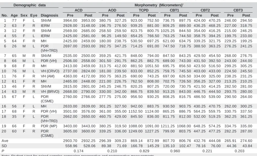
In conclusion, UBM demonstrated that there is no significant difference of the anterior segment morphometric parameters bet-Table 1. Anterior segment morphometry in the pre- and late postoperative period of pars plana vitrectomy
Demographic data Morphometry (Micrometers)†
ACD AOD TCPD CBT1 CBT2 CBT3
No. Age Sex Eye Diagnosis Pre Post Pre Post Pre Post Pre Post Pre Post Pre Post
01 77 F L SNVM 3964.00 3953.00 380.75 327.25 923.00 752.50 736.75 897.75 424.00 470.25 246.00 294.50
02 63 F R IERM 2928.00 3148.00 196.75 276.50 939.25 881.00 809.25 889.00 436.25 468.25 227.00 318.75
03 12 F R SNVM 2569.00 2685.00 258.50 259.50 823.75 800.75 1025.25 844.50 354.00 416.25 215.00 246.25
04 55 F L IERM 2425.00 2581.00 096.25 149.50 654.25 766.50 763.25 744.50 358.75 394.25 199.75 233.00
05 64 F R MH 2425.00 2459.00 180.00 238.75 530.50 697.25 621.00 634.75 339.25 321.75 229.25 199.25
06 26 M L PDR 2697.00 2593.00 392.75 347.25 714.25 691.00 747.50 718.75 388.50 363.25 276.25 241.25 (VH+TRD)
07 65 M R SNVM 2535.00 2500.00 359.25 421.75 848.00 794.00 847.50 843.25 429.50 454.50 268.00 276.75
08 66 M L PDR (VH) 2506.00 2558.00 301.50 291.75 862.25 882.75 689.00 743.00 431.50 392.50 243.00 244.00
09 68 F R MH 2413.00 2459.00 313.75 412.00 881.50 1051.50 695.75 856.50 423.50 516.50 299.25 305.25 10 76 M L VH (CRVO) 2737.00 2824.00 181.00 239.50 833.00 852.25 759.75 745.50 403.00 433.00 224.50 245.50 11 76 F R VH (AM) 4363.00 4172.00 350.75 363.25 690.00 743.25 697.00 626.50 334.00 325.00 238.25 231.25 12 61 F R MH 2465.00 2448.00 221.00 226.75 762.50 808.00 702.75 726.50 356.25 327.00 213.25 210.25 13 46 F R SNVM 2815.00 2801.00 245.25 246.75 820.25 807.25 720.00 730.75 421.50 414.25 282.50 281.00 14 63 M R VH (BRVO) 2668.00 2790.00 330.00 342.00 866.75 839.50 913.25 843.00 446.75 444.50 293.75 280.00 15 67 F L PDR 2760.00 2766.00 277.75 275.00 956.50 930.25 808.25 816.75 486.50 539.00 290.50 264.00
(CSME)
16 56 F L MH 2633.00 2639.00 301.25 327.50 942.00 883.75 930.50 903.75 430.25 470.75 262.00 300.25 17 68 F R PDR (VH) 3501.00 3576.00 361.00 355.00 1152.50 1124.00 865.25 886.75 564.25 559.75 330.75 337.50 18 35 F L PDR 2662.00 2650.00 460.75 429.00 845.50 836.00 811.75 812.00 532.00 519.25 362.25 361.25
(VH+TRD)
19 66 F R PDR (MP) 3403.00 3443.00 380.25 319.50 1088.00 1091.00 1211.25 1068.00 648.25 574.25 334.75 335.00 20 60 F R PDR 3605.00 3600.00 339.25 336.00 1249.00 1227.25 799.00 803.75 447.25 477.25 282.25 287.00
(CSME)
Ave 2903.70 2932.25 296.39 309.23 869.14 872.99 807.70 806.76 432.76 444.08 265.91 274.60 SD 0558.96 0528.06 089.38 071.69 166.78 145.29 135.10 102.47 078.16 076.00 044.36 043.84
P 0.174 0.210 0.829 0.960 0.221 0.203
Note: Student t test for paired samples comparing preoperative and postoperative measurements.
†For all parameters (except of ACD), numbers represent the average of measurements taken at the 3:00, 6:00, 9:00 and 12:00 o’clock positions.
Arq Bras Oftalmol. 2006;69(6):919-22
922 Ultrasound biomicroscopy in the comparison of the anterior segment morphometry before and after pars plana vitrectomy
ween the preoperative and postoperative periods after PPV. Ba-sed on these findings, the normal long-term pattern to be expected after PPV is the conservation of the preoperative morphometry.
RESUMO
Objetivos: O objetivo do presente estudo foi determinar, por
meio da biometria ultra-sônica (UBM), se a vitrectomia via “pars plana” pode induzir alterações permanentes na ana-tomia do segmento anterior. Métodos: Foi realizado estudo prospectivo, analisando-se uma série consecutiva de pacien-tes, encaminhados para um centro de referência terciário para serem submetidos a vitrectomia via “pars plana” como único procedimento. Vinte olhos de 20 pacientes a serem submeti-dos a vitrectomia como único procedimento foram estudasubmeti-dos pela biomicroscopia ultra-sônica. Óleo de silicone ou introfle-xão escleral não foram usados em nenhum dos casos incluí-dos. Os seguintes parâmetros morfométricos foram compara-dos antes e após 3 meses da cirurgia: profundidade da câmara anterior, abertura do ângulo a 500 µm do esporão escleral, distância trabéculo-processos ciliares, espessura do corpo ciliar a 1, 2 e 3 milímetros do esporão escleral e medida da espessura do espaço supraciliar, quando fluido foi detectado.
Resultados: Não foram encontradas diferenças
estatistica-mente significativas entre os períodos pré e pós-operatório para os parâmetros morfométricos estudados. Conclusões: A vitrectomia via “pars plana” não-complicada não induz altera-ções permanentes na morfometria do segmento anterior. Com base nestes achados, o padrão normal esperado após a vitrec-tomia via “pars plana” é a conservação da morfometria pré-operatória.
Descritores: Segmento anterior do olho/ultra-sonografia;
Ultra-som; Anatomia seccional/métodos; Corpo ciliar/ultra-sonografia; Vitrectomia; Microscopia/instrumentação
REFERENCES
1. Hikichi T, Ohnishi M, Hasegawa T. Transient shallow anterior chamber indu-ced by supraciliary fluid after vitreous surgery. Am J Ophthalmol. 1997;124 (5):696-8.
2. Genovesi-Ebert F, Rizzo S, Chiellini S, Gabbrielinni G, Laddaga F, Nardi M, et al. Ultrasound biomicroscopy in the assessment of secondary glaucoma after vitreoretinal surgery and silicone oil injection. Ophthalmologica. 1998;212 Suppl:4-5.
3. Minamoto A, Nakano KE, Tanimoto S, Mizote H, Takeda Y. Ultrasound biomicroscopy in the diagnosis of persistent hypotony after vitrectomy. Am J Ophthalmol. 1997;123(5):711-3.
4. Liu W, Wu Q, Huang S, Tang S. Ultrasound biomicroscopic features of anterior proliferative vitreoretinopathy. Retina 1999;19(3):204-12.
5. Chen WL, Yang CM, Chen YF, Yang CH, Shau WY, Huang JS, et al. Ciliary detachment after pars plana vitrectomy: an ultrasound biomicroscopic study. Retina. 2002;22(1):53-8.
6. Pavlin CJ, Harasiewicz K, Foster FS. Ultrasound biomicroscopy of anterior segment structures in normal and glaucomatous eyes. Am J Ophthalmol 1992; 113(4):381-9. Comment in: Am J Ophthalmol. 1992;114(4):516-7. 7. Pavlin CJ, Rutnin SS, Devenyi R, Wand M, Foster FS. Supraciliary effusions
and ciliary body thickening after scleral buckling procedures. Ophthalmology. 1997;104(3):433-8.
8. Tello C, Liebmann J, Potash SD, Cohen H, Ritch R. Measurement of ultra-sound biomicroscopy images: intraobserver and interobserver reliability. Invest Ophthalmol Vis Sci. 1994;35(9):3549-52.
9. Urbak SF, Pedersen JK, Thorsen TT. Ultrasound biomicroscopy. II. Intraobser-ver and interobserIntraobser-ver reproducibility of measurements. Acta Ophthalmol Scand. 1998;76(5):546-9.
10. Urbak SF. Ultrasound biomicroscopy. I. Precision of measurements. Acta Oph-thalmol Scand. 1998;76(11):447-55.
11. Urbak SF. Ultrasound biomicroscopy. III. Accuracy and agreement of measure-ments. Acta Ophthalmol Scand. 1999;77(3):293-7.
12. Marchini G, Pagliarusco A, Toscano A, Tosi R, Brunelli C, Bonomi L. Ultrasound biomicroscopic and conventional ultrasonographic study of ocular dimensions in primary angle-closure glaucoma. Ophthalmology. 1998;105(11): 2091-8.
13. Maruyama Y, Yuuki T, Kimura Y, Kishi S, Shimizu K. Ciliary detachment after retinal detachment surgery. Retina. 1997;17(1):7-11.
14. Yoshida S, Sasoh M, Arima M, Uji Y. Ultrasound biomicroscopic view of detachment of the ciliary epithelium in retinal detachment with atopic dermati-tis. Ophthalmology. 1997;104(2):283-7.
15. Kawahara S, Nagai Y, Kawakami E, Ida RY, Takeuchi M, Uyama M. Cilio-choroidal Detachment Following Scleral Buckling Surgery for Rhegmatogenous Retinal Detachment. Jpn J Ophthalmol. 2000;44(6):692-3.