Transcranial Doppler for paTenT
foramen ovale screening
Is there a good correlation with transesophageal echocardiography?
Marcos Christiano Lange
1, Viviane Flumignan Zétola
1, Admar Moraes de Souza
2, Élcio Juliato Piovesan
1,
Juliano André Muzzio
1, Francisco Manoel Branco Germiniani
1, Lineu César Werneck
1Abstract – Right-to-left shunt (RLS) can be identified by contrast-enhanced transcranial Doppler (cTCD) in patent foramen ovale (PFO) patients. Aim: To evaluate cTCD for PFO screening comparing it to cTEE. Method:
45 previous cTCD performed for PFO diagnosis and correlated its findings with cTEE. Patients were submitted to a cTCD standardized technique and were divided in two groups according to RLS: Group 1, patients with a positive RLS and Group 2 when RLS was negative. Results: 29 (65%) patients were included in group 1 and 16 (35%) in group 2. PFO confirmation by cTEE was performed in 28 (62%) patients. cTCD had a 92.85% sensitivity, 82.35% specificity, 89.65% positive predictive value and 87.5% negative predictive value when compared to cTEE for PFO diagnosis. Conclusion: Standardized technique cTCD allows for RLS visualization in PFO patients with a good correlation with cTEE and can be used as a screening test before cTEE.
KEy wORDS: patent foramen ovale, transesophageal echocardiography, transcranial Doppler, ultrasonography.
Doppler transcraniano contrastado para triagem de forame oval patente: existe boa correlação com ecocardiograma transesofágico?
Resumo – A comunicação direita-esquerda (CDE) pode ser identificada por Doppler transcraniano contrastado (DTCc) em pacientes com forame oval patente (FOP). Objetivos: Analisar o DTCc para triagem de FOP comparado a ecocardiografia transesofágica (ETEc). Método: Realizamos 45 exames de DTCc para diagnóstico de FOP e correlacionamos com os achados do ETEc. Os pacientes foram submetidos a técnica padronizada e divididos em dois grupos conforme a positividade do exame. Resultados: 29 (65%) pacientes foram incluídos no grupo 1 (CDE positiva) e 16 (35%) no grupo 2 (CDE negativa). A confirmação do FOP pelo ETEc ocorreu em 28 (62%) pacientes. O DTCc apresentou sensibilidade de 92,85%, especificidade de 82,35%, valor preditivo positivo de 89,65% e valor preditivo negativo de 87,5% comparado ao ETEc para o diagnóstico de FOP. Conclusão: A técnica padronizada de DTCc possibilita a visualização de CDE em pacientes com FOP com boa correlação com o ETEc.
PALAvRAS-ChAvE: forame oval patente, ecocardiograma transesofágico, Doppler transcraniano, ultra-sonografia.
1Cerebrovascular Diseases, Neurology Division and 2Echocardiography, Cardiology Division, Department of Internal Medicine, hospital de Clínicas, Federal University of Paraná, Curitiba PR, Brazil. The authors declare they do not have any conlict of interest related to this article.
Received 25 July 2008. Accepted 29 September 2008.
Dr. Marcos Christiano Lange – Hospital de Clínicas / Serviço de Neurologia - Rua General Carneiro 181 / 4º andar - 80060-900 Curitiba PR - Brasil. E-mail: [email protected]
Patent foramen ovale (PFO) is a congenital heart dis-ease characterized by an opening between the right and left atria resulting from incomplete closure of the ostium secundum by the septum secundum1. Recent studies have
found an increase prevalence of PFO in women with mi-graine with aura and young adults (less than 55 years old) with so-called “cryptogenic” ischemic stroke2-6. Emboli
from the venous system can cross the PFO reaching the arterial circulation through a right-to-left shunt (RLS) and
thus leading to a stroke. PFO diagnosis is done by using a contrast-enhanced technique (by injecting saline solu-tion in a peripheral vein) while performing a transesopha-geal echocardiography (cTEE) and, when positive, it shows a high correlation with necropsy studies7. In spite of both
ad-vantage of this method is the direct visualization of inter-atrial septum and inter-atrial septal aneurysm (ASA) identiica-tion. ASA is an abnormally redundant septum primum lap that extends across the atria9. when ASA is associated with
PFO, this combined pathology leads to an increase in the risk factor for recurrence of embolic “cryptogenic” stroke6.
On the other hand, contrast-enhanced transcranial Doppler (cTCD) is a low cost, non-invasive method, which is easy to perform and to interpret as a screening method for PFO diagnosis. Even though cTCD is known as a diag-nostic tool with high sensibility, techniques for perform-ing TCD vary accordperform-ing to some authors2,5,10-12.
The primary aim of this study was to standardize cTCD technique for RLS as a screening method for PFO. Second-arily we tried to establish the sensibility and sensitivity of this method when compared with cTEE.
meThoD
we retrospectively analyzed 45 cTCD and cTEE studies for PFO investigation from April 2005 to May 2007. All studies were done after a thorough clinical and neurological evaluation and all patients gave their written, informed consent. Clinical indi-cation for RLS investigation was stroke on 41 patients and mi-graine on the remaining four.
Contrast-enhanced transcranial Doppler ultrasound
All cTCD studies were performed with the patient in a supine position in a controlled temperature environment (24 to 28ºC) by a trained neurologist (Doctors MCL, vFZ, JAM). The equip-ments used were a RIMED – Smart Lite or a DwL – Doppler Box, both with two 2-Mhz transducers. Bilateral middle cerebral ar-teries (MCA) were insonated through the temporal window at a depth of 50 to 60 mm and ixed with a helmet, as described else-where13. Contrast consisted of 10 mL air-mixed saline solution (9
mL of normal saline solution + 1 mL of air) injected as a bolus in-to a large right antecubital vein while resting (resting phase) and before valsalva maneuver (vM). The valsalva maneuver was per-formed ive seconds after intravenous contrast injection and its effectiveness was monitored by a 25% decrease of MCA low ve-locity. Both studies (resting phase and vM phase) were repeat-ed three times, with each test lasting one minute. A right-to-left shunt (RLS) was considered positive (Group 1) when at least one air microbubble was detected on the spectral display of at least
one of the monitored MCA. Conversely, RLS was negative (Group 2) when during the next 60 seconds following contrast injection there was no identiied microbubble in either MCA. Patients with a positive test were classiied in two grades: small RLS (≤ 10 bub-bles) and large RLS (>10 bubbles), the latter subgroup was further labeled as a “curtain” RLS if uncountable signals passed during MCA monitoring. In addition, we separated Group 1 in two other subgroups: positive only during vM phase and positive at rest and with the vM. Finally we compared results from cTCD with cTEE.
Contrast-enhanced transesophageal echocardiography
All patients underwent cTEE, which was performed by a car-diologist trained in this technique (Dr. AMS). All exams were done with a hewlett Packard Sonos 5500 imaging system and a 5Mhz wide-band multiplane transducer. Patients were examined in the fasting state and received only local pharyngeal anesthe-sia (topical lidocaine spray). For the diagnosis of a RLS, contrast consisted of 10 mL air-mixed saline solution (9 mL of normal sa-line solution + 1 mL of air) injected as a bolus into a large ante-cubital vein during resting and after valsalva maneuver. Patients were trained in performing the vM before the procedure with a ive seconds’ duration. The effectiveness of the vM was veriied by observing the bulging of the interatrial septum into the left atrium. The presence of a PFO was assumed if at least one mi-crobubble passed from the right to the left atrium on the irst three cardiac cycles after contrast injection. An ASA was pre-sented if interatrial septum moved more than 10 mm in either atrium side during systole.
Statistical analysis was performed with SPSS 12.0 software (SPSS Inc.). Statistical signiicance was assessed by t-Student test for parametric variables and Chi-Square or Mann whitney tests were used for non-parametric variables. Correlation tests were done for etiological and risk factors with RLS grades by cTCD. Statistical signiicance was determined at p<0.05.
resulTs
A total of 29 (65%) patients had positive RLS (Group 1): mean age was 38±14.6 years and 17 (57%) were females. The other 16 (35%) patients had a negative RLS (Group 2), with a mean age of 37±11 years. In this group 11 (68%) pa-tients were females. There was no statistical difference between groups for demographic variables (age and gen-der distribution) (Table 1).
Table 1. Demographic data.
Group 1 (positive RLS) Group 2 (negative RLS) p
n (%) Mean age±sd n (%) Mean age±sd
Gender distribution Female
Male Total
17 (58.62) 12 (41.38) 29 (64.44)
39.7±12.49 36.75±17.65 38.48±14.62
11 (68.75) 5 (31.25) 16 (35.56)
36.9±11.68 37.2±10.7 37±11.02
0.706* 0.792* 0.734*
Clinical indication for RLS study in Group 1 was stroke in 26 (90%) patients and migraine in the remaining three (10%). For Group 2, 15 (93%) patients were evaluated for stroke and one (7%) for migraine. There was no statistical difference between the two groups (Table 2).
In Group 1 four (14%) patients were cigarette smokers, three (10%) had high blood pressure, three (10%) had hy-percholesterolemia and one (3.5%) had diabetes. In Group 2 two (12.5%) patients were smokers, six (37%) had high blood pressure, one (7%) had hypercholesterolemia and one (7%) had diabetes. There was no statistical differ-ence between groups for any of the risk factors, except for high blood pressure that was more common in Group 2 (p=0.031) (Table 2).
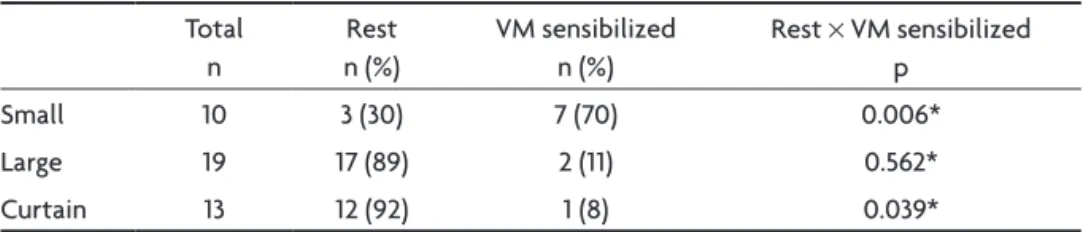
In relation to RLS grade in Group 1, ten (34%) patients had a small RLS and 19 (66%) had a large RLS, of the latter 13 (68%) had a “curtain” effect on cTCD (Table 3). In Group 1, 20 (69%) patients had a positive RLS during both phases (resting and vM), nine (31%) had a positive RLS only dur-ing vM phase, seven (77%) presented with a small RLS and two (22%) with a large RLS; of those, only one (11%) pre-sented with a curtain effect (Table 3).
when vM was performed we could recognized a posi-tive RLS increase in 45%, which was more signiicant in the small RLS subgroup (a 200% increase) than in the large RLS subgroup (11% increase). No patient in the study had posi-tive test only while resting.
After comparing RLS during both the resting phase and vM versus RLS triage only in the vM phase we found the following results: for the small grade RLS subgroup (n=10) there is a statistical signiicance for vM test (p=0.006), however for the large grade subgroup (n=19) there is no statistical signiicance (p=0.562) between the two tech-niques. Also, when the “curtain” RLS subgroup was stud-ied, we found that for those patients undergoing the com-bined the resting test and vM phases there was a signii-cant inding for RLS when compared with the isolated vM phase (p=0.039).
Contrast-enhanced transesophageal echocardiogra-phy, the so-called gold-standard technique for PFO identi-ication, was positive in 28 (62%) patients and negative on
Table 2. Clinical indication for RLS investigation and stroke risk factors.
Group 1 (n=29) n (%)
Group 2 (n=16) n (%)
EP+ × EP– p Indication
Ischemic stroke Migraine
26 (90) 3 (10)
15 (93) 1 (7)
0.432* 0.616** Stroke risk factors
hBP DM hCh CS
3 (10) 1 (3.5) 3 (10) 4 (14)
6 (37) 1 (6.25) 1 (6.25) 2 (12.5)
0.031* 0.666* 0.648* 0.904*
hBP, high blood pressure; DM, diabetes mellitus; hCh, hypercholesterolemia; CS, cigarette smoking; *Mann-whitney test; **Chi-square test.
Table 3. Bubble indings in Group 1 (n=29).
Total n
Rest n (%)
vM sensibilized n (%)
Rest × vM sensibilized p
Small 10 3 (30) 7 (70) 0.006*
Large 19 17 (89) 2 (11) 0.562*
Curtain 13 12 (92) 1 (8) 0.039*
Small: ≤10 bubble; large: >10 bubble; curtain- uncountable signals; rest represents patients with positive RLS study both at rest and during vM study; vM sensibilized represents patients with RLS study positive only during vM test; *Mann-whitney test.
Table 4. Contrast-enhanced TCD versus contrast-enhanced TEE for PFO identiication.
cTEE+ cTEE– Total
cTCD + 26 3 29
cTCD – 2 14 16
Total 28 17 45
Sensibility: 92.85%; Speciicity: 82.35% Positive predictive value: 89.65%; Negative predictive value: 87.50%
the others 17 (38%). when we compared cTCD versus cTEE, we could identify two patients from group 2 with a posi-tive cTEE and three from group 1 with a negaposi-tive cTEE (one with a small RLS and two with a large RLS) (Table 4). Thus, cTCD for PFO diagnosis had a 92.85% sensibility, 82.35% speciicity, 89.65% positive predictive value and 87.5% neg-ative predictive value when compared to cTEE (Table 4). In addition, three patients from group 1 had a positive ASA on cTEE, all of which had a large RLS by cTCD (two of them with a “curtain” effect). This corresponded to 15% of all large RLS grade cTCDs. Conversely, none of the pa-tients in group 2 had ASA.
we found a good correlation between headache and “curtain” RLS (p=0.013) and stroke and large RLS (p=0.039), but not for other risk-factors as high blood pressure, dia-betes, hypercholesterolemia and cigarette smoking.
Discussion
Our study conirmed that cTCD can be safely performed as a screening method for suspected PFO in patients with either stroke or migraine prior to a cTEE study, with a high sensibility (92.85%) and speciicity (82.35%). A standard-ized technique was important for these results with a vM test leading to a 45% increase the positive results.
cTCD is a non-invasive, low cost test, which also can be easily repeated and is well tolerated by the patients. Time and again cTCD was proved to be a valuable toll in the evaluation of stroke and others neurological dis-eases5,14. Our study showed similar results of cTCD when
compared to cTEE for PFO evaluation as previously pub-lished in both national and international studies with a sensibility ranging from 66% to 100% and a speciicity of 62% to 100%2,5,10-12,15-19.
we highlight that the inding of positive RLS by cTCD with negative cTEE, as in three of our cases, can corre-spond to a cTEE false-negative. This can be due to several factors, such as an inadequate transesophageal window, negative contrast effect at right atrium and high pressure levels in the left atrium without low inversion crossing the PFO from the right atrium to the left one20-23. This can
also occur in the setting of an extracardiac shunt, such as a pulmonary arteriovenous istula24. Using cTCD, the
tim-ing from contrast injection until identiication of the irst bubble on the MCA can be used to differentiate between a cardiac and an extracardiac shunt: if the irst bubble is identiied in up to 11 seconds after contrast injection, the RLS is considered cardiac; on the other hand, if the time until identiication of the irst bubble is over 14 seconds, the shunt can be considered to be extracardiac in origin. however, this remains a controversial topic in the litera-ture and there is no consensus regarding this criterium, which we could not conirm in our study10.
A positive cTEE with a negative cTCD for the evalu-ation of PFO, as found in two of our patients, can occur if the PFO is a small one, thus impairing cTCD sensitivity when it is performed by insonating only two brain vessels or if there is some kind of limitation that prevents the patient from performing the vM correctly.
vM evaluation led to an increase of 45% in RLS iden-tiication. This inding is more signiicant in patients with a small RLS. In order to avoid misdiagnosis, a negative resting test should be complemented by a vM test. vM increases the pressure in the right atrium causing a low inversion across the PFO that cannot be observed in the resting phase. we cannot overstress the signiicance of the vM in the diagnosis of RLS, as several strokes result from embolization occurring in similar high-pressure set-tings such as the cuff maneuver and physical activity25.
Incorrectly performed vM studies are due to uncooper-ative patients who fail to perform the maneuver prop-erly, in ICU patients who are intubated and in assisted mechanical ventilation, if sedatives were used prior or concurrently with the cTCD and in patients with cogni-tive impairment.
It is also important to emphasize that the majority of patients with a positive RLS had a large RLS (66%), with a curtain pattern occurring in 68% of these patients and in 45% of all patients. Previous studies have already estab-lished the importance of quantitative evaluation related to stroke reccurrence5.
Only three patients with PFO plus ASA were identi-ied, all of whom had a large RLS. we hypothesize that the association of ASA and PFO has a high probability of RLS, which can be identiied by cTEE in those patients with a large shunt. This dual pathology could increase RLS and recurrence of stroke as showed in previous studies6.
Finally, we concluded that cTCD performed with a standardized technique is an excellent method for PFO identification, with both high sensibility (92.85%) and speciicity (82.35%) when compared to cTEE. It is impor-tant to perform either test both while resting and under vM in order to increase these values. Availability, low cost and a less invasive technique are important features that allow the neurologist to perform a cTCD study prior to cTEE when investigating for PFO. In addition, cTCD ind-ings can be used when performing a follow-up test after surgical or percutaneous closure of PFO.
references
3. Sztajzel R, Genoud D, Roth S, Mermillod B, le Floch-Rohr J. Patent fo-ramen ovale, a possible cause of symptomatic migraine: a study of 74 patients with acute ischemic stroke. Cerebrovasc Dis 2002;13:102-106. 4. Lechat P, Mas JL, Lascault G, et al. Prevalence of patent foramen ovale
in patients with stroke. N Engl J Med 1988;318:1148-1152.
5. Serena J, Segura T, Pérez-Ayuso MJ, Bassaganyas J, Molins A, Dávalos A. The need to quantify right-to-left shunt in acute ischemic stroke: a case-control study. Stroke 1998;29:1322-1328.
6. Overell JR, Bone I, Lees KR. Interatrial septal abnormalities and stroke: a meta-analysis of case-control studies. Neurology 2000;55:1172-1179. 7. Schneider B, Zienkiewicz T, Jansen V, Hofmann T, Noltenius H,
Mei-nertz T. Diagnosis of patent foramen ovale by transesophageal echo-cardiography and correlation with autopsy findings. Am J Cardiol 1996;77:1202-1209.
8. Cabanes L, Coste J, Derumeaux G, et al. Interobserver and intraobserver variability in detection of patent foramen ovale and atrial septal aneu-rysm with transesophageal echocardiography. J Am Soc Echocardiogr 2002;15:441-446.
9. Pearson AC, Magelhout D, Castello R, Gomez CR, Labovitz AJ. Atrial septal aneurysm and stroke: a transesophageal echocardiography study. J Am Coll Cardiol 1991;18:1223-1229.
10. Angeli S, Del Sette M, Beelke M, Anzola GP, Zanette E. Transcranial Doppler in the diagnosis of cardiac patent foramen ovale. Neurol Sci 2001;22:353-356.
11. Droste DW, Silling K, Stypmann J, et al. Contrast transcranial Doppler ultrasound in the detection of right-to-left shunts: time window and threshold in microbubble numbers. Stroke 2000;31:1640-1645. 12. Droste DW, Lakemeier S, Wichter T, et al. Optimizing the technique of
contrast transcranial Doppler ultrasound in the detection of right-to-left shunts. Stroke 2002;33:2211-2216.
13. Newell DW, Aaslid R. Transcranial Doppler. New York: Raven Press, 1992:145-151.
14. Zétola VF, Lange MC, Muzzio JA, Marchioro I, Novak EM, Werneck LC. Transcranial Doppler in the neurological practice. Arq Neuropsiquiatr 2006;64:100-103.
15. Negrão EM, Brandi IV, Nunes SV, Beraldo PS. Abnormalities of inter-atrial septum and ischemic stroke in young people. Arq Neuropsiquiatr 2005;63:1047-1053.
16. Droste DW, Kriete JU, Stypmann J, et al. Contrast transcranial Doppler ultrasound in the detection of right-to-left shunts: comparison of differ-ent procedures and differdiffer-ent contrast agdiffer-ents. Stroke 1999;30:1827-1832. 17. Zanette EM, Mancini G, Castro S, Solaro M, Cartoni D, Chiarotti F. Pat-ent foramen ovale and transcranial Doppler: comparision of differPat-ent procedures. Stroke 1996;27:2251-2255.
18. Anzola GP, Renaldini E, Magoni M, Costa A, Cobelli M, Guindani M. Validation of transcranial Doppler sonography in the assessment of pat-ent foramen ovale. Cerebrovasc Dis 1995;5:194-198.
19. Devuyst G, Despland PA, Bogousslavsky J, Jeanrenaud X. Complemen-tarity of contrast transcranial Doppler and contrast transesophageal echocardiography for the detection of patent foramen ovale in stroke patients. Eur Neurol 1997;38:21-25.
20. Hamann GF, Schätzer KD, Fröhlig G, et al. Femoral injection of echo contrast medium may increase the sensivity of testing for a patent fo-ramen ovale. Neurology 1998;50:1423-1428.
21. Lindeboom JE, van Deudekom MJ, Visser CA. Traditional contrast echo-cardiography may fail to demonstrate a patent foramen ovale: negative contrast in the right atrium may be a clue. Eur J Echocardiogr 2005;6:75-78. 22. Gin KG, Huckell VF, Pollick C. Femoral vein delivery of contrast me-dium enhances transthoracic echocardiography detection of patent fo-ramen ovale. J Am Coll Cardiol 1993;22:1994-2000.
23. Movsowitz HD, Movsowitz C, Jacobs LE, Kotler MN. Negative air-contrast test does not exclude the presence of patent foramen ovale by transesophageal echocardiography. Am Heart J 1993;126:1031-1032. 24. Aguirregomozcorta M, Ustrell X, Ramió-Torrentà LL, Serena J.
Diag-nosis of isolated pulmonary arterio-venous istula using contrast tran -scranial Doppler. Neurologia 2006;21:40-43.