298
REVIEW ARTICLE
INTRODUCTION
Nephrolithiasis is a common condition affecting the population with a peak incidence around the third to fourth decade of life (1). The lifetime risk of urolithiasis in the general popula-tion is 13% in men and 7% in women (2).
The preferred treatment modalities for ureteric calculi include shock wave lithotripsy
(SWL) or ureteroscopy (URS) (3,4). With the ad-vancement in technology of fi bre optics and the production of smaller calibre ureteroscopes, ure-teroscopic extraction has led to a higher stone free rate than SWL and is recommended as fi rst line management for ureteric calculi (5-8).
However, despite the advancements made in the instrumentation, urologists have always opted to correct coagulopathy before
undertak-Vol. 38 (3): 298-306; May - June, 2012
Introduction and Objectives: The management of urolithiasis in patients on anti-coagulants presents a challenge to the endourologist. Due to multiple comorbidi-ties, it may be impossible to safely discontinue the anticoagulant treatment. Other modalities such as shock wave lithotripsy and PCNL are contraindicated in these patients, so ureteroscopic treatment may be the only option. We conducted a sys-tematic review of the literature to look at the safety and effi cacy of ureteroscopic management in these patients.
Methods: Systematic review and quantitative meta-analysis was performed using studies identifi ed by a systematic electronic literature search from January 1990 to August 2011. All articles reporting on treatment for stones in patients with a bleeding diathesis using ureteroscopy and a Holmium:YAG laser were included. Two reviewers independently extracted the data from each study. The data was included into a meta-analysis and discussed.
Results: Three studies were identifi ed reporting on 70 patients (73 procedures). All patients had stone fragmentation using Holmium laser. The mean stone size was 13.2mm with a range of 5-35mm. The quality of the included studies was modest. Stone free status was achieved in sixty-four patients (87.7%). There were no major complications and only 11% of the patients developed minor complications with only 4% rate of minor bleeding.
Conclusions: Retrograde stone treatment using ureteroscopy and holmium laser lithotripsy can be safely performed in patients with bleeding diathesis with a low complication rate.
Flexible Ureteroscopy and Holmium:YAG Laser Lithotripsy
for Stone Disease in Patients with Bleeding Diathesis: a
Systematic Review of the Literature
_______________________________________________
Omar M Aboumarzouk, Bhaskar K. Somani, Manoj Monga
Department of Urology, Wales Deanery, Cardiff, Wales (OMA), University Hospitals Southampton NHS Trust, Southampton (BKS), United Kingdom and Glickman Urological & Kidney Institute, Cleveland
Clinic, Department of Urology (MM), Cleveland, Ohio, USA
ABSTRACT ARTICLE INFO
_______________________________________________________________ _____________________
Key words:
Ureteroscopy; Laser Therapy; Lithotripsy; Bleeding time; Blood Coagulation Disorders; Urinary Calculi
Int Braz J Urol. 2012; 38: 298-306
________________
Submitted for publication: October 13, 2011
________________
ing endourological procedures (7). This poses a controversial question concerning the manage-ment of patients who are anticoagulated or have a coagulopathy (9). SWL and percutaneous neph-rolithotomy are contraindicated in these patients and correction of coagulopathy is recommended before endoscopic procedures (9,10). However, despite the use of low molecular weight heparin for thromboembolic protection, patients can still develop organ or life threatening clots (10). Con-versely, if coagulopathy was not reversed, the pro-cedures run the risk of causing continual bleeding or haematoma formation (10).
In view of all these facts, we aimed to con-duct a systematic review to assess the safety and efficacy of ureteroscopic procedures in patients with bleeding diathesis.
MATERIALS AND METHODS
Search strategy and study selection
The systematic review was performed ac-cording to the Cochrane diagnostic accuracy re-views guidelines. The search strategy was con-ducted to find relevant studies from MEDLINE (1990- March 2011), EMBASE (1990- March 2011), Cochrane Central Register of Controlled Trials - CENTRAL (in The Cochrane Library - Issue 1, 2011), CINAHL (1990- March 2011), Clinicaltrials.gov, Google Scholar and Individual urological journals.
Terms used included: ‘ureteroscopy’, ‘co-agulopathy‘, ‘anticoagulant‘, ‘warfarin‘, ‘bleeding‘, ‘urolithiasis‘, ‘aspirin’, ‘coumarin’, ‘clopidogrel’, ‘thrombocytopenia’, and ‘calculi‘.
Mesh phrases included: (“Ureteroscopy”[Mesh]) AND “Blood Coagulation Disorders”[Mesh], (“Anticoagulants”[Mesh]) AND “Ureteroscopy”[Mesh], (“Ureteroscopy”[Mesh]) AND “Hemorrhage”[Mesh], (“Anticoagulants”[Mesh]) AND ( “Lasers”[Mesh] OR “Laser Therapy”[Mesh] ), (“Lasers”[Mesh]) AND “Calculi”[Mesh]) AND “Anticoagulants”[Mesh], (“Anticoagulants”[Mesh]) AND “Calculi”[Mesh], (“Ureteroscopy”[Mesh]) AND “Aspirin”[Mesh], (“Ureteroscopy”[Mesh]) AND “clopidogrel” [Supple-mentary Concept], (“Ureteroscopy”[Mesh]) AND “Coumarins”[Mesh], and (“Ureteroscopy”[Mesh]) AND “Thrombocytopenia”[Mesh], (“Kidney Calculi”[Mesh] OR “Ureteral Calculi”[Mesh]) AND
“Aspirin”[Mesh], (“Coumarins”[Mesh]) AND ( “Kidney Calculi”[Mesh] OR “Ureteral Calculi”[Mesh] ), (“Kidney Calculi”[Mesh] AND “Ureteral Calculi”[Mesh]) AND “Coumarins”[Mesh], and (“Thrombocytopenia”[Mesh]) AND ( “Kidney Calculi”[Mesh] OR “Ureteral Calculi”[Mesh]).
Reference lists of previous reviews and previous trials were included; papers in languag-es other than English were included, referenclanguag-es of searched papers were evaluated for potential inclu-sion, and recently published versions were included if the publication was duplicated. Authors of the included studies were contacted whenever the data was not available or not clear.
Two reviewers (OA and BS) identified all studies that appeared to fit the inclusion criteria for full review. Each reviewer independently selected studies for inclusion in the review. Disagreement between the two extracting authors was resolved by consensus. If consensus between the two review-ers could not be reached, a third author (MM) was deferred to for arbitration and consensus.
Data extraction and analysis
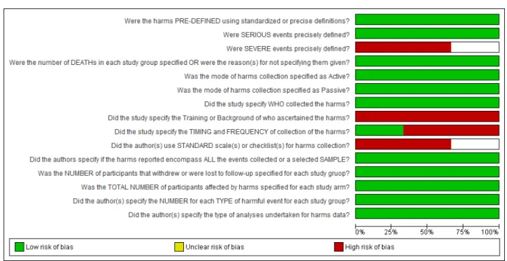
Studies reporting on the treatment of pa-tients with a bleeding diathesis with flexible ure-teroscopy and laser lithotripsy were included. Patients included were adults with a bleeding diathesis who had urinary stones. The following variables were extracted from each study: period of the study; country of origin of the study; study population demographics; type of anticoagulant used or coagulopathy; stent insertion; stone free rates; follow up; and complications. The data of each study was grouped into a meta-analysis to allow a numerical representation of the results. A quality assessment of harms using the McHarm scale was conducted for each included study (11). We used Review Manager (RevMan 5.0.23) to plot the quality assessment of harms tables.
RESULTS
300
IBJU|FLEXIBLE URETEROSCOPY AND HOLMIUM: YAG LASER LITHOTRIPSY
studies were then retrieved for further assess-ment, of which three were included in the review (7,8,12). All the included studies were published between 1998 and as recent as 2008, reflecting the continued debate of how to treat stones in patients with bleeding diathesis.
All 3 studies were retrospective studies; however, Turna et al. also compared the antico-agulated group to a similar group of patients as a control group. All the studies reported on the variables indicated in the ‘data extraction section’ and were plotted into Table-1. Wherever data was not available in the reports or there was not
enough clarification, lead authors were contacted to get the raw data.
Five articles were excluded after reading the full manuscript. One study was not included since the authors looked at all treatment modali-ties for urinary stones and though mentioned that 8 patients were ureteroscopically treated only 2 were holmium laser treated (10). Furthermore, the authors had not provided demographic, co-agulopathy, or stone details separately for these patients and therefore could not be extracted. Attempts at contacting the author were unsuc-cessful. The remaining four studies did not look Figure 1 - Flowchart for article selection process of the review.
Flowchart for article selection process of the review
Literature Search (No. = 199)
Articles excluded after screening of the Title (No. = 165)
Potential Articles for evaluation of Abstract (No. = 34)
Articles excluded after screening
Abstracts (No. = 26) Potential Articles for evaluation of Full Manuscript (No. = 8)
Articles excluded after screening
3
01
IBJU
|
FLEXIBLE URETEROSCOPY AND HOLMIUM: Y
A
G LASER LITHO
TRIPSY
ce (range) mean (range) Location (mm)
mean (range)
Kuo Urology 1998 1997-1998 USA 8 58.3 (42-74) Coumadin (INR: 2.1
(1.6-2.9)): 5 Thrombocytopaenia: 2
vWd: 1
Flexi Hol (3-15)
Ureteric (proxi-mal: 1; middle: 1; distal: 1)
Renal: 5
All 6/7 (one 2mm
residual frag (1 patient had
no stone on URS)
2 – (epistaxis: 1; post-op urinary
retention: 1)
Watterson Journal of
Urology
2002 1996-2001 USA 25
(28)
(42-84) Warfarin (INR 2.3): 17
Thrombocytopaenia: 4 vWd: 1 Liver dysfunction: 3
Flexi Hol 11.9 (6-25)
Ureteric (proxi-mal: 9; middle: 3; distal: 7)
Renal: 9
87% (26/30)
96% (27/28) 2 patients had electrohydrau-lic lithotripsy treatment
2 – (renal colic: 1; AF: 1)
Turna Journal of
Urology
2008 2001-2007 USA 37 58.2 (35-86) Coumadin (INR 1.8
(1.1-3.3)): 14 Clopidogrel: 5 Low dose (81mg) Aspirin:
13 High dose (325 mg)
Aspirin: 5
Flexi Hol 13.2 (5-35)
Ureteric: 8 Renal: 29
All 81.1% (30/37) 4 – (transient
macroscopic haematuria: 3;
UTI: 1)
302
IBJU|FLEXIBLE URETEROSCOPY AND HOLMIUM: YAG LASER LITHOTRIPSY
at patients with bleeding diathesis and therefore were excluded (2,4,9,13).
Characteristics of the included studies
All the studies were conducted in the USA (Table-1). Seventy patients who underwent 73 pro-cedures were included in this review. The study population was composed of patients with 35 to 86 years old. All patients had some sort of coagulopa-thy including 36 patients on warfarin, 6 patients with thrombocytopaenia, 2 with von Willebrand disease, 3 had liver dysfunction, 5 on clopidogrel, 13 on low dose aspirin, and 5 patients on a high dose aspirin. None of the patients had their coag-ulopathy reversed, except for 2 patients who had thrombocytopaenia and had recently had chemo-therapy; both were given 2 units of platelets for fear of the platelet count dropping further (7,12). The mean international normalization ratio (INR) for the patients on warfarin was 2.1 with a range of 1.1-3.3. Turna et al. had included patients on coumadin; however, their INR was 1.1, and there was no mention of how many patients with sub-therapeutic INR levels were included. All patients were treated with a flexible ureteroscopy and a holmium:YAG laser. The stone sizes ranged from 3-35mm with 43 renal stones and 30 ureteric, of which 10 were proximal, 4 middle and 8 distal ure-teric. Turna et al. had not mentioned the location of the ureteric stones.
Two studies routinely stented their patients after ureteroscopy and holmium:YAG laser frag-mentation. However, the third study (by Watterson et al.) did not differentiate between the patients who had holmium treatment and those that had electro-hydraulic lithotripsy and stent insertion, therefore their data was not included.
With regards to stone free rate, 87.7% (64/73) of the patients were stone free. In this re-view, none of the patients developed any major complications and 11% (8/73) of the patients de-veloped minor complications; however, five of the patients had complications unrelated to their coagulopathy. This brought the complication rate that could be attributed to an anticoagulated state, i.e. bleeding, to 4.1% (3/73). These three patients developed transient macroscopic haematuria for at least 3 days but did not require continuous
blad-der irrigations, secondary procedures or blood transfusions (8). The five other complications in-cluded one patient who developed a post-operative urinary retention, one patient developed worsen-ing renal colic attributed to stone passage, another developed atrial fibrillation, another developed a urinary tract infection and the last developed an epistaxis. The epistaxis was attributed to ketorolac; however, there was no mention of how they were certain that ketorolac was the cause rather than the coagulopathy. All patients were routinely followed up, however each study varied in the length of fol-low up. Kuo et al. folfol-lowed up their patients for 4-6 weeks, while Turna et al. followed up for 4 weeks, and Watterson et al. for 1-2 weeks only. All the patients were stone free and complication free after follow up discharge.
Methodological quality assessment of the includ-ed studies
Overall, the quality of the reported studies was modest as two of the studies were reported as retrospective while one was unclear; however it seemed to be retrospective from the methodology. All the included studies might have been subjected to bias as their method of recruitment of patients consisted of recruiting patients from databases; this could lead to selection as well as reporting bias. None of the studies were randomized, blinded (7,12), and only one study had a control group (8). However, the study group (coagulopathy patients) was significantly older than the control group, which poses the question to whether or not these groups could be compared. Furthermore, there was no mention on how the control group patients were selected from the authors’ database of 692 patients. This again could be construed as selection bias.
The quality assessment of harms indicates that the studies generally have a low risk of bias concerning reporting the harms that could poten-tially be caused by the procedure (Figures 2 and 3).
DISCUSSION
sur-Figure 2 - Risk of bias graph: review authors’ judgements about each risk of bias item presented as percentages across all included studies.
304
IBJU|FLEXIBLE URETEROSCOPY AND HOLMIUM: YAG LASER LITHOTRIPSY
geons with the haematologists and anaesthetists (12). However, the risk of thromboembolic events during perioperative bridging with heparin is 1-2% (14). Furthermore, treating the coagulopa-thy is significantly more costly when compared to patients without coagulopathy undergoing similar procedures (10).
Though other modalities exist for the treat-ment of large urinary stones, such as SWL, PCNL, and open or laparoscopic surgery, these are contra-indicated if the bleeding diathesis is not corrected (8,10). This only leaves ureteroscopic management for these patients (8).
Advancements in endoscope engineering and laser technologies allow an operator to vi-sualise and treat stones in the whole upper uri-nary system, including the renal calyxes with a reported long-term complication rate of less than 1% (12,15,16). Holmium lasers provides effective and efficient intracorporeal lithotripsy for even hard stones such as cysteine and calcium oxalate monohydrate stones, and can also be used to ab-late upper urinary tract tumours (7,12). Further-more, holmium lasers offer haemostatic capabili-ties during the procedure, which gives an additive benefit to patients with bleeding diathesis (12). Lastly, holmium laser energy is rapidly absorbed by water, leading to a minimal risk of ureteric in-jury if the laser fibre is at least 0.5mm away from the ureter and no risk of ureteric perforation if the distance is more than 1mm (12).
This review found that the use of flexible ureteroscopes and holmium lasers on patients with bleeding diathesis is not only safe but also efficient, with an overall stone free rate of 87.7%, a minor complication rate of 11%, but only a 4% rate of mi-nor bleeding, and a major complication rate of 0%. The validity of the results of systematic re-view depends on the quality of included studies including selection of participants and inclusion criteria. The studies included seemed to all be ret-rospective reports of a larger database. Therefore at most this review has a level 2a Levels of Evi-dence according to CEBM (17). No study evaluated cost analyses.
The other limitation of this review is re-lated to the patient population; the majority of pa-tients were on warfarin. However, the remaining
had various other causes for coagulopathy, wheth-er the hetwheth-erogeneity of the study sample would im-pact outcomes is not known. However, we aimed at reviewing all patients with coagulopathy and did not target one group. Furthermore, due to the lim-ited number of patients, we did not see a need of conducting sub-groups analysis which would have reduced the cohort even further.
Furthermore, though the level of evidence is considered a 2a, this review has a small cohort of patients (70) from case series basing this evidence on. In addition, no trial or study was found in the literature. This reflects the need for further larger participant studies to further explore the safety and efficacy of ureteroscopy in these patients.
Despite the limitation, grouping of the data was possible and revealed the safety and ef-ficacy of the combined studies. Furthermore, this review opens possibility for further research into the question.
This review has shown that it is not only safe but also efficient to treat patients suffering with urinary stones and afflicted with a bleeding diathesis with ureteroscopy and holmium laser. This can have cost benefits in practice as patients on anticoagulants need not undergo reversal and most patients with coagulopathy need further man-agement to support their coagulation system.
Future research efforts should be concen-trated on higher quality, more rigorous evaluation of ureteroscopic treatment in these groups of pa-tients. Studies should be multi-institutional and protocol driven, preferably peer reviewed before the start. Studies should be prospectively evaluated and include a control group of patients who are not anticoagulated for comparison. A detailed evalua-tion of the different types of bleeding diathesis such as patients on warfarin, clopidogrel, thrombocyto-penia or haemophilia should be analyzed individu-ally rather than as a whole. Furthermore, health economic outcome measures should be analyzed.
CONCLUSIONS
Further-more, these patients do not need their coagulopa-thy reversed, which leads to reduction the risk of thromboembolism with very minimal short-term complications and no long term consequence.
CONFLICT OF INTEREST
None declared.
REFERENCES
1. Ramello A, Vitale C, Marangella M: Epidemiology of neph-rolithiasis. J Nephrol. 2000; 13(Suppl 3): S45-50.
2. Argyropoulos AN, Tolley DA: SWL is more cost-effective than ureteroscopy and Holmium:YAG laser lithotripsy for ureteric stones: A comparative analysis for a tertiary refer-ral centre. British Journal of Medical & Surgical Urology. 2010; 3: 65-71.
3. Hendrikx AJ, Strijbos WE, de Knijff DW, Kums JJ, Does-burg WH, Lemmens WA: Treatment for extended-mid and distal ureteral stones: SWL or ureteroscopy? Results of a multicenter study. J Endourol. 1999; 13: 727-33.
4. Verze P, Imbimbo C, Cancelmo G, Creta M, Palmieri A, Mangiapia F, et al.: Extracorporeal shockwave lithotripsy vs ureteroscopy as first-line therapy for patients with single, distal ureteric stones: a prospective randomized study. BJU Int. 2010; 106: 1748-52.
5. Marchant F, Storme O, Osorio F, Benavides J, Palma C, Os-sandón E: Prospective trial comparing shock wave litho-tripsy and ureteroscopy for management of distal ureteral calculi. Actas Urol Esp. 2009; 33: 869-72.
6. Lee YH, Tsai JY, Jiaan BP, Wu T, Yu CC: Prospective ran-domized trial comparing shock wave lithotripsy and ure-teroscopic lithotripsy for management of large upper third ureteral stones. Urology. 2006; 67: 480-4.
7. Kuo RL, Aslan P, Fitzgerald KB, Preminger GM: Use of ure-teroscopy and holmium:YAG laser in patients with bleeding diatheses. Urology. 1998; 52: 609-13.
8. Turna B, Stein RJ, Smaldone MC, Santos BR, Kefer JC, Jackman SV, et al.: Safety and efficacy of flexible ureterore-noscopy and holmium:YAG lithotripsy for intrarenal stones in anticoagulated cases. J Urol. 2008; 179: 1415-9. 9. Williams SB: Is continuing warfarin in the perioperative
pe-riod safe for patients undergoing urologic procedures? Eur Urol. 2011; 59: 372-3.
10. Klingler HC, Kramer G, Lodde M, Dorfinger K, Hofbauer J, Marberger M: Stone treatment and coagulopathy. Eur Urol. 2003; 43:75-9.
11. Santaguida P, Raina P, Ismaila A: McMaster Quality As-sessment Scale of Harms (McHarm) for primary studies: Manual for use of the McHarm. Available from: http://hiru. mcmaster.ca/epc/mcharm.pdf
12. Watterson JD, Girvan AR, Cook AJ, Beiko DT, Nott L, Auge BK, et al.: Safety and efficacy of holmium: YAG laser litho-tripsy in patients with bleeding diatheses. J Urol. 2002; 168: 442-5.
13. Sofer M, Watterson JD, Wollin TA, Nott L, Razvi H, Den-stedt JD: Holmium:YAG laser lithotripsy for upper urinary tract calculi in 598 patients. J Urol. 2002; 167: 31-4. 14. Kaatz S, Paje D: Update in bridging anticoagulation. J
Thromb Thrombolysis. 2011; 31: 259-64.
15. Grasso M, Beaghler M, Bagley DH, Strup S: Actively de-flectable, flexible cystoscopes: no longer solely a diagnos-tic instrument. J Endourol. 1993; 7: 527-30.
16. Harmon WJ, Sershon PD, Blute ML, Patterson DE, Segura JW: Ureteroscopy: current practice and long-term compli-cations. J Urol. 1997; 157: 28-32.
17. Jeremy Howick BP, Chris Ball, Dave Sackett, Doug Bad-enoch, Sharon Straus, Brian Haynes, et al.: Centre for Ev-idence-Based Medicine. Available from: http://www.cebm. net/index.aspx?o=1025.
______________________ Correspondence address:
Dr. Manoj Monga
Glickman Urological & Kidney Institute, Cleveland Clinic,
306
IBJU|FLEXIBLE URETEROSCOPY AND HOLMIUM: YAG LASER LITHOTRIPSY
EDITORIAL COMMENT
It has been recently published that uroli-thiasis is an entity associated with metabolic syn-drome, which is characterized by hypertension, obesity, diabetes and abnormal lipid levels (1).
As the world drags its way towards obe-sity, urologists of all around the globe have no-ticed that, not only kidney stones have become more frequent, but also those patients who pre-sent them have more often other co-morbidi-ties. One particular instance is the drug-indu-ced blood diathesis, which is characterized by the use of “blood thinners” for cardiovascular protection.
These phenomena (obesity, metabolic syndrome, kidney stones, blood thinners) have brought upon the endourologist a current and challenging topic that every kidney stone specia-list needs to be up-to-date on: Stone treatment versus bleeding diathesis.
The present study reports on what has been published in the literature that could ser-ve as foundation to our decision making process while counseling a stone patient with any kind of bleeding diathesis. Surprisingly, the authors very well presented the lack of prospective (high evidence levels) studies on this matter; however, based on what has been judiciously selected in the literature, stone free and complication rates
of flexible ureteroscopy with holmium: YAG laser lithotripsy for patients with blood diathesis are similar to healthy individuals.
It is important to emphasize that if one considers doing a retrograde endoscopic stone treatment in a patient with bleeding diathesis, it is strongly advised, based on evidence level 2a, following analogous surgical technique to what has been described in the selected studies of this systematic review:
A) Energy/lithotripsy - there is no scientific support for using other energy than holmium:YAG laser;
B) Scopes - flexible ureteroscopes were used in all cases;
C) Double J - stenting seems to be a wise routine.
D) General anesthesia might be safer (rou-tine in USA), given the obvious risks of spinal puncture in such patients.
In conclusion, due to a pandemy of obesi-ty and its metabolic consequences, kidney stone patients will more often present co-morbidities and also some kind of bleeding diathesis (aspirin, warfarin, clopidogrel), thus, they must be infor-med that flexible ureteroscopy and holmium:YAG laser lithotripsy is safe and efficient for treating their ureteral/renal stones.
REFERENCE
1. Lange JN, Mufarrij PW, Wood KD, Holmes RP, Assimos DG: The association of cardiovascular disease and metabolic syn-drome with nephrolithiasis. Curr Opin Urol. 2012; 22: 154-9.
Dr. Renato Nardi Pedro