Outcomes of miniaturized percutaneous nephrolitotomy in
infants: single centre experience
_______________________________________________
Eyyup Sabri Pelit
1, Bülent Kati
1, Cengiz Çanakci
2, Süleyman Sa
ğ
ir
1, Halil Çiftçi
11 Department of Urology, Harran University Faculty of Medicine, Sanliurfa, Turkey; 2 Bolvadin State Hospital, Istanbul, Turkey
ABSTRACT
ARTICLE
INFO
______________________________________________________________ ______________________
Objectives: The present study was aim to evaluate the safety and efficacy of Mini-PNL to treat kidney stones in patients aged <3 years. This is the one of the largest series in the literature in this age group of patients.
Material and methods: From May 2012 to April 2016, the medical records of 74 infant patients who underwent mini-PNL for renal stones were reviewed retrospectively. All infants were evaluated with the plain abdominal radiograph, urinary ultrasound, non-contrast computerized tomography and/or intravenous urogram. Pre-operative, intra-operative and post-intra-operative data were analyzed.
Results: A total of 74 infant (42 male, 32 female) with a mean age 21.5±8.2 (10-36) months were included in this study. The mean size of the stones was 22.0±5.9 (14-45) mm. A 17 Fr rigid pediatric nephroscope with a pneumatic intracorporeal lithotripsy were used through 20-22 Fr access sheath. The stone-free rate was 84.7% at 1 month after the operation. Mean operative time was 74.0 (40-140) min. Mean fluoroscopy screening time was as 4.3(3.1-8.6) min. Average hospitalization time was 3.8 (2-9) day. Auxiliary procedures were performed to 11(15.3%) patients (7 extracorporeal shock wave lithotripsy, 3 re- percutaneous nephrolitotomy, 1 retrograde intrarenal surgery). No major complication classified as Clavien IV-V observed in study group.
Conclusions: Mini-PNL with pneumatic intracorporeal lithotripsy can be performed safely and effectively to manage kidney stones in infants with high stone free rate and low complications.
Keywords:
Nephrostomy, Percutaneous; Kidney Calculi; Infant
Int Braz J Urol. 2017; 43: 932-8
_____________________
Submitted for publication: December 01, 2016
_____________________
Accepted after revision: March 12, 2017
_____________________
Published as Ahead of Print: June 12, 2017
INTRODUCTION
Childhood urolithiasis is a major heal-th problem in developing countries especially in endemic regions. The incidence of childhood uro-lithiasis ranges between 4-6% and the incidence increases up to 14.8% in endemic regions of the World such as Turkey (1, 2). Pediatric patients ge-nerally have underlying metabolic, anatomical, functional abnormalities and/or recurrent urinary tract infections that cause urinary stone. Children with kidney stone are classified as high-risk
pa-tients for recurrence and requirement of multiple interventions (3).
first treatment option for stones smaller than 2cm. Although it is the least invasive method it may re-quire more auxiliary procedures than the other me-thods and most of the pediatric patients requires anesthesia during the ESWL procedure (6). PCNL should be the preferred treatment method in stones larger than 2cm and ESWL resistant hard stones (7). Since the first series of mini-PCNL technique was published in 1998, mini-PCNL has reached higher stone free rates (SFR) in pediatric patients with lo-wer complication rates (8).
In published literature, there are few stu-dies about mini-PCNL in infancy and number of patients included in these studies are limited. The present study aims to evaluate the safety and effi-cacy of mini-PNL to treat kidney stones in patients aged <3 years. To our best knowledge, this study is one of the largest series in the literature in this age group of patients.
MATERIALS AND METHODS
From May 2012 to April 2016, the medi-cal records of 72 infant (42 boys, 30 girls) patients who underwent mini-PNL for renal stones were re-viewed retrospectively in a referral tertiary institu-tion in Turkey. The presence of renal stones larger than 2cm, history of previous unsuccessful ESWL and ESWL resistant stones smaller than 2cm were accepted as indications for mini-PNL. ESWL resis-tance was accepted as two ESWL session failures. Patients who have stones with renal congenital anomaly such as ureteropelvic junction obstruction were excluded from the study. Serum biochemis-try, complete blood count, urine analysis and urine culture were performed for all patients prior to sur-gery. All infants were evaluated with plain abdomi-nal radiography (KUB), urinary ultrasound (USG), non-contrast computerized tomography (NCCT) and/or intravenous urogram (IVP). All patients had sterile urine culture prior to surgery. Urinary tract infection was treated according to bio-sensitivity result of the urine culture. Stone size was accepted as the longest axis measured on NCCT and if multi-ple stones exists, stone burden was assessed as the sum of longest diameter of each stone.
All patients received intravenous antibio-tic prophylaxis 1 hour before the surgery. Patients
All statistical analyses were conduc-ted by using SPSS statistical software (version 15.0; SPSS, Inc., Chicago, IL, USA). A probabi-lity value (p value) of <05 was considered sta-tistically significant.
RESULTS
A total of 72 infants (42 male, 30 female) with a mean age 21.5±8.2 (10-36) months were included in this study. The mean size of the sto-nes was 22.0±5.9 (14-45) mm. Renal stosto-nes were located in renal pelvis (n=20), lower pole (n=17), middle pole/upper pole (n=11), all calyx (n=24). Patients had no hydronephrosis (n=12), grade 1(n=15), grade 2 (n=37) and grade 3 hydrone-phrosis (n=8). All intrarenal access was perfor-med in the prone position and under fluoroscopic guidance. Mean operative time was 69.0 (40-140) min. Mean fluoroscopy screening time was 4.3 (3.1-8.6) min. The stone-free rate was 84.7% at 1 month after the operation. Nephrostomy tube was not inserted postoperatively in 6 (8.4%) patients with no residual stone, extravasation and periope-rative hemorrhage and especially in single access, short-running procedures. Auxiliary procedures were performed to 11(15.3%) patients (7 ESWL, 3 re- PCNL, 1 RIRS). Seven out of these 11 patients were completely stone free and these additional procedures increased the overall success rate from 84.7% to 94.4%. The stone size was 15±4.2mm in the clinically successful procedures and 22±4.9 in the failed procedures (P=0.004). Additionally, renal pelvis or single calyceal location was 73.7% in the clinically successful procedures, whereas it was 27.2% in the failed procedures (P=0.002). Four patients were followed via ultrasonography for insignificant fragments. Average hospitalization time was 3.0 (2-9) days. Complications classified as Clavien IV-V were not observed, however one major complication classified as Clavien III was observed in the study group. Five patients requi-red blood transfusions. Extravasation of urine to the retroperitoneum then pleura after withdrawal of the nephrostomy tube was observed in 1 infant and in this patient spontaneous resolution was observed after DJ stent insertion. Bowel perfora-tion was seen in 1 patient which was diagnosed
when colonic content was seen in nephrostomy tube and perforated area of descending colon was primarily repaired on postoperative day 3; 1 pa-tient had hydrothorax due to pleural injury during the upper pole access and thorax tube was inser-ted. Seven patients developed urinary infections and they were treated according to antibiogram results of the urinary culture.
Nine uric acid stones, 11 cystine stones, 16 calcium oxalate-calcium phosphate stones and 7 struvite stones were detected during the pos-toperative stone analysis Stone composition was not known in 29 patients because of the discon-tinuation of the follow-up. Demographics, preo-perative, intraopreo-perative, postoperative findings of patients, stone composition and factors that affect the stone free status of the patients are summari-zed in Tables 1-4.
DISCUSSION
The high risk of recurrence of the stones in the pediatric age group and necessity of multiple surgical interventions has led to development of
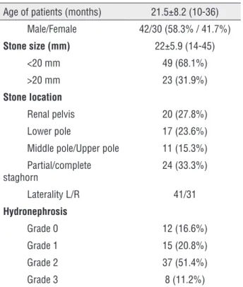
Table 1 - Patients’ demographics and preoperative data.
Age of patients (months) 21.5±8.2 (10-36)
Male/Female 42/30 (58.3% / 41.7%)
Stone size (mm) 22±5.9 (14-45)
<20 mm 49 (68.1%)
>20 mm 23 (31.9%)
Stone location
Renal pelvis 20 (27.8%)
Lower pole 17 (23.6%)
Middle pole/Upper pole 11 (15.3%)
Partial/complete staghorn
24 (33.3%)
Laterality L/R 41/31
Hydronephrosis
Grade 0 12 (16.6%)
Grade 1 15 (20.8%)
Grade 2 37 (51.4%)
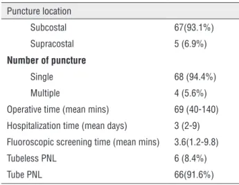
Table 2 - Intraoperative data.
Puncture location
Subcostal 67(93.1%)
Supracostal 5 (6.9%)
Number of puncture
Single 68 (94.4%)
Multiple 4 (5.6%)
Operative time (mean mins) 69 (40-140)
Hospitalization time (mean days) 3 (2-9)
Fluoroscopic screening time (mean mins) 3.6(1.2-9.8)
Tubeless PNL 6 (8.4%)
Tube PNL 66(91.6%)
Table 3 - Postoperative data.
Initial Stone free rate(after 1 month) 61 (84.7%)
Stone free rate after additional therapy 68(94.4%)
Additional procedures
ESWL 7
Re-PNL 3
URS 1
Minor (Clavien I-II) complications 14 (19.4%)
Major (Clavien III-V) complications 1 (1.3%)
Preoperative hemoglobin level 12.3 (9.8-15.3)
Postoperative hemoglobin level 11.3 (8.9-13.9)
Stone composition
Uric acid 9 (12.5%)
Cystine 11 (15.3%)
CaOx-CaP 16 (22.3%)
Struvite 7 (9.7%)
Unknown 29 (40.2%)
minimally invasive treatment methods with maxi-mal efficiency. ESWL, RIRS, PCNL (micro-, mini-) and laparoscopic surgery are the standard minimal invasive procedures to treat renal stones in pediatric patients.
ESWL is the least invasive and first-line treatment method that is used for the management of pediatric renal stones. Despite its widespread use in adults, ESWL for pediatric renal stones was first
performed in 1986 by Newman (9). Reports have showed the safety and efficacy of the ESWL even in low birth weight infants (10). Limited reports about ESWL in infancy exist because of the rarity of renal stones in infancy. The success rate of ESWL in infant patient was 84.6-100% (11, 12). Despite these high stone free rate of the procedure, ESWL has some disadvantages such as requirement of anesthesia, ureteral obstruction in high volume renal stones and higher additional intervention rate (13). In cur-rent literature ESWL is a safe method of treatment from the point of view of development of new onset diabetes mellitus and hypertension; however, long term detrimental effect of ESWL on kidney which can cause diabetes mellitus and hypertension is still controversial (12, 14).
With the advancement of small caliber f--URS, management of renal stones in childhood have become possible even in infant patients. Can-non et al. published the first series of the RIRS in pediatric patient (15). Stone free rate in pediatric RIRS studies varies between 76-99%. However age of patient in these series was generally greater than 3 years old (15, 16). Li et al. published first series of RIRS in infant patient with SFR of 94.6%. Ten out of 55 infants underwent simultaneous bilateral RIRS (17). One of the major problems during the procedure in this age group of patient was urete-ral access sheath (UAS) insertion. The younger the child was, the harder the insertion of UAS. In re-ported series of pediatric RIRS, UAS was inserted in 43.7-61.5% of patients (16-18). Li et al. did not use UAS, rather they inserted DJ stent in all patients before the procedure and after a while 8Fr f-URS was advanced over the hydrophilic guidewire (17). Generally, major complications were not observed in these procedures. Urinary system infection, pos-toperative hematuria, ureteral mucosal injury and ureteral perforation were the most commonly obser-ved complications in pediatric RIRS series (16, 17).
posto-perative 24 hour and 81.2% at the end of the first postoperative week in 48 infant mini-PNL proce-dures (20). Bo Xiao et al. performed all mini-PCNL with ultrasound-guidance in 67 renal units of 56 patients aged <3 years. They found that SFR during the hospital discharge was 92.5% (21). In the retros-pective study of Brodie et al. which was conduc-ted in 46 patients under age of 16, 76% of patients achieved 100% stone clearance after a single session of mini-PCNL and 100% of patients achieved stone clearance of greater than or equal to 80% (22). SFR was 76.9% in a prospective study of Kareem Daw et al. and SFR increased to 85% and 92.3% after ESWL and auxiliary therapy respectively (23). Pelit et al. reported a SFR of 84.4% and 91.1% after initial and additional treatment respectively (18). In our study, we reached SFR of 84.7% and 94.4% after single and additional session respectively. Further analysis of SFR revealed that stone size and stone location were factors that affected the stone free status of the patients. In our observation, although hydronephro-sis grade did not affect the success of the procedure it facilitated the access to the collecting systems and shortened the operation time. We believe that an SFR of 94.4% for mini PNL procedure in infants is acceptable. In parallel to the literature on mini PCNL in infants, stone free rates of our series after initial
and additional therapy is higher than some studies in infant patients. This is because we have an incre-ased experience in childhood urolithiasis due to the location of our hospital in an endemic stone area.
Holmium: yttrium-aluminum-garnet (h--YAG) laser and pneumatic intracorporeal lithotrip-sy tecniques were both used for stone fragmentation in infant mini-PCNL. As it was in our study, Bodak-ci et al. used only pneumatic lithotoripsy for frag-mentation and SFR was 81.2% in their series (20). Brodie and Bo Xiao et al. used both pneumatic and laser lithotripsy for fragmentation depending on the surgeon’s preference and SFR was 76% and 92.5% in their studies,respectively (21, 22). Kareem Daw et al. used only h-YAG laser for stone fragmentation and they obtained a 76.9% of SFR (23). However in current litarature, there was no study that compare the effect of lithotripsy techniques on SFR in infant mini-PCNL.
Although the stone-free rates were higher after mini PNL, complications were not uncommon in infancy, such as bleeding, urosepsis, colon perfo-ration, hypothermia and urinary leakage. Pediatric patients were more likely to bleed during system dilatation because of the fragile renal parenchyma and delicate collecting system. In mini PNL series of infancy, blood transfusion rates were lower than the
Table 4 - Factors that affect the stone free status of the patients.
Initional stone-free status (84.7%)
Residual stone without additional treatment (15.3%)
P value
Gender 0.820
Male 36 (59.1%) 6 (54.5%)
Female 25 (40.9%) 5 (45.5%)
Laterality 0.223
Right side 27 (44.2%) 4 (36.3%)
Left side 34 (55.8%) 7 (63.7%)
Stone size (mm) 15±4.2 22±4.9 0.004
Stone location 0.002
Renal pelvis or single calyx 45 (73.7%) 3 (27.2%)
Partial/complete staghorn 16 (26.3%) 8 (72.8%)
Hydronephrosis Grade 0.065
Grade 0-1 22 (36.1%) 5 (45.4)
pediatric age patients and varied between 0% and 7.5% (20, 21). Studies showed that blood transfu-sion rates were higher with the use of 24-26Fr she-aths than with ≤18Fr (5.9%) sheaths (24). We dilate the renal tract up to 20Fr or 22Fr according to the stone size and age of patients. In our opinion, the 22Fr sheath should be kept for stones larger than 2cm to reduce the bleeding if the patient’s age and physical development is appropriate.
Children can easily become hypothermic due to the long-running operations, cold irrigation fluids and operation room (25). Roberts et al. sho-wed that hypothermia is directly related with the duration of the operation and they also stated that preoperative preparation, anesthesia induction and patient positioning contribute the fall of the body temperature as much as the surgical procedure itself (26). In our patients, we did not observe any hypo-thermic complications. We think that irrigation flui-ds at body temperature and heater blanket prevent infants from being hypothermic and shortening the duration of all surgical steps including preoperati-ve procedures, anesthesia induction and positioning preserve the core body temperature of the patients.
One of the important issues of mini-PCNL in infants is radiation exposure. Some methods have been tried to minimize radiation exposure. Bo Xiao et al. punctured the collecting system under USG guidance to reduce radiation exposure (21). Bodak-ci et al. achieved US-guided intrarenal access in 7 of 40 to decrease the fluoroscopy time (22). In all series, lead aprons were used to protect patient’s go-nads like our studies.
Bowel perforations are also rare complica-tions, however carry high morbidity and mortality risk. It is observed as 0.2% to 0.3%. Dilated collec-ting tubules, horseshoe kidney, and retro-renal co-lon are risk factors for coco-lon injury (27). To avoid colon perforation in PCNL, some techniques have been proposed. Ultrasound-guided puncture or CT--guided puncture of the pelvicaliceal system in pa-tients with anatomic abnormalities could prevent colon perforation (28, 29). However, these access techniques were not routinely applied during PCNL procedures. For this reason, especially the left re-nal lower pole access with other retro-rere-nal colon risk factors, the bowel injury should be considered during PNL. Conservative treatment with drainage
of urinary system and gastrointestinal system se-parately is generally the first choice of method (30). In our patient, we observed colonic content in ne-phrostomy tube at postoperative 3 day and we de-cided to repair the colon primarily with pediatric surgeons. The patient was discharged uneventfully at postoperative 9 day following laparotomy.
The overall incidence of hydrothorax after PCNL procedures was between 0% and 3.3%. The risk of injury increased with the supracostal access due to the position of the pleura (31). As in our case that we have performed supracostal puncture, respi-ratory distress developed on postoperative day 1 and hydrothorax was diagnosed. This patient was mana-ged successfully by intercostal chest tube drainage.
Our study is limited by its retrospective na-ture. On the other hand, it is strengthened by the high number of patients.
CONCLUSIONS
Mini-PNL with pneumatic intracorporeal li-thotripsy can be performed safely and effectively to manage kidney stones with high stone free rate and low complications in patients under the age of 3. Exposure of infant patients to hypothermia and ra-diation must be kept in mind during the operation. This method of treatment provides an acceptable SFR in experienced center.
CONFLICT OF INTEREST
None declared.
REFERENCES
1. Tasian GE, Ross ME, Song L, Sas DJ, Keren R, Denburg MR, et al. Annual Incidence of Nephrolithiasis among Children and Adults in South Carolina from 1997 to 2012. Clin J Am Soc Nephrol. 2016;11:488-96.
2. Akinci M, Esen T, Tellaloğlu S. Urinary stone disease in Turkey: an updated epidemiological study. Eur Urol. 1991;20:200-3. 3. Baştuğ F, Gündüz Z, Tülpar S, Poyrazoğlu H, Düşünsel R.
4. Sen H, Seckiner I, Bayrak O, Erturhan S, Demirbağ A. Treatment alternatives for urinary system stone disease in preschool aged children: results of 616 cases. J Pediatr Urol. 2015;11:34.e1-5.
5. Straub M, Gschwend J, Zorn C. Pediatric urolithiasis: the current surgical management. Pediatr Nephrol. 2010;25:1239-44.
6. Rodrigues Netto N Jr, Longo JA, Ikonomidis JA, Rodrigues Netto M. Extracorporeal shock wave lithotripsy in children. J Urol. 2002;167:2164-6.
7. Etemadian M, Maghsoudi R, Shadpour P, Mokhtari MR, Rezaeimehr B, Shati M. Pediatric percutaneous nephrolithotomy using adult sized instruments: our experience. Urol J. 2012 Spring; 9:465-71.
8. Jackman SV, Hedican SP, Peters CA, Docimo SG. Percutaneous nephrolithotomy in infants and preschool age children: experience with a new technique. Urology. 1998;52:697-701. 9. Newman DM, Coury T, Lingeman JE, Mertz JH, Mosbaugh
PG, Steele RE, et al. Extracorporeal shock wave lithotripsy experience in children. J Urol. 1986;136(1 Pt 2):238-40. 10. Shukla AR, Hoover DL, Homsy YL, Perlman S, Schurman S,
Reisman EM. Urolithiasis in the low birth weight infant: the role and efficacy of extracorporeal shock wave lithotripsy. J Urol. 2001;165(6 Pt 2):2320-3.
11. El Nashar AM, Metwally AH, Abd El Kader O, Ali EE, Abdelbaseer M: Efficacy of shock wave lithotripsy in management of kidney stones in infants.2013; j.afju 11.002.
12. Turna B, Tekin A, Yağmur İ, Nazli O. Extracorporeal shock wave lithotripsy in infants less than 12-month old. Urolithiasis. 2016;44:435-40.
13. Nazli O, Cal C, Ozyurt C, Günaydin G, Cüreklibatir I, Avcieri V, et al. Results of extracorporeal shock wave lithotripsy in the pediatric age group. Eur Urol. 1998;33:333-6.
14. Krambeck AE, Gettman MT, Rohlinger AL, Lohse CM, Patterson DE, Segura JW. Diabetes mellitus and hypertension associated with shock wave lithotripsy of renal and proximal ureteral stones at 19 years of followup. J Urol. 2006;175:1742-7.
15. Cannon GM, Smaldone MC, Wu HY, Bassett JC, Bellinger MF, Docimo SG, et al. Ureteroscopic management of lower-pole stones in a pediatric population. J Endourol. 2007;21:1179-82.
16. Erkurt B, Caskurlu T, Atis G, Gurbuz C, Arikan O, Pelit ES, et al. Treatment of renal stones with flexible ureteroscopy in preschool age children. Urolithiasis. 2014;42:241-5.
17. Li J, Xiao J, Han T, Tian Y, Wang W, Du Y. Flexible ureteroscopic lithotripsy for the treatment of upper urinary tract calculi in infants. Exp Biol Med (Maywood). 2017;242:153-159. 18. Pelit ES, Atis G, Kati B, Akin Y, Çiftçi H, Culpan M, et al.
Comparison of Mini-percutaneous Nephrolithotomy and Retrograde Intrarenal Surgery in Preschool-aged Children. Urology. 2017;101:21-25.
19. Jackman SV, Docimo SG, Cadeddu JA, Bishoff JT, Kavoussi LR, Jarrett TW. The “mini-perc” technique: a less invasive alternative to percutaneous nephrolithotomy. World J Urol. 1998;16:371-4.
20. Bodakci MN, Daggülli M, Sancaktutar AA, Söylemez H, Hatipoglu NK, Utangaç MM, et al. Minipercutaneous nephrolithotomy in infants: a single-center experience in an endemic region in Turkey. Urolithiasis. 2014;42:427-33. 21. Xiao B, Hu W, Zhang X, Chen S, Li Y, Li J. Ultrasound-guided
mini-percutaneous nephrolithotomy in patients aged less than 3 years: the largest reported single-center experience in China. Urolithiasis. 2016;44:179-83.
22. Brodie KE, Lane VA, Lee TW, Roberts JP, Raghavan A, Hughes D, et al. Outcomes following ‘mini’ percutaneous nephrolithotomy for renal calculi in children. A single-centre study. J Pediatr Urol. 2015;11:120.e1-5.
23. Daw K, Shouman AM, Elsheemy MS, Shoukry AI, Aboulela W, Morsi HA, et al. Outcome of Mini-percutaneous Nephrolithotomy for Renal Stones in Infants and Preschool Children: A Prospective Study. Urology. 2015;86:1019-26. 24. Unsal A, Resorlu B, Kara C, Bozkurt OF, Ozyuvali E. Safety
and efficacy of percutaneous nephrolithotomy in infants, preschool age, and older children with different sizes of instruments. Urology. 2010;76:247-52.
25. Vorrakitpokatorn P, Permtongchuchai K, Raksamani EO, Phettongkam A. Perioperative complications and risk factors of percutaneous nephrolithotomy. J Med Assoc Thai. 2006;89:826-33.
26. Roberts S, Bolton DM, Stoller ML. Hypothermia associated with percutaneous nephrolithotomy. Urology. 1994;44:832-5. 27. El-Nahas AR, Shokeir AA, El-Assmy AM, Shoma AM,
Eraky I, El-Kenawy MR, et al. Colonic perforation during percutaneous nephrolithotomy: study of risk factors. Urology. 2006;67:937-41.
28. Alken P, Hutschenreiter G, Günther R, Marberger M. Percutaneous stone manipulation. J Urol. 1981;125:463-6. 29. Matlaga BR, Shah OD, Zagoria RJ, Dyer RB, Streem SB,
Assimos DG. Computerized tomography guided access for percutaneous nephrostolithotomy. J Urol. 2003;170:45-7. 30. Noor Buchholz NP. Colon perforation after percutaneous
nephrolithotomy revisited. Urol Int. 2004;72:88-90.
31. Yadav R, Aron M, Gupta NP, Hemal AK, Seth A, Kolla SB. Safety of supracostal punctures for percutaneous renal surgery. Int J Urol. 2006;13:1267-70.