Pituitary tumor aPoPlexy
Claudia V. Chang
1, Andre C. Felicio
2, Andrea Cecilia Toscanini
1,
Manoel Jacobsen Teixeira
3, Malebranche Berardo Carneiro da Cunha-Neto
1abstract – Pituitary tumor apoplexy is a medical emergency due to acute infarction or hemorrhage in the pituitary gland. In this review, the authors discuss the sellar anatomy, the pituitary gland and adenomas’ vascularization and the general aspects of the syndrome such as its ethiopatogenesis, predisposing factors, clinical features, treatment and prognosis.
Key words: pituitary tumor apoplexy, pituitary, pituitary adenomas.
apoplexia em tumor hipofisário
resumo – A apoplexia em tumor hipofisário é uma emergência médica decorrente do infarto agudo ou hemorrágico na glândula hipófise. Nesta revisão os autores discutem a anatomia da região selar, a vascularização da hipófise e adenomas hipofisários, e demais aspectos da síndrome como etiopatogenia, fatores predisponentes, quadro clínico, tratamento e prognóstico.
PAlAvrAs-chAve: apoplexia hipofisária, hipófise, adenoma hipofisário.
1Unidade de Neuroendocrinologia da divisão de Neurocirurgia do hospital das clínicas da Faculdade de Medicina da Universidade de são Paulo, são
Paulo sP, Brazil (hc-FMUsP); 2departamento de Neurologia da Universidade Federal de são Paulo, são Paulo sP, Brazil; 3departamento de Neurologia
do hc-FMUsP.
received 2 september 2008, received in inal form 15 december 2008. Accepted 9 March 2009.
Dr. Claudia Veiga Chang – Av. Bosque da Saude 834 / 193 04142-081 São Paulo SP - Brasil. E-mail: [email protected]
Pituitary apoplexy is a potentially life-threatening disor-der due to acute ischemic infarction or hemorrhage of the pituitary gland. The main clinical features of this syndrome are headache, nausea, vomiting, visual impairment, altered
mental status, and even coma1-4
. As the primary event
in-volves the adenoma, the syndrome should be referred to as pituitary tumor apoplexy and not as pituitary apoplexy5.
Notwithstanding pituitary apoplexy occurs within pituitary adenomas, it may also occur in non-adenomatous or even
the normal pituitary gland6, especially during pregnancy.
Although the “subclinical pituitary apoplexy”7-9
ter-minology is widely used to describe anatomophatologi-cal evidence of asymptomatic pituitary ischemia or hem-orrhage, it is meaningful to note that pituitary apoplexy is a clinical syndrome rather than an anatomopathologi-cal deinition10,11.
anatomy of the sellar region
The pituitary gland is circumscribed by the sphenoid bone among its whole surface and enclosed with the dia-phragma sellae. It relies on the sella turcica under the hy-pothalamus and optic chiasma. In the adult, it measures
approximately 12×9×6mm diameters and weights 0,6g,
and during pregnancy its volume may double. The
pitu-itary gland can be divided into two major parts: one sit-uated at the anterior site corresponding to 80% of the gland (adeno-pituitary) and the remaining 20% on the posterior site of the gland (neuro-pituitary)12.
Vascularization of hyPothalamus and Pituitary gland
There are up to ten portal pituitary vessels that orig-inate dorsally at capillaries from the median eminence. These vessels move alongside the ventral surface of the anterior pituitary stalk and drain to the adeno-pituitary making anastomosis with the neuro-pituitary capillaries. The circulation is predominantly through hypothalamus to pituitary gland and allows pituitary to be the most ir-rigated region of the body (0.8 ml/g/min). It is still not known whether the anterior lobe of the pituitary gland receives its blood supply exclusively from the portal cir-culation or if there is some additional arterial blood sup-ply5. It is believed that 70 to 90% of adeno-pituitary
arteri-al supply for the adeno-pituitary could be capsular arter-ies, originated from inferior pituitary arteries which are branches from the internal carotid artery. on the other side, venous drainage takes place within adjacent venous sinus to the jugular veins12.
To sum up, pituitary gland is one of the greatest irri-gated structures of our organism with a complex
vascu-lar system13, and therefore pituitary adenomas have a 5.4
greater chance to bleed than any other brain tumor14.
Vascularization of Pituitary adenomas
The number and size of tumoral vessels are variable. Generally they are lesser than the normal pituitary vessels and are divided into irregular islets. Under electronic mi-croscopy they have incomplete maturation, low fenestra-tion, and fragmented basal membranes with sized perivas-cular spaces illed with plasmatic proteins or red cells15,16.
Adenomas receive a mixed arterial or portal blood, but in contrast to the normal pituitary gland, the arterial source
is generally more dominant in pituitary adenomas17.
The angiographic characteristic of tumoral vessels that supply adenomas implies that they come from the inferi-or pituitary artery, and are under the inluence of system-ic arterial pressure12,17.
etioPathology
The etiopathology of pituitary apoplexy is still a mat-ter of debate. some authors believe pituitary apoplexy occurs due to rapid tumoral growth that outstrips
arte-rial supply18-20. It is uncertain whether the pathological
process is a primary hemorrhage or whether the event is really a hemorrhagic infarction. The size of the adenoma
appears to be a major factor21, but it is known that even
smaller adenomas can bleed. Another rationale for pitu-itary apoplexy could be the tumoral tissue growing inside the narrow space situated between the pituitary stalk and diaphragma sellae leading to constriction of the thin vas-cular net adjacent to the stalk, and inally ischemia, ne-crosis and hemorrhage on the anterior lobe and
tumor-al tissue22. Taking into account that the adenoma is
sup-plied by the inferior pituitary artery, the compression of the superior pituitary artery and its branches against the diaphragma sellae could lead to ischemia of the adeno-pituitary and not the adenoma. If this is true the hemor-rhage would begin within the normal pituitary gland in-stead of the adenoma itself22.
The intentional (surgical) occlusion in animals or hu-mans of portal pituitary vessels leads to ischemic infarc-tion and not hemorrhage. The process begins with the vol-ume reduction of acinar cells and its splitting from the
basal membranes, culminating to necrosis23. Patients who
have tumor infarction have generally less severe clinical symptoms and longer course before presenting for
medi-cal care than those who have hemorrhage or hemorrhag-ic infarction of the adenoma24.
The hypothesis of tumoral “intrinsic” factors ending to hemorrhage is also suggested. There is a statistically sig-niicant relationship between the aggressive and invasive
tumoral behavior and hemorrhage11,23. The role of
vasos-pasm was also implicated on the etiophatology of pitu-itary infarction. Intracranial aneurisms are more often as-sociated to pituitary adenomas than in the general
pop-ulation or with other brain tumors5,12. Additionally,
pitu-itary adenomas bleed much more than any other central nervous system tumor. Thus, one might speculate of an “intrinsic” factor such as a kind of tumoral-vasculopathy underlying the mechanism of aneurism development and pituitary hemorrhage25.
PredisPosing factors
Numerous conditions have been linked to pituitary apoplexy but in the majority of cases it happens
with-out any predisposing factor but pituitary adenoma5,25.
The main predisposing factors associated to pituitary
ap-oplexy (~25% of cases) are: medication (bromocriptine
and cabergoline)26-28, radiotherapy29, pituitary function
tests30,31, diabetes mellitus, trauma, thrombocytopenia or
recent surgery32-36. high blood pressure is also considered
as a risk factor for pituitary apoplexy although this asso-ciation is not always seen1,6.
The prevalence of apoplexy according to different subtypes of pituitary tumors is homogeneous with a small trend for non-functioning adenomas and prolactinomas to develop apoplexy. Gh–, AcTh–, Tsh–, and
gonado-trophin-secreting tumors shares similar prevalence5,21. It
is believed that between non-functioning adenomas and prolactinomas the prevalence is slightly greater in the for-mer, perhaps because the peak prevalence of pituitary apoplexy is similar to that of non-functioning adenomas, which is the iftieth decade of life12.
clinical features
Altered visual ield or visual acuity means involvement of the optic nerves, chiasma, or optic tracts. Motor ocular nerves (III, Iv, and vI) are vulnerable at the cavernous si-nus and therefore in the majority of patients that do not show altered mental status headache is associated with
diplopia5,12. The medial aspect of the cavernous sinus
cor-responds to the lateral aspect of the pituitary fossa and for that reason an acute hemorrhage or necrosis within this region can shift the oculomotor nerves. Thus, visual impairment and the presence of motor ocular involvement do not necessarily means supra or para-sellar growth of pituitary tumors. Ipsilateral mydriasis and ptosis are owing to IIIrd nerve involvement. Facial numbness may also take
place due to the vth nerve irst division involvement12.
Altered mental status is the most severe neurological
inding in patients with pituitary apoplexy5. Its mechanism
remains elusive and might be related to sub-arachnoid hem-orrhage, increased intracranial pressure, obstructive hydro-cephalus, adrenal insuficiency leading to arterial hypoten-sion and hypoglycemia, and hypothalamic compreshypoten-sion.
Nausea and vomiting may occur due to adrenal in-suficiency, meningeal irritation, and hypothalamic dys-function or raised intracranial pressure. Neck stiffness is observed in patients with pituitary apoplexy and should
raise the attention for sub-arachnoid hemorrhage3.
Focal signs such as loss of muscle strength or aphasia are less common and are attributed to internal carotid
ar-tery compression or vasospasm6.
The majority of patients do not have the diagnosis of pituitary adenoma at the time of pituitary apoplexy, but a retrospective and careful evaluation of these patients can show previous evidence of endocrine dysfunction. The most clinically important deicit is that of AcTh because it leads to acute glucocorticoid insuficiency occurring at the time of severe physical stress. The majority of patients present with, at least, partial hypopituitarism. reviewing a series of patients that had pituitary apoplexy, veldhuis
and hammond38 found multiple adeno-pituitary
hormon-al deiciencies such as: Gh deicit (88%), AcTh hyposecre-tion (66%), hypothyroidism (42%) and hypogonadothroic hypogonadism (85%).
Transient diabetes insipidus is not a common feature
of pituitary apoplexy and is reported in only 4% of cases39.
rarely, inappropriate Adh secretion occurs, perhaps due to sparing of neuro-pituitary or pituitary stalk10.
neuroimaging
Although it is in disuse, plain radiography of the skull is a quick and inexpensive method for evaluating pituitary apoplexy showing enlargement of the pituitary fossa and destruction of sellar dorsum4.rarely, a fracture of sellar
dorsum is seen, and this is considered an speciic sign of pituitary apoplexy. A normal plain radiography does not exclude pituitary apoplexy.
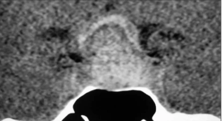
A recent hemorrhage can appear as a single or mul-tiple lesion with hyperdense sign on brain comput-er tomography (cT) (Fig 1) and no one or tiny contrast
enhancemet4,12,40. on subsequent days after hemorrhage
a progressive reduction of lesion hyperdensity occurs and after iodine contrast injection a peripheral ring is seen
around the lesion22. At this time (approximately four days
after bleeding) the hemorrhage can be misinterpreted as cystic degeneration, abscess, or local infarction, since all
these conditions have lower density sign on scans25. Brain
cT can also demonstrate sub-arachnoid hemorrhage and whether there is involvement of brain and ventricles. sometimes the differentiation between a non-complicat-ed and a blenon-complicat-eding adenoma can be dificult or even impos-sible because the observed densities on cT may be variable and there is no hallmark to differentiate both conditions. occasionally, serial scans are recommended but it should
Fig 1. Note on this sellar computer tomography (coronal view) the ad-enoma located at the sella turcica with an acute hemorrhage seen as an hyperdense lesion.
be taken into consideration that the irst three days after
pituitary apoplexy are the best period for cT handling34.
Unless speciic techniques of imaging acquisition are
obtained (diffusion-weighted)4 brain magnetic resonance
imaging (MrI) is less eficient than cT in the acute stage of pituitary apoplexy. on the sub-acute and chronic stages of pituitary apoplexy brain MrI is considerably better than cT25. one of the advantages of MrI is the possibility of
es-timating the onset of bleeding. In the acute stage of pitu-itary apoplexy (irst seven days) it is seen on MrI charac-teristic hypo- or isointense lesions on T1- and T2-weigthed
images; between seven to fourteen days on the sub-acute stage there is marginal signal reinforcement although the hematoma core remains isointense; on the chronic stage there is an overall increase on T1- and T2- signal (Fig 2). The
hyperintense lesion on MrI may last much longer than cT (up to one year after pituitary apoplexy)12.
hypersignal on T1-weighted sequences is seen on
hem-orrhagic tissues, fat, and lesions with high protein or mel-anin content. Thus, intra-sellar tumors that may present
with T1 hyperintense signal are craniopharingiomas,
lipo-mas, dermoid cysts, metastatic melanomas or any
oth-er hemorrhagic tumor34. clinical characteristics together
with other brain MrI abnormalities may help to elucidate
diagnosis. Unlike pituitary apoplexy on T2-weighted
imag-es sub-acute or chronic hemorrhagic tissuimag-es, lipomas, der-moid cysts, and melanomas are often hypointense.
cran-iopharingiomas may be hyperintense either on T1 or T2
im-ages due to its high protein content within cysts, becom-ing a dificult differential diagnosis with pituitary tumors
that bleed or show necrosis12. craniopharingiomas,
lipom-as, and dermoid cysts generally presents with slow clini-cal deicits opposing to hemorrhagic or metastatic lesions that presents with acute neurological manifestations.
Angiography may also be helpful in the management of pituitary apoplexy as it shows the presence or absence of concomitant aneurism (7% of cases), and vasospasm. If there are no associated abnormalities the tumoral mass can be seen with contrast enhancement. Angiography is obligatory especially in patients with neck stiffness ac-companied to focal neurological signs or inconclusive cT or MrI scans4.
differential diagnosis
clinical conditions frequently misdiagnosed as pitu-itary apoplexy are sub-arachnoid hemorrhage due to rup-tured intracranial aneurism and meningitis. other diseases that may share similar clinical characteristics are basilar ar-tery occlusion, hypertensive encephalopathy, brain abscess or cyst, cavernous sinus thrombosis, intracerebral hemato-ma, encephalitis, retrobulbar neuritis, temporal arteritis, and ophthalmoplegic migraine. some key points described
below are recommended to elucidate diagnosis1,4,41 .
clinical presentation of pituitary apoplexy may resem-ble ruptured intracranial aneurism due to sudden onset headache, ocular palsy and altered mental status. Prompt diagnosis of pituitary apoplexy is important in patients with ocular palsy (ophthalmoplegia is more likely to be seen in patients with tumor apoplexy) because decom-pressive surgery of the optic pathway may alleviate symp-toms. In patients with aneurismal subarachnoid hemor-rhage the symptoms develop more rapidly after the onset of headache than would be expected in patients with
tu-mor apoplexy5. diagnostic differentiation using only
clini-cal data is not reliable. Frequently time lag between head-ache and onset of altered mental status is shorter on sub-arachnoid hemorrhage than on pituitary apoplexy. recur-rent bleeding may occur not only due to aneurism but pi-tuitary apoplexy as well.
In the past, cerebrospinal luid (csF) red cell counting took great interest for the differentiation between aneu-rismatic or pituitary sub-arachnoid hemorrhage. Altered mental status together with small red cell counting on csF was ordinarily attributed to pituitary apoplexy. high-er red cell counting howevhigh-er may be seen in pituitary ap-oplexy especially if there is rupture of diaphragma sell-ae. currently, csF is used only to detect if there is con-comitant sub-arachnoid hemorrhage not seen on cT but not as a tool for differentiating pituitary apoplexy from bleeding aneurisms32.
treatment
Treatment of pituitary apoplexy in the acute stage is still controversial with regard to surgical intervention7,42.
regardless the severity of clinical presentation the course of pituitary apoplexy is unpredictable. The choice for con-servative or surgical interventions is generally individual-ly assessed. Untreated patients with apoplexy have high-er morbidity and mortality. Althigh-ered consciousness, hy-popituitarism, and intercurrent illness account for the in-creased mortality of untreated patients.
If there is altered mental status without recovery af-ter neurological and endocrinological treatment surgical intervention is required. visual ield and visual acuity im-pairment are also better assessed with surgical decom-pression after supportive clinical management. Although stereotactic aspiration is currently ongoing, open surgery via trans-sphenoidal allows a better chance for complete tumor removal. The stereotactic method however may be required for unstable or high risk patients in spite of open surgical access. craniotomy is considered in cases of non-aired sphenoidal sinus, small sella turcica, great supra-sell-ar mass, and short diaphragma sellae12,23.
can be performed without dissection of chiasmatic cis-terna and lower risk of damaging the superior irrigation of optic chiasm. This is relevant since the inferior blood supply for optic chiasm is already affected due to
adeno-ma or its expansion due to apoplexy7.
In cases of internal artery carotid compression sur-gery is urgently required and cannot be postponed un-less brain infarction is already evident. we believe treat-ment of pregnant women with pituitary apoplexy should be identically managed12.
ocular palsy has to be evaluated with caution and it is recommended that a conservative procedure is better than an invasive one in patients with small deicits or with recovering symptoms. however early detection of ocu-lar palsy and severe involvement of extra-ocuocu-lar muscles
should be surgically and promptly assessed5.
As soon as diagnosis of pituitary apoplexy is made and after collecting blood sample for hematological, bio-chemistry, and hormonal analysis, glucocorticoids should be administered in supraphysiological doses to serve not only as replacement for endogenous hormone deicien-cy but also to help control the effect of edema. dose rec-ommended is between 8 to 16 mg dexametasone, or hy-drocortisone 50 mg intravenously every 6 hours during the irst 48 hours4,5. occasionally, patients are clinically or
biochemically hypothyroid at presentation. This aspect of endocrine function should be recognized before surgical intervention. however, hypothyroidism is not a contrain-dication for surgery. Unless the hypothyroidism is severe, the surgical decompression need not be delayed, but it is important to avoid medications and procedures that are particularly deleterious and that can potentially worsen
clinical symptoms, such as depressants and narcotics5.
neurosurgical outcome
Unless neurosurgery is postponed the results of micro-surgery decompression after pituitary apoplexy are satis-factory. The degree of visual ield recovery depends much on the time for neurosurgery approach than the severity of visual deicit. It was seen that surgery within one week after pituitary apoplexy lead to a greater visual acuity
re-covery than patients operated later37. The likelihood for
visual acuity to recover is lower than ocular palsy. It is important to note that endocrinological follow-up after surgery is necessary since many patients need hormonal replacement for a long-term basis. Patients with sub-acute or chronic pituitary apoplexy should have se-rial (control) neuroimaging and interestingly it is not un-common to see tumor reduction or even tumor vanishing due to apoplexy or necrosis avoiding unnecessary neuro-surgical removal.
In conclusion, although pituitary apoplexy is an un-common complication of pituitary tumors it should be
recognized as a neuroendocrinological and neurosurgical emergence that requires prompt diagnosis and treatment individualizing each subject to direct for conservative or more invasive therapies.
references
1. Randeva HS, Schoebel J, Byrnet J, Esiri M, Adams CB, Wass JA. Classical pituitary apoplexy: clinical features, management and outcome. Clin Endocrinol 1999;51:181-188.
2. Sibal L, Ball SG, Connolly V, et al . Pituitary apoplexy: a re-view of clinical presentation, management and outcome in 45 cases. Pituitary 2004;7:157-163.
3. Elsässer Imboden PN, De Tribolet N, Lobrinus A, et al. Apo-plexy in pituitary macroadenoma: eight patients presenting in 12 months. Medicine 2005;84:188-196.
4. Dubuisson AS, Beckers A, Stevenaert A. Classical pituitary tu-mour apoplexy: clinical features, management and outcomes in a series of 24 patients. Clin Neurol Neurosurg 2007;109:63-70. 5. Nawar RN, AbdelMannan D, Selma WR, Arafah BM. Pituitary
tumor apoplexy: a review. J Intensive Care Med 2008;23:75-89. 6. Biousse V, Newman NJ, Oyesiku NM. Precipitating factors in pi-tuitary apoplexy. J Neurol Neurosurg Psychiatry 2001;71:542-545. 7. Onesti ST, Wisniewski T, Post KD. Clinical versus subclinical
pituitary apoplexy: presentation, surgical management, and outcome in 21 patients. Neurosurgery 1990;26: 980-986. 8. Miranda M, Barros L, Knopfelmacher M, et al. Pituitary
apo-plexy followed by endocrine remission. Report of two cases. Arq Neuropsiquiatr 1998;56:449-452.
9. Pinheiro MM, Cukiert A, Salgado LR, et al. Subclinical apo-plexy in pituitary tumors. Arq Neuropsiquiatr 1999;57:74-77. 10. Fraioli B, Esposito V, Palma L, Cantore G. Hemorrhagic pitu-itary adenomas: clinicopathological features and surgical treat-ment. Neurosurgery 1990;27:741-747.
11. McFadzean RM, Doyle D, Rampling R, Teasdale E, Teasdale G. Pituitary apoplexy and its effect on vision. Neurosurgery 1991;29:669-675.
12. Cunha-Neto MBC, Musolino NRC, Toscanini AC. Síndrome da
sela vazia e apoplexia hipoisária. In Saad MJA, Maciel RMB,
Mendonça BB (Eds). Endocrinologia. São Paulo: Editora Ath-eneu, 2007:47-62.
13. Reid RL, Quigley ME, Yen SS. Pituitary apoplexy. A review. Arch Neurol 1985;42:712-719.
14. Wakai S, Fukushima T, Teramoto A, Sano K. Pituitary apo-plexy: its incidence and clinical significance. J Neurosurg 1981;55:187-193.
15. Hirano A, Tomiyasu U, Zimmerman HM. The ine structure
of blood vessels in chromophobe adenoma. Acta Neuropathol (Berl) 1972;22:200-207.
16. Schechter J. Ultrastructural changes in the capillary bed of hu-man pituitary tumors. Am J Pathol 1972;6:333-342.
17. Baker HL Jr. The angiographic delineation of sellar and para-sellar masses. Radiology 1972;104:67-78.
changes in adenomas of the pituitary body – with special ref-erence to pituitary apoplexy. J Neurosurg 1950;7:421-439. 19. Uihlein A, Balfour WM, Donovan PF. Acute haemorrhage into
pituitary adenomas. J Neurosurg 1957;14:140-151.
20. Wakai S, Yamakawa K, Manaka S, Takakura K. Spontaneous Intracranial hemorrhage caused by brain tumor: its incidence
and clinical signiicance. Neurosurgery 1982;10:437-444.
21. Arafah BM, Ybarra J, Tarr RW, Madhun ZT, Selman WR. Pitu-itary tumor apoplexy: pathophysiology, clinical manifestations and management. J Intensive Care Med 1997;12:123-134. 22. Rovit RL, Fein JM. Pituitary apoplexy: a review and
reapprais-al. J Neurosurg 1972;37:280-288.
23. Fraioli B, Esposito V, Palma L, Cantore G. Hemorrhagic pitu-itary adenomas: clinicopathological features and surgical treat-ment. Neurosurgery 1990;27:741-747.
24. Semple PL, De Villiers JC, Bowen RM, Lopes MB, Laws ER.
Pi-tuitary apoplexy: do histological features inluence the clinical
presentation and outcome? J Neurosurg 2006;104:931-937. 25. Cardoso ER, Peterson EW. Pituitary apoplexy: a review.
Neu-rosurgery 1984;14:363-373.
26. Shirataki K, Chihara K, Shibata Y, Tamaki N, Matsumoto S, Fu-gita T. Pituitary apoplexy manifested during a bromocriptine test in a patient with a growth hormone- and prolactina-pro-ducing pituitary adenoma. Neurosurgery 1988; 23:395-398. 27. Vella A, Young Jr WF. Pituitary apoplexy. The Endocrinologist
2001;11:282-288.
28. Knoepfelmacher M, Gomes MC, Melo ME, Mendonça BB. Pi-tuitary apoplexy during therapy with carbegoline in an ado-lescent male with prolactin-secreting macroadenoma. Pituitary 2004;7:83-87.
29. Ostrov SG, Quencer RM, Hoffman JC, Davis PC, Hasso NA, David NJ. Hemorrhage within pituitary adenomas: how of-ten associated with pituitary apoplexy syndrome? AJR Am J Roentgenol 1989;153:153-160.
30. Miranda M, Barros L, Knopfelmacher M, et al. Apoplexia
pitu-itária seguida de remissão endócrina relato de dois casos. Arq
Neuropsiquiatr 1998;56:449-452.
31. Dometas HS, Selcuklu A, Colak R, Unluhizarci K, Bayram F, Kelestimur F. Pituitary apoplexy probably due to TRH and GnRH stimulation tests in a patient with acromegaly. J Endo-crinol Invest 1999;23:698-700.
32. Bonicki W, Kasperkik-Zluska A, Koszewski W, Zgliczynski W, Wilawski J. Pituitary apoplexy: endocrine, surgical and on-cological emergency. Incidence, clinical course and treatment with reference to 799 cases of pituitary adenomas. Acta Neu-rochir 1993;120:118-123.
33. Bhattacharrya A, Tymms DJ, Naqvi N. Asymptomatic pituitary apoplexy after aortocoronary bypass surgery. Int J Clin Pract 1999;53:394-395.
34. Motta LA, Mello PA, Lacerda CM, Neto AP, Motta LD, Filho MF. Pituitary apoplexy. Clinical course, endocrine evaluations and treatment analysis. J Neurosurg Sci 1999;43;25-36. 35. Wongpraparut N, Pleanboonlers N, Suwattee P, et al.
Pitu-itary apoplexy in a patient with acute myeloid leukaemia and thrombocytopenia. Pituitary 2000;3:113-116.
36. Mukhida K, Kolyvas G. Pituitary apoplexy following cardiac surgery. Can J Neurol Sci 2007;34:390-393.
37. Bills DC, Meyer FB, Laws ER, et al. A retrospective analysis of pituitary apoplexy. Neurosurgery 1993;33:602-609.
38. Veldhuis JD, Hammond JM. Endocrine function after sponta-neous infarction of the human pituitary: report, review and re-appraisal. Endocr Rev 1980;1:100-107.
39. dos Santos Silva CM, Lima GA, Machado EO, Van Haute FR, Gadelha MR. Transient central diabetes insipidus followed by pituitary apoplexy treated in a conservative way. Arq Neurop-siquiatr 2008;66:415-417.
40. Post MJ, David NJ, Glaser JS, Safran A. Pituitary apoplexy: di-agnosis by computed tomography. Radiology 1980;134:665-670 41. Kleinschmidt-DeMasters BK, Lillehei KO, Stears JC. The path-ological, surgical and MR spectrum of Rathke cleft cysts. Surg Neurol 1995;44:19-27.