Mean intrasellar pressure, visual ield,
headache intensity and quality of life
of patients with pituitary adenoma
Aldo Pereira-Neto1, André Meireles Borba1,Paulo Andrade de Mello1, Luciana Ansanelli Naves2, Antônio Santos de Araújo Jr1, Luis Augusto Casulari2
ABSTRACT
Intrasellar pressure (ISP) measurement technique has recently opened a new line of research in neuroendocrinology. The absolute and mean ISP values were investigated in 25 patients consecutively operated at the Brasilia University Hospital (DF). These data were correlated with serum prolactin levels, number of visual quadrant affected, tumor size, quality of life (measured through the SF-36 scale) and graded headache (measured through the HIT-6 scale). No correlation was observed. The p values were 0.887; 0.137; 0.892; 0.812 and 0.884; respectively. The HIT-6 values were inversely and moderately correlated with total RAND SF-36 and its mental and physical dimensions.
Key words: pituitary adenoma, intrasellar pressure, headache.
Pressão intra-selar média, campo visual, intensidade de cefaléia e qualidade de vida em portadores de adenoma hipofisário
RESUMO
A técnica da medida da pressão intra-selar (PIS) abriu, recentemente, uma nova linha de pesquisa em neuroendocrinologia. O objetivo deste estudo foi aferir os valores absolutos da PIS e calcular a pressão intra-selar média (PIM) em uma população de 25 pacientes operados consecutivamente no Hospital Universitário de Brasília (DF). Não se observou correlação significativa entre a PIM e o número de quadrantes visuais comprometidos (p=0,137), área do tumor (p=0,892), nível de qualidade de vida mensurado pela escala SF-36 (p=0,812) e a presença e a intensidade da cefaléia mensurada pela escala HIT-6 (p=0,884). Contudo, o HIT-6 correlacionou-se de forma inversa e intensidade moderada com os valores de HIT-6 e suas dimensões mental e física.
Palavras-chave: adenoma hipofisário, pressão intraselar, cefaléia.
Correspondence
Paulo Andrade de Mello SMDB conjunto 7/casa 1 71680-070 Brasília DF - Brasil E-mail: [email protected]
Received 25 June 2009
Received in final form 16 November 2009
Accepted 26 November 2009 University Hospital of Brasília, Brasília DF, Brazil: 1Neurosurgery Service; 2Endocrinology Service.
Pituitary adenomas are benign and fre-quent neoplasms, accounting for 10-14% of all intracranial tumors1. They
mani-fest clinically through syndromes of hy-per or hypo hormone secretion associat-ed or not to the compression of peri-sellar structures. heir blood is supplied main-ly by vessels in a portal system in the pi-tuitary stalk, although there are evidences that under special conditions an addition-al arteriaddition-al supply occurs2. he interruption
of the low determines the suspension on the hypothalamic control on the
anteri-or pituitary gland, which explains the hy-popituitarism and hyperprolactinemia ob-served in several patients with macroade-nomas3. Also, the growth of a tumor inside
the conined rigid limits of the sella turci-ca increases the intrasellar pressure (ISP), an additional factor for ischemia and ne-crosis within the gland4.
in all cases, as with patients with microadenomas, for ex-ample5. he headache and the hormonal disturbance are
among the factors that contribute to worsen the quali-ty of life in a large number of patients6. he relationship
between increased ISP, visual changes and headache was object of studies in recent years4,7-15, including in Brazil16.
However, none of these evaluations used scales for assess-ing the quality of life and severity of headache in patients with pituitary adenomas.
his study measured ISP in 25 consecutive patients operated at the Brasília University Hospital - DF (HUB) from August 2002 to July 2004. he ISP values were stud-ied according to the lesion area, number of visual quadrant afected, quality of life presence and severity of headaches.
Method
he inclusion criteria were: origin at the same Institu-tion (Brasília University Hospital - HUB); conjoint assess-ment by endocrinology and neurosurgery teams, diagnos-tic conirmation, severity of illness and the secretory pro-ile of the tumor was assessed by clinical, laboratorial and radiological exams (sella turcica CT scan) in addition to visual campimetry. It should be the primary transphenoi-dal adenomectomy and it was followed by histopatholog-ical conirmation of the diagnosis.
he number of visual quadrant afected was deined through preoperative visual campimetry using the same appliance to all patients: Humprhey System - HFA II 730-1163-A12.3, Chaatawapha, MI, USA).
he headache severity was assessed by the Headache Impact Test scale (HIT-6)17. he quality of life was
evaluat-ed with the Research and Development Short Form Health Survey (RAND SF-36)18, both by means of the total score
and by its mental and physical dimensions. Both evalua-tions were performed during the hospitalization period. he anesthetic procedure was carefully taken as to avoid interference in the intracranial pressure that could impact on the ISP. All patients were operated and mon-itored by the same team. In cases of macroadenomas, the loor of the sella turcica and the dura were minimal-ly opened (2 × 1 mm). he iberoptic catheter was intro-duced for 2 mm through the opening. When in microade-nomas, the fenestration of the sella turcica was performed right at the site of lesion, based on image exams, although its borders could be minimally extended to obtain better identiication of the tumor area. Prior to the measure-ment, a 60 seconds period was given as to ensure that the pressure wave stabilized. he iberoptic tip then re-mained in the tumor for the next ten minutes. During this interval, ISP values were recorded every 20 seconds. At three, six and nine minutes, a Valsalva maneuver was performed for a period of ive seconds. he mean intra-sellar pressure corresponded to the average 30 values
re-corded for each patient. After the measurement and es-tablishment of the mean intrasellar pressure, surgery fol-lowed the usual planning, with the removal of the lesion and closure by plans.
he project was approved by the Ethics Committee of the Medical School of the University of Brasília (CEP/ FM-028/02) according to the ethical standards for scien-tiic research involving humans. A post-informed consent form in accessible language explaining the procedure was signed prior to surgery. he normality distribution anal-ysis was veriied by the Kolmogorov-Smirnov test. Pear-son correlation (r), Spearman correlation (rs), analysis of
variance (ANOVA) and Student t tests were used. he data were processed with the software Statistical Pack-age for the Social Sciences - SPSS 13.0 (SPSS Inc., Chica-go, IL, USA) and p was considered signiicant if less than 0.05, always in bicaudal analyses. Averages are presented as mean ± standard deviation (SD).
Results
Twenty-eight patients with pituitary adenoma were operated from August 2002 to July 2004 at the Brasília University Hospital (DF). hree patients were excluded due to reoperation, while 25 met the inclusion criteria. Ten patients (60%) were female and ifteen (40%) were male. he mean age was 36.4±14.7 years. Sixteen patients had non-secreting adenomas (64%), ive had Cushing's disease (20%) and four had acromegaly (16%). he size of the tumors ranged from 0.1 and 25.4 cm2, with average
of 5±5.8 cm2. Eight (32%) lesions were microadenomas
and 12 (48%) had extrasellar extension of the lesion. he average ISP of all cases was 33.3±12.1 mmHg, ranging from 13.9 to 67.1 mmHg (Table 1). he average ISP val-ue was 81±9 mmHg.
Fifteen patients (60%) had changes in the visual ield: one patient had changes in three quadrants, three patients had four quadrants afected, one patient had ive and an-other had six, three in seven and, inally, six patients had changes in all the eight quadrants. On average, 3.8±3.5 vi-sual quadrants per patient were found afected. From the ten patients with no visual ield changes, four had mac-roadenomas.
he questionnaire for assessing quality of life RAND SF-36 showed values from 16 to 91 (average of 53±21.4). he assessment of the mental factors through RAND SF-36 showed results from 11 to 93 (average of 49±24.1). On the other hand, the physical dimension through RAND SF-36 ranged from 16 to 95 (average of 53±21.7).
and for eight patients (32%) the headache produced slight impact (49 points or less) in their lives.
In Table 2 are shown correlations among ISP and the number of visual quadrant afected, tumor area, RAND SF-36 (total, mental, and physical), and HIT-6 scores. he normality distribution was found in all variables, ex-cept for the “area of the tumor” and the “number of visual quadrant afected”, in which the nonparametric test was applied (Table 2). No analysis reached signiicance pow-er (p<0.05). he highest and nearest correlation about to
reject the null hypothesis was found between ISP and the number of visual quadrant afected (rs=0.355, p=0.08).
Stratiication according to the severity of headache impact (HIT-6) showed no signiicant diference in their respective mean ISP values. here is no value for nor-mality of the RAND SF-36 scale with which the studied population could have been stratiied as normal, abnor-mal or any other degrees, nevertheless, by dividing those patients into groups according to quartiles or percentiles no signiicant diference was found among them. Even the comparison of the mean ISP between these subgroups (p<0.05) was not signiicant (data not presented).
he analysis of possible diferences in quality of life (RAND-SF36 total, mental and physical) and in headache severity according to the hormonal proile of the tumor also showed no signiicant results (Table 3).
he HIT-6 values, and thus the headache severity, was inversely correlated to the quality of life assessed by total RAND SF36 (r= –0.516; p=0.008), including its mental (r= –0.547; p=0.005) and physical dimensions (r= –0.473; p=0.01).
discussion
here are other published series of patients that had its ISP monitored4,7-16. he diiculty with this subject is,
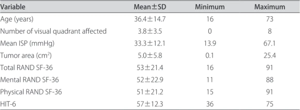
Table 1. Age, number of visual quadrant afected, mean intrasellar pressure, tumor area and RAND SF-36 (total, mental and physical) and HIT-6 scores, n=25.
Variable Mean±SD Minimum Maximum
Age (years) 36.4±14.7 16 73
Number of visual quadrant afected 3.8±3.5 0 8
Mean ISP (mmHg) 33.3±12.1 13.9 67.1
Tumor area (cm2) 5.0±5.8 0.1 25.4
Total RAND SF-36 53±21.4 16 91
Mental RAND SF-36 52±22.9 11 88
Physical RAND SF-36 51±21.2 15 91
HIT-6 57±12.3 36 75
HIT-6: Headache Impact Test; RAND SF-36: Research and Development Short Form Health Survey, SD: Standard Deviation.
Table 2. Correlations between intrasellar pressure and the number of visual quadrant afected, tumor area, RAND SF-36 (total, mental and physical) and HIT-6 scores, n=25.
Variable
Correlation with ISP
r or rs p
Number of visual quadrant afected 0.355S 0.08
Tumor area (cm2) 0.081S 0.70
Total RAND SF-36 –0.050P 0.81
Mental RAND SF-36 –0.027P 0.90
Physical RAND SF-36 –0.066P 0.75
HIT-6 –0.031P 0.88
HIT-6: Headache Impact Test; RAND SF-36: Research and Development Short Form Health Survey; PPearson correlation, r; SSpearman’s rank correlation, r
s.
Table 3. Headache severity measured by the HIT-6 score and quality of life evaluated by the total RAND SF36 and its mental and physical dimensions, according to the hormonal proile of the tumor.
Variable (n)
Type of tumor (n) Cushing’s disease (5)
mean±SD (range) mean±SD (range)Acromegaly (4) Non-secreting (16)mean±SD (range)
Total RAND SF36 37±18 (19-64) 48±31.9 (16-91) 59±17.8 (33-89) Mental RAND SF36 32±16.4 (11-55) 45±38.1 (12-88) 60±16.3 (26-86) Physical RAND SF36 37±16.9 (17-61) 46±32.3 (15-91) 57±18.1 (34-89) HIT-6 58±12.5 (44-72) 55±18.5 (36-75) 57±11.4 (36-70)
among other factors, the lack of an appropriate animal model and the need for surgical indication due to pa-thology in the sellar region. he intervention in healthy patient with the aim of measuring the pressure would be ethically reprehensible and highly questionable, consid-ering the current evidence from literature.
he ISP values obtained in diferent studies have var-ied. he irst 24 published values were in patients that showed an average of 25 mmHg7. In the largest series, an
ISP average of 20 mmHg, ranging from 11 to 30 mmHg, was found9. Yet, the irst work using iberoptic catheter
(which is technically simpler and does not require reposi-tioning and injection of a solution to ensure the patency of the system) showed mean ISP value of 28.8 mmHg. In the present series, an average value of 33.3±12.1 mmHg was found, which is higher than those previously published. To our knowledge, up to the present moment this is the only study in which the HIT-6 assessment scale was used to deine the presence and severity of headache in patients with intrasellar adenoma. his scale has been ap-plied in 2537 primary medical care patients with various pathologies17. A distribution according to the class of pain
of 6% mild, 14% moderate, 14% substantial and 66% in-tense was observed. he prevalence of any type of head-ache in the general population is considered as 80%19 and
in patients with pituitary adenoma it is 69%16.
Consider-ing it as frequent complaint, it would be possible to sup-pose that there were no diferences between headache in patients with pituitary adenomas and within the gener-al population.
Some features, however, could deine subpopulations of patients with adenoma where headache was more im-portant. In 64 patients, macroadenomas (diameter ≥10 mm) were more associated to the presence of headache16.
In another analysis involving ISP, 56.6% of 30 patients with adenoma had headache. In these, the pressure was higher (19.2±9.8 against 15±9.2 mmHg) and so was the volume (18.3±32.3 against 5.2±6.1 cm3)13. In the present
study, such indings could not be conirmed. hat could be because stratiication by the severity of headache re-duces the number of individuals in subgroups and, hence, limits the analysis. he sizes of the tumor were evaluat-ed as micro and macroadenomas and by extent of the area, but not by volume. On the other hand, and in ac-cordance with our inding of no correlation between ISP and headache severity through HIT-6, there are other fac-tors that may be more important than ISP in headache genesis. Cavernous sinus invasion by the pituitary ade-noma has already been described as a condition associ-ated with the presence of headache16. In our series, six
patients had such inding, and the mean HIT-6 value for these cases was 64.5, against 56.7 for the others. he pres-ence of headache in patients with microadenoma, with
intact sella and no cavernous invasion, addresses to the likely involvement of inlammatory or hormonal factors of local action20.
Headache, characteristic or not of patients with pitu-itary adenomas, has substantial impact on quality of life. he World Health Organization deines quality of life as “the individual’s perception of his position in life, in the context of culture and system of values in which he lives and in relation to his goals, expectations, standards and concerns”. his concept is complex and emphasizes pa-rameters that are wider than the control of symptoms, re-duction in the mortality rate or increase in life expectan-cy21. he use of standardized instruments to assess
func-tional status and well-being dates back over 300 years18.
he SF-36 is an abbreviated questionnaire resulting from RAND (Research and Development Corporation) health batteries based on a multidimensional deinition of health and created for the Health Insurance Experiment and lat-er for the Medical Outcomes Study18. An Italian study
in-cluding 150 patients with headache showed a total and subgroup score at the SF-36 scale well below Italian gen-eral population22. he relationship between headache
se-verity and the level of quality of life was also conirmed by this study, once the total RAND SF-36 measurement, and also its mental and physical dimensions, had inverse cor-relations of moderate and signiicant intensities with the HIT-6 values. his is probably due to the impact of pain on the daily life and limitations in individual capabilities.
he quality of life may vary according to the endo-crine function of the tumor. In 168 patients with pituitary adenomas, it was noted that patients with acromegaly had scores that were lower in the physical than in the men-tal dimension. However, patients with Cushing’s disease showed lower-than-average scores compared to the con-trol population. he same authors found a RAND SF-36 average of less than 50 for the group of non-secreting tu-mors. Patients with Cushing’s disease seem to have the worst quality of life, compared with other types of tu-mors23. In this series, both in the total assessment and in
the mental and physical dimensions, patients with Cush-ing’s disease also presented the worst quality of life, fol-lowed by acromegaly (Table 3), according to the RAND SF-36. It is worth mentioning that the procedure for cal-culating each dimension results from the average of ive diferent scales, from the eight present in the question-naire, with superposition of measures of general health and vitality24.
he prevalence of visual ield changes in pituitary ade-nomas has been variable throughout history. In 200 con-secutive patients, only 9% of visual ield defects could be found25. he authors of that report also reviewed four
se-ries (total of 1307 patients) and examined the presence of visual ield changes in patients with pituitary adenomas. he prevalence of this association ranged from 31 to 86%. In another study, 93 patients with non-secreting pituitary adenomas were examined26. Eighty-eight patients (94.6%)
had visual ield deicits. In 74.2% of the times, changes were typical (bitemporal hemianopsia). All patients with lesions larger than 2 cm2 had visual changes, and the
se-verity was proportional to the tumor volume. Gondim13
found 36.7% of visual changes in 30 patients and associat-ed16 the displacement of the chiasm to headache, and the
ISP for the group with visual deicits (19.0 mmHg) was similar to the group without the deicit (18.1 mmHg). It is reasonable to believe that the increase in intrasellar pres-sure produces a force vector directed to the least resis-tant regions, in this case, the upper portion of the sella, near the diaphragm. his alteration would injure the optic chiasm15. However, the increased pressure does not
oc-cur steadily with the increase in volume, since it is high-ly dependable on sellar integrity14. If any sellar limit is
ruptured, increased intrasellar mass volume is no longer necessarily raising the ISP. Individual factors such as vol-ume and local inlammatory reaction may have similar or greater impact in determining visual loss, as well as head-ache20, and may be object of future researches. he
con-comitant assessment of quality of life would also be as im-portant to analyze the full extent of these and other con-ditions in individuals with pituitary adenoma.
In conclusion, the mean ISP measured was 33.3±12.1 mmHg. here was no correlation between ISP values and the number of visual quadrant afected. here was no cor-relation between the ISP values and the tumor area. here was no correlation between the ISP values and the scores on the total RAND SF-36 scale and with its physical and mental dimensions. here was no correlation between the ISP values and the scores obtained through the HIT-6 scale. he headache severity measured by the HIT-6 scale showed moderate and inverse correlation with quality of life, i.e., with total, mental and physical values of the RAND SF36 scale.
RefeRences
1. Yamada S. Epidemiology of pituitary tumors. In: Thapar K, Kovacs K, Scheithauer BW, Lloyd RV (Eds). Diagnosis and management of pituitary tumors. New Jer-sey: Humana Press 2001:57-70.
2. Gorczyca W, Hardy J. Arterial supply of the human anterior pituitary gland. Neurosurgery 1987;20:369-378.
3. Arafah BA, Nekl KE, Gold RS, Selman WR. Dynamics of prolactin secretion in patients with hypopituitarism and pituitary macroadenomas. J Clin Endo-crinol Metab 1995;80:3507-3512.
4. Arafah BA, Prunty D, Ybarra J, Hlavin ML, Selman WR. The dominant role of increased intrasellar pressure in the pathogenesis of hypopituitarism, hyper-prolactinemia and headaches in patients with pituitary adenomas. J Clin En-docrinol Metab 2000;85:1789-1793.
5. Abe T, Matsumoto K, Kuwazawa J, Toyoda I, Sasaki K. Headache associated with pituitary adenomas. Headache 1998;38:782-786.
6. Baird A, Sullivan T, Zafar S, Rock J. Quality of life in patients with pituitary tu-mors: a preliminary study. Qual Manag Health Care 2003;12:97-105. 7. Lees PD, Pickard JD. Hyperprolactinemia, intrasellar pituitary tissue pressure
and the pituitary stalk compression syndrome. J Neurosurg 1987;67:192-196. 8. Lees PD. Intrasellar pressure. Acta Neurochir (Wien) 1990;47(Suppl):S68-S70. 9. Lees PD, Fahlbusch R, Zrinzo A, Pickard JD. Intrasellar pituitary tissue
pres-sure, tumor size and endocrine status: an international comparison in 107 patients. Br J Neurosurg 1994;8:313-318.
10. Kruse A, Astrup J, Cold GE, Hansen HH. Pressure and blood low in the pitu-itary adenomas measured during transsphenoidal surgery. Br J Neurosurg 1992;6:333-341.
11. Kruse A, Astrup J, Gyldensted C, Cold GE. Hyperprolactinemia in patients with pituitary adenomas. The pituitary stalk compression syndrome. Br J Neuro-surg 1995;9:453-457.
12. Zayour DH, Selman WR, Arafah BM. Extreme elevation of intrasellar pressure in patients with pituitary tumor apoplexy: relation to pituitary function. J Clin Endocrinol Metab 2004;89:5649-5654.
13. Gondim JA. Pressão intra-selar nos tumores da hipóise. Estudo da relação: pressão intra-selar versus função hipotalâmica-hipoisária. Dissertação, Uni-versidade Federal de São Paulo. São Paulo, 2004.
14. Gondim JA, Tella Jr OI, Schops M. Intrasellar pressure and tumor volume in pituitary tumor: relation study. Arq Neuropsiquiatr 2006;64:971-975. 15. Pereira-Neto A. Pressão intra-selar média, cefaléia, qualidade de vida e
pro-lactina sérica: estudo prospectivo em 25 pacientes operados consecutiva-mente no hospital universitário de Brasília. Tese, Universidade de Brasília. Bra-sília (DF):2006.
16. Gondim JA, Almeida JP, Albuquerque LA, Schops M, Gomes E, Ferraz T. Head-ache associated with pituitary tumors. J HeadHead-ache Pain 2009;10:15-20. 17. Nachit-Ouinekh F, Dartigues JF, Henry P, et al. Use of the headache impact
test (HIT-6) in general practice: relationship with quality of life and severity. Eur J Neurol 2005;12:189-193.
18. Meneses RF, Ribeiro JP, Silva AM. Revisão da literatura sobre avaliação da qual-idade de vida (QDV) de adultos com epilepsia: II. Facilqual-idades na abordagem do tema. Psicologia, Saúde Doenças 2002;3:119-139.
19. Dahlöf CGH. Measuring disability and quality of life in migraine. Drugs Today (Barc) 2003;39(Suppl D):S17-S23.
20. Borba AM. Relação entre função hipoisária, síndrome metabólica e pressão intra-selar média em pacientes com adenoma de hipóise. Dissertação, Uni-versidade de Brasília. Brasília (DF), 2008.
21. Yeng LT, Teixeira MJ, Romano MA, Greve JMD, Kaziyama HHS. Avaliação fun-cional do doente com dor crônica. Rev Med (São Paulo) 2001;(ed. esp. pt. 1): 443-473.
22. Bussone G, Usai S, Grazzi L, Rigamonti A, Solari A, D’Amico D. Disability and quality of life in diferent primary headaches. Results from italian studies. Neurol Sci 2004;25(Suppl 3):S105-S107.
23. Johnson MD, Woodburn CJ, Vance ML. Quality of life in patients with pitu-itary adenoma. Pitupitu-itary 2003;6:81-87.
24. Kalantar-Zadeh K, Kopple JD, Block G, Humphreys MH. Association among SF36 quality of life measures and nutrition, hospitalization, and mortality in hemodialysis. J Am Soc Nephrol 2001;12:2797-2806.
25. Anderson D, Faber P, Marcovitz S, Hardy J, Lorenzetti D. Pituitary tumors and the ophtalmologist. Ophthalmology 1983;90:1265-1270.