Validation of a Brazilian quantitative
sensory testing (QST) device for the
diagnosis of small iber neuropathies
Pedro Schestatsky1, Luciana Cadore Stefani2,
Paulo Roberto Sanches3, Danton Pereira da Silva Júnior3,
Iraci Lucena Silva Torres2, Letizzia Dall-Agnol2,
Luciane Fachin Balbinot4, Wolnei Caumo2,4,6
ABSTRACT
Quantitative sensory testing (QST) is defined as the determination of thresholds for sensory perception under controlled stimulus. Our aim was to validate a new QST device for Brazilian sample. In 20 healthy adults, thermoalgesic thresholds were assessed using a QST prototype (Heat Pain Stimulator-1.1.10; Brazil). A 30 × 30 mm2 thermode with a 1°C/s stimulus change rate were applied. Thresholds of three consecutive stimuli were averaged in two different sessions separated by at least two weeks. Additionally long thermal heat pain stimulus was performed. To evaluate the consistency of our method we also analyzed 11 patients with small fiber neuropathy. Results showed good reproducibility of thermal perception thresholds in normal individuals and plausible abnormal thresholds in patients. We conclude that our QST device is reliable when analyzing the nociceptive pathway in controls and patients.
Key words: quantitative sensory testing, validation, nociceptive pathway, psychophysics, heat pain stimulation.
Validação de um aparelho brasileiro de teste de quantificação sensitiva brasileiro para o diagnóstico de neuropatia de fibras finas
RESUMO
Teste de quantificação sensitiva (TQS) significa determinação de limiares de percepção sensitiva frente a um estímulo de intensidade controlada. Nosso objetivo foi validar um novo equipamento de TQS adaptado à população brasileira. Em 20 adultos saudáveis, limiares termoalgésicos foram avaliados, utilizando um aparelho protótipo do TQS (Heat Pain Stimulator-1.1.10; Brazil). Foi utilizado um termodo de 30 × 30 mm2, com estímulo térmico de 1°C/s. A média dos limiares de três estímulos consecutivos foi obtida em duas sessões diferentes, separadas por pelo menos 2 semanas. Adicionalmente, foram aplicados estímulos térmicos dolorosos de longa duração. Para avaliar a consistência do nosso método, foram também analisados 11 pacientes com neuropatia de fibras finas. Os resultados mostraram boa reprodutibilidade dos limiares de percepção nos indivíduos saudáveis, assim como limiares anormais nos pacientes. Em conclusão, nosso aparelho de TQS apresentou boa confiabilidade ao analisar a via nociceptiva de controles e pacientes.
Palavras-Chave: teste de quantificação sensitiva, validação, via nociceptiva, estímulo termoalgésico.
Correspondence
Pedro Schestatsky
Hospital de Clínicas de Porto Alegre Neurology Department, EMG Unit Rua Ramiro Barcelos 2350 90035-003 Porto Alegre RS - Brasil E-mail: [email protected]
Support
This study was supported by the “Fundo de Incentivo à Pesquisa e Eventos (FIPE/HCPA)”
Received 2 June 2011
Received in final form 18 July 2011 Accepted 25 July 2011
1Neurology Department, EMG Unit from Hospital de Clínicas de Porto Alegre (HCPA), Porto Alegre RS, Brazil; 2Post-Graduation
Program of Medical Sciences, Universidade Federal do Rio Grande do Sul (UFRGS), Porto Alegre RS, Brazil; 3Biomedical Engineering
of HCPA; 4Post-Graduation Program of Neuroscience, UFRGS; 5Anesthetist, Pain and Palliative Care Service at HCPA; 6Pharmacology
Department, Instituto de Ciências Básicas da Saúde da UFRGS, Porto Alegre RS, Brazil. Quantitative sensory testing (QST) is
a widely used psychophysical method for
informa-tion on large iber status, the term “QST” is normally used for small iber and nociceptive pathway assessment, using controlled thermal, instead of tactile or vibratory stimuli3-6. his method is important for the diagnosis of neuropathies for two main reasons. First, because small iber dysfunc-tion is usually the irst alteradysfunc-tion in axonal neuropathies (i.e., diabetic) the QST could aid in its early diagnosis7. Second, contrary to QST, conventional nerve conduc-tion studies are unable to assess small iber funcconduc-tion1. Diferently than nerve conduction studies, QSTs are psychophysical in nature, requiring cooperation from the patient2. While the sensory stimulus is controlled by the examiner during QST, the response represents the subjective perception of thermoalgesic stimulus. If abnormal, the QST result may signalize dysfunction anywhere along the sensory pathway between thermal receptors, small ibers, spinothalamic tract and other ce-rebral areas i.e, pain matrix8.
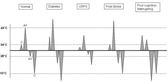
But how is perception measured? By means of con-trolled thermal stimuli, the patient is asked to press a button when he (she) feels diferent thermal perceptions from a thermode device in contact with the skin. Warm perception thresholds are used as a parameter relecting the function of unmyelinated C-ibers, whereas heat pain and cold perception thresholds indicate Aδ-iber function; and to a lesser extent also the function of subgroups of C-ibers9,10. However, in clinical routine, because cold thresh-olds are more variable, warm and heat pain threshthresh-olds are measured preferentially2. Figure 1 shows the most common QST abnormalities in different clinical conditions8.
In the last few years QST has been one of the main methods used in human experimental pain models and in the early diagnosis of neuropathies6,11. However, al-though considered an important tool in Neurology, QST devices are not easily available in most neurophysiologic laboratories worldwide. One of the reasons for such a
lack of availability is the high cost and the complexity of the grading sensory system. In Brazil, few centers have been developing QST devices12,13, but no validation studies have been reported so far. Our aim is to show our experience in the development of a new computer-controlled thermal stimulation device, demonstrate its reliability in repetitive sessions within controls and pa-tients and propose standardized QST verbal instructions for Brazilian patients.
METHOD Subjects
We selected 20 healthy volunteers (10 men) aged from 22 to 44. We excluded subjects with peripheral ner-vous system diseases or using medication that could af-fect the sensory perception, such as psychotropic and an-algesic medications. All subjects assigned the informed consent that was approved by the ethics committee of the Hospital de Clínicas de Porto Alegre, Brazil.
Patients
We selected 11 age- and sex-matched patients with painful neuropathies from the Neuromuscular Ambu-latory from our institution, six of them with diabetes melittus, four with leprosy and two with human immu-nodeiciency virus. All of them complaining of typical neuropathic pain over distal extremities.
Equipment
he system for heating/cooling of the thermal stim-ulator was based on Peltier principle. his module is a thermoelectric element which generates a temperature diference between the two sides of the component, cou-pled to an aluminum polished surface (Fig 2A) that gets in contact with the patient’s skin (Fig 2B). he aluminum plate temperature is monitored by a temperature sensor,
which has a response time of tenths of a second. he an-alog temperature after conditioning was sampled with 100 Hz and recorded with a 10 bit resolution. his sign is displayed in real time on the computer screen and is used to control the module temperature. his control is performed by the microcontroller that generates a pulsed signal applied to the power stage, which provides the current levels for the Peltier module excitation. he tem-perature of the thermode rised in a constant rate or sta-bilized in a pre-deined temperature during the experi-ment. To ensure this constant rising of temperature, a digital controller was implemented through software de-veloped in visual basic platform on personal a computer. he patient can stop the heat at any time if he/she feels discomfort. In this case, temperature and time interval are monitored. Apart from the button that is used to mark the temperature in which the subject feels warm and heat pain sensations, the system also provides a linear analog input to inform, by means of a manual lever (Fig 2C), the intensity of discomfort on a visual analogue scale ranging from 0 (no discomfort) to 10 (intolerable pain). he system has an additional safety device, that automatically turns of the module when temperature reaches 52°C in order to avoid skin damage. he temper-ature and pain analog scale curves were stored in iles for later analysis. Figure 3 shows a scheme of QST set up.
Experimental procedure
he evaluation was performed in a quiet, semidark room, at a temperature between 23°C and 24°C. Subjects were always addressed by the same researcher (LCS), who systematically read the instructions and explained the standardized experimental procedure, using a previ-ously published QST protocol orientation14 adapted to Brazilian portuguese language.
hermoalgesic stimuli were delivered through a Pel-tier thermode of a surface of 30 × 30 mm2 (Heat Pain Stimulator-1.1.10, Brazil). Baseline temperature was al-ways set at 30°C and ramp rate was ixed at 1°C/s, to a maximum at 52°C. Subjects were seated on a comfort-able chair with arms on the armrest, and had the Pelt-ier’s thermode attached with a velcro strip to the ven-tral aspect of their mid forearm Figure 2B. We changed slightly the exact site of the skin where the thermode was applied between three consecutive trials. Subjects had an available button to press to immediately stop the ther-moalgesic stimuli, when necessary. For all tests, we used the same software to apply a controlled change in the thermode temperature. Subjects were requested to pay attention to the thermal sensation and avoid speaking, coughing or breathing deeply during the experiment. In order to conirm that the thermode was homogeneously heated we also perform termography (Eletrophysics,
PV320T) in some subjects. All signals were represented on a screen out of the subject’s visual ield, for on-line monitoring and of-line analysis.
We assessed warm and heat pain thresholds, as well as sensory perception during long 45°C thermal stimula-tion, in two diferent occasions with an at least two-week interval for each subject.
Warm and pain thresholds
Warm and pain thresholds were assessed with the method of limits2. he thermode was placed on the non-dominant upper arm. After a warning signal, the temper-ature rose from an adaptation tempertemper-ature of 30°C with a ramp rate of 1°C/s. he participant was asked to press as quickly as possible a button at the moment the stimu-lation became warm or painful. hree assessments were taken with an interstimulus interval of 40 seconds15 and thresholds were calculated by taking the average temper-ature of the three assessments.
Fig 2. Quantitative sensory testing devices: [A] Thermode; [B] Position of the thermode in the arm; [C] Electronic visual ana-logue scale.
Long painful thermal stimulation
he thermode temperature was rapidly increased up to 45°C. hen, this temperature was maintained for 60 seconds. During this time, subjects marked their per-ception using an electronic visual analogue scale (VAS), with six diferent levels of perception:15 [1] no temper-ature perception; [2] light warm; [3] medium warm; [4] light pain; [5] medium pain and [6] high pain perception. We considered levels 2 and 4 as thresholds for warm and pain, respectively.
Statistical analyses
All statistical analyses were done using the Statistical Package for the Social Sciences (SPSS) for Windows ver-sion 16. Correlations between the indices of two separate measurements of warm threshold and heat pain thresh-olds were tested by Pearson coeicient. Also, the repro-ducibility of assessments was tested by intraclass coei-cient. Student’s t test was used for threshold comparisons between patients and controls. All analyses were per-formed with conidence interval of 95%.
RESULTS
Twenty healthy volunteers (10 females) participated in this study. Average age was 28±6.6 years. he correla-tion indices and intraclass coeicient average for warm and pain thresholds are presented in Table. No difer-ences were found between thermal thresholds obtained with button versus electronic VAS (Student’s t test; p>0.05 for all comparisons). When analyzing three pa-tients with small iber diseases and pain we observed el-evated warm and heat thresholds in all of them. Figure 4 shows illustrative examples of normal (4A) and abnormal (4B) thresholds obtained from illustrative control sub-ject and patient, respectively. hermography recordings showed homogenous increment of skin temperature in a circumscribed area that exactly matched with the ther-mode dimensions (Fig 5). Although skin redness was ob-served in the vast majority of the subjects, no major skin injuries occurred in any of them. Subjective perception
during long painful stimulation followed a stereotyped pattern in controls. Most subjects moved the lever very rapidly; right after thermode had started to cool down. Diferently, patients maintained the lever in pain sensa-tion in the electronic VAS even after the thermode was cold. Figure 6 illustrates such diferences showing an-other control subject (Fig 6A) and patient (Fig 6B) sub-mitted to long painful stimulus.
Fig 4. Thermal thresholds in normal subjects [A] and patients with small iber disease [B]. Note higher thresholds for warm and pain sensation in a illustrative patient.
Fig 5. Thermography recording after heat pain stimulation. Note the size of the thermode imprinting in the skin and the actual achieved temperature.
Warm and heat pain thresholds were significantly higher in patients than in controls (39.4±1.4°C vs. 35.6±1.3°C, for warm and 49.9±3°C vs. 44.5±2.5°C, for pain perception; Student’s t test; p<0.001 for all comparisons).
DISCUSSION
Our study has four main findings: [1] All thermal thresholds values were compatible with normal thermo-algesic transmission from warm and pain receptors to the brain; [2] All values were highly reproducible among time-separated measurements; [3] hermal thresholds obtained with the button were the same obtained with electronic VAS and [4] As expected, abnormal thermal thresholds were observed in patients with small fiber dysfunction.
Several algorithms have been suggested to determine thermal perception thresholds3,9,16. Two general schemes have emerged: the method of limits and the method of levels2. In the method of limits, a subject is required to indicate as soon as an increasingly thermal stimulus is detected. herefore this method is considered “time re-action inclusive”. In the method of levels, stimuli of de-ined intensity levels are tested with the subject signaling whether a speciic level is detected. In this case, thresh-olds are not dependent on reaction time and they are usually higher than the method of limits. Both tests are reliable, however the method of levels is time consuming (6 times longer than the method of limits) and no difer-ences in sensitivity between the two methods were seen in diabetic patients17.
It is important to stress that QST does not measure pain itself. Actually QST measure sensory deicit that could be or not related to pain complaints. In a patient with chronic pain, lower thermal thresholds point to hy-peralgesia, whereas elevated warm and heat pain thresh-olds point to a small iber dysfunction which sometimes leads to neuropathic pain6,8,11,18.
he thresholds for warm and heat pain stimuli ob-tained in this study are in agreement with previously published reports4,15,19,20. In addition, psychophysical re-sponses to long thermal stimulation were also similar to previous results21. An interesting inding was the greater variability of heat pain thresholds in comparison to warm
thresholds. his is in line with previous study20 and may be explained by the fact that pain is greatly inluenced by modulatory mechanisms22. he long thermal stimulation protocol also brought some interesting indings. Normal subjects almost always have their perception decreased after a few seconds of steady pain stimulation. his can be explained by the refractoriness of thermal receptors after prolonged excitation making the brain equivocally think that there is a transient reduction of sensation. An-other curious inding was the maintenance of pain per-ception such as seen in an illustrative patient with small iber disease. his can be assumed to be an after sensa-tion phenomenon, a very typical inding in patients with neuropathic pain due to spinal central sensitization after peripheral lesions23. hese indings highlight the useful-ness of an electronic VAS instead of a conventional and static button pressed when relevant perceptions are per-ceived. Besides, this strategy also serve not only as con-irmation of the subject’s ability to detect and perceive pain, but also as a method for monitoring the possibility that innocuous stimuli are painful in sporadic cases of allodynia phenomenon20. hus, incorporating simulta-neous electronic VAS during thermal stimulation would make the QST more complete as a diagnostic procedure.
Apart from the accuracy of thermal thresholds we have also observed a very good reproducibility within subjects, which is in accordance with several authors24-27. In fact, the QST reproducibility in normal subjects is probably better than that of patients with neuropathy28.
Our study has two main limitations. First we do not use thermodes with diferent ramp rates in diferent body sites. In the same way we only used a large thermode and this could not be sensitive enough to detect mild neurop-athy because of spatial summation29. However, the ramp rate and body location used in this study was mainly em-ployed by others2 and large thermode allows sensation to be more easily discriminated in normal skin30 and provide more reproducible thresholds26. Indeed, small thermodes should be used only at rounded body surfaces5. Second, we do not perform electrophysiological studies in order to conirm the absence of subclinical small iber disease i.e., laser or contact heat-evoked potentials. However none of our subjects complained of any sensory disturbances neither have some risk factor for small iber neuropathy.
Table. Mean and standard deviation values for thermoalgesic thresholds and correlation coeicients between quantitative sensory testing sessions.
Thresholds
Assessment
Pearson coeicient Intraclass coeicient
First Second
Warm (°C) 35.6±1.3 35.3±1.4 0.8* 0.88*
Heat Pain (°C) 44.5±2.5 43.3±2.9 0.91* 0.92*
Despite the limitations of our study, our QST device showed good accuracy and reproducibility in both con-trols and patients. Using QST, sensory deicits may be quantiied and the data can be used in parametric statis-tical analysis. herefore, this tool can reliably be used for research and clinical purposes adapted to Brazilian por-tuguese language. Further studies are needed using dif-ferent body regions and thermode sizes.
REFERENCES
1. Zaslansky R, Yarnitsky D. Clinical applications of quantitative sensory testing (QST). J Neurol Sci 1998;153:215-338.
2. Chong PS, Cros DP. Technology literature review: quantitative sensory testing. Muscle Nerve 2004;29:734-747.
3. Fruhstorfer H, Lindblom U, Schmidt WC. Method for quantitative esti-mation of thermal thresholds in patients. J Neurol Neurosurg Psychiatry 1976;39:1071-1075.
4. Yarnitsky D, Sprecher E, Zaslansky R, Hemli JA. Heat pain thresholds: nor-mative data and repeatability. Pain 1995;60:329-332.
5. Hilz MJ, Stemper B, Axelrod FB, Kolodny EH, Neundörfer B. Quantitative thermal perception testing in adults. J Clin Neurophysiol 1999;16:462-471. 6. Arendt-Nielsen L, Yarnitsky D. Experimental and clinical applications of quantitative sensory testing applied to skin, muscles and viscera. J Pain 2009;10:556-572.
7. Jensen TS, Bach FW, Kastrup J, Dejgaard A, Brennum J. Vibratory and thermal thresholds in diabetics with and without clinical neuropathy. Acta Neurol Scand 1991;84:326-333.
8. Schestatsky P, Nascimento OJ. What do general neurologists need to know about neuropathic pain? Arq Neuropsiquiatr 2009;67:741-749. 9. Verdugo R, Ochoa JL. Quantitative somatosensory thermotest: a key
method for functional evaluation of small calibre afferent channels. Brain 1992;115:893-913.
10. Campero M, Serra J, Ochoa JL. C-polymodal nociceptors activated by noxious low temperature in human skin. J Physiol 1996;497:565-572. 11. Cruccu G, Anand P, Attal N, et al. EFNS guidelines on neuropathic pain
assessment. Eur J Neurol 2004;11:153-162.
12. Villarroel MF, Orsini MB, Lima RC, Antunes CM. Comparative study of the cutaneous sensation of leprosy-suspected lesions using Semmes-Weinstein monofilaments and quantitative thermal testing. Lepr Rev 2007;78:102-109.
13. Azevedo E, Silva A, Martins R, Andersen ML, Manzano GM, Tufik S. Acti-vation of C-fiber nociceptors by low power diode laser (extracted from Doctoral Thesis of Eduardo Azevedo Pacheco entitled “Estudo do limiar
nociceptivo durante a privação de sono utilizando o potencial evocado por laser“ – UNIFESP, 2010).
14. Rolke R, Baron R, Maier C, et al. Quantitative sensory testing in the German Research Network on Neuropathic Pain (DFNS): Standardized protocol and reference values. Pain 2006;123:231–243.
15. Schestatsky P-a, Algaba R, Pérez D, et al. Transient decrease of sensory perception after thermoalgesic stimuli for quantitative sensory testing. Muscle Nerve 2007;36:466-470.
16. Dyck PJ, O’Brien PC, Kosanke JL, Gillen DA, Karnes JL. A 4, 2, and 1 step-ping algorithm for quick and accurate estimation of cutaneous sensation threshold. Neurology 1993;43:1508-1512.
17. Claus D, Hilz MJ, Neundorfer B. Thermal discrimination thresholds: a com-parison of different methods. Acta Neurol Scand 1990;81:533-540. 18. Merskey H, Bogduk N. Classification of chronic pain. Seattle: IASP Press
1994.
19. Yarnitsky D, Sprecher E. Thermal testing: normative data and repeatability for various test algorithms J Neurol Sci 1994;125:39-45.
20. Kelly KG, Cook T, Backonja MM. Pain ratings at the thresholds are nec-essary for interpretation of quantitative sensory testing. Muscle Nerve 2005;32:179-184.
21. Schestatsky P, Callejas MA, Valls-Solé J. Abnormal modulation of electro-dermal activity by thermoalgesic stimuli in patients with primary palmar hyperhidrosis. J Neurol Neurosurg Psychiatry 2011;82:92-96.
22. Fields HL. Pain modulation: expectation, opioid analgesia and virtual pain. Prog Brain Res 2000;122:245-253.
23. Jensen TS, Baron R. Translation of symptoms and signs into mechanisms in neuropathic pain. Pain 2003;102:1-8.
24. Bertelsmann FW, Heimans JJ, Weber EJ, van der Veen EA, Schouten JA. Thermal discrimination thresholds in normal subjects and in patients with diabetic neuropathy. J Neurol Neurosurg Psychiatry 1985;48:686-690. 25. Arezzo JC, Schaumburg HH, Laudadio C. Thermal sensitivity tester
de-vice for quantitative assessment of thermal sense in diabetic neuropathy. Diabetes 1986;35:590-592.
26. Hilz MJ, Claus D, Neundörfer B. Early diagnosis of diabetic small fiber neu-ropathy by disturbed cold perception. J Diabet Complications 1988;2:38-43. 27. Dyck PJ, Kratz KM, Lehman KA, et al. The Rochester diabetic neuropathy study: design, criteria for types of neuropathy, selection bias, and repro-ducibility of neuropathic tests. Neurology 1991;41:799-807.
28. Bertelsmann FW, Heimans JJ, van Rooy JC, Heine RJ, van der Veen EA. Reproducibility of vibratory perception thresholds in patients with dia-betic neuropathy. Diabetes Res 1986;3:463-466.
29. Defrin R, Urca G. Spatial summation of heat pain: a reassessment. Pain 1996;66:23-29.

![Fig 6. Long painful stimulation. Note the lower thermal thresh- thresh-olds of illustrative patient [B] in comparison with a control sub-ject [A]](https://thumb-eu.123doks.com/thumbv2/123dok_br/15433086.595219/4.955.472.843.820.1123/painful-stimulation-thermal-thresh-illustrative-patient-comparison-control.webp)
