Alzheimer’s disease
Relationship between cognitive aspects and
power and coherence EEG measures
Lineu C. Fonseca1, Gloria M.A.S. Tedrus1, Larissa R. Prandi2, Adriana M. Almeida3, Danilo S. Furlanetto2
ABSTRACT
Objective: To evaluate the relationship between specific cognitive aspects and quantitative EEG measures, in patients with mild or moderate Alzheimer’s disease (AD). Method: Thirty-eight AD patients and 31 controls were assessed by CERAD neuropsychological battery (Consortium to Establish a Registry for AD) and the electroencephalogram (EEG). The absolute power and coherences EEG measures were calculated at rest. The correlations between the cognitive variables and the EEG were evaluated. Results: In the AD group there were significant correlations between different coherence EEG measures and Mini-Mental State Examination, verbal fluency, modified Boston naming, word list memory with repetition, word list recall and recognition, and constructional praxis (p<0.01). These correlations were all negative for the delta and theta bands and positive for alpha and beta. There were no correlations between cognitive aspects and absolute EEG power. Conclusion: The coherence EEG measures reflect different forms in the relationship between regions related to various cognitive dysfunctions.
Key words: dementia, cognition, Alzheimer’s disease, electroencephalography.
Doença de Alzheimer: relação entre aspectos cognitivos e medidas de potência e coerência no eletrencefalograma
RESUMO
Objetivo: Avaliar as relações entre aspectos cognitivos específicos e medidas quantitativas do EEG em pacientes com doença de Alzheimer (DA) leve e moderado. Método: Trinta e oito pacientes com DA e 31 controles foram avaliados com a bateria neuropsicológica CERAD (Consortium to Establish a Registry for AD) e o eletroencefalograma. Foram realizadas medidas de potência absoluta e coerência do EEG, durante repouso, e avaliadas suas relações com variáveis do CERAD. Resultados: No grupo DA houve correlações significativas entre diferentes medidas de coerência e o mini-exame do estado mental, fluência verbal, teste de nomeação de Boston modificado, memória de lista de palavras com repetição, recordação e reconhecimento de lista de palavras e praxia construtiva (p<0,01). Essas correlações foram negativas para as faixas delta e teta e positivas para alfa e beta. Conclusão: As medidas de coerência do EEG em repouso refletem diferentes formas de organização nas relações entre regiões, relacionadas a várias disfunções cognitivas.
Palavras-Chave: demência, cognição, doença de Alzheimer, eletroencefalograma. Correspondence
Lineu Corrêa Fonseca Rua Juçara 134
13013-910 Campinas SP - Brasil E-mail: [email protected]
Support FAPIC, FAPESP
Received 29 December 2010 Received in final form 20 July 2011 Accepted 27 July 2011
1Professor of Neurology, Faculdade de Medicina da Pontifícia Universidade Católica de Campinas (PUC-Campinas), Campinas SP, Brazil; 2Placement Scholarship Student (FAPIC)/Reitoria, PUC-Campinas; 3Placement Scholarship Student (FAPESP), PUC-Campinas.
he EEG has been used as an aid in the evaluation of patients with cognitive dei-cits, especially when the diagnosis remains open after the initial clinical evaluations1,2.
which the most used are the EEG band power and coher-ence analyses. In the EEG band power analysis, various band power ranges that constitute the electrical cerebral activity are calculated. In the analysis of coherence, the relationship between two regions with respect to their compositions in electrical cerebral activity is assessed, which makes it possible to evaluate the connectivity be-tween them. his analysis relects functional interactions between neural networks represented in the cortex3.
In comparison with normal controls, there is in-creased activity in the theta band in subjects with Al-zheimer’s disease (AD), which is accompanied by an in-crease in delta activity and dein-creases in the alpha and beta bands4. In studies on coherence in AD, the most
consistent inding has been a reduction in coherence in the alpha band5-7.
There is still insufficient scientific evidence of the diagnostic utility of qEEG to establish this method for the initial evaluation of subjects with cognitive impair-ment in routine clinical practice8. here is thus a need
for studies on other approaches using qEEG in order to improve its diagnostic value.
he literature has cited correlations between qEEG measures, such as increases in theta and delta power and alterations in the global scores of tests used in cognitive assessments, such as the Mini-Mental State Examination (MMSE)9-13. Cross-sectional studies with the coherence
measures of the qEEG failed to show correlation with the MMSE, although only global coherence measures or those restricted to the alpha and beta bands were used in these studies7,14,15.
On the other hand, there are a few studies assessing correlations between qEEG measures and speciic cogni-tive aspects15,16, but in these studies the coherence
mea-surements were also limited with respect to the bands and locations studied. A study of relationships between the variables of the resting qEEG and specific cogni-tive aspects could contribute to knowledge concerning their physiopathology, and also contribute to the choice of variables to be used in qEEG studies during speciic tasks with a view to diagnostic utility.
he objective of this study was to assess relationships between speciic cognitive aspects and easy-to-use vari-ables on the qEEG, such as EEG power and coherence analyses, in patients with slight and moderate AD, in-cluding not only the alpha and beta bands, but also the delta and theta bands and regional measurements.
METHOD
hirty-eight patients were included, all attending the outpatient’s clinic at the Department of the Neurology Clinic - PUC/Campinas and all with dementia according to the Diagnostic and Statistical Manual of Mental
Dis-orders17, and diagnosis of AD (mild or moderate stages)
according to the criteria of the NINCDS/ADRDA (Na-tional Institute of Neurological and Communicative Dis-orders and Stroke and Alzheimer’s disease)18. A control
group was also set up, including 31 subjects with no his-tory of cognitive decline or previous neurological or psy-chiatric disorders and of similar gender and age range. hese were also submitted to the EEG and the same clin-ical-neurological assessments and cognitive tests as the AD patients.
The following exclusion criteria were applied: co-morbidity with signiicant reduction in life expectancy; treatment with acetylcholinesterase inhibitors or any other modulator of cognitive functions or drugs with the potential to alter them.
he patients were submitted to the following proce-dures: a clinical-neurological assessment; routine labora-tory testing and neuro-imaging to rule out other causes of dementia; CERAD neuropsychological battery (Con-sortium to Establish a Registry for Alzheimers Disease),
Pfefer questionnaire; Clinical Dementia Rating - CDR
and the qEEG. The cognitive and behavioral assess-ments followed the recommendations of the National Consensus19.
The standard CERAD neuropsychological battery used in the cognitive assessment was composed of a semantic verbal fluency assessment, abridged Boston Naming Test (15 items), the MMSE, word list memory with repetition, recall and recognition, and construc-tional praxis with copy and recall. Its applicability to the Brazilian population was previously veriied20 and the
scores correspond to the right answers in all items. he Pfefer questionnaire is a functional and cognitive assess-ment scale applied to the informants.
Electroencephalography
he EEG was recorded with a resolution of 12 bits, 0.5 and 35 Hz ilters and 200 samples per second, using
the Braintech 4.0 equipment (EMSA Equipamentos
Médicos). Impedance was maintained below 10k. he electrodes were placed according to the International 10-20 System, with the use of an additional two elec-trodes placed 1 cm below (left side) and above (right side) the external angle of the eyelid, with the objective of evaluating eye movements. he inter-connected ear lobe electrodes served as the reference. he data were recorded during approximately 12 minutes, alternating resting periods with the eyes closed with awake periods when the eyes were open, each period lasting 2 minutes.
Fourier Transform, the absolute powers of 17 electrodes were studied (Fp2, Fp1 and Oz were not included) in the following frequency bands: delta (0.8 to 3.9 Hz), theta (4.29 to 7.8 Hz), alpha 1 (8.2 to 9.8 Hz), alpha 2 (10.1 to 12.5 Hz) and beta (12.8 to 19.9 Hz). To obtain the normal distribution, the values for absolute power were substi-tuted by their logarithms. he absolute power was ana-lyzed for the various bands at all the individual electrode positions. he averages were also calculated for the elec-trodes of the left (F7, T3, T5, F3, C3, P3 & O1) and right (F8, T4, T6, F4, C4, P4 & O2) hemispheres and the global average of all the electrodes.
To analyze coherence, this was deined as:
Cohxy (f ) = [Rxy (f )]2 = [Gxy (f )] 2
Gxx (f ) (Gyy (f )
where G denotes the spectral estimate of two EEG sig-nals x and y for a given frequency band f. he numerator contains the cross-spectrum for x and y (Gxy), while the denominator contains the respective spectra for x (Gxx) and y (Gyy). A result of 0 corresponds to no coherence and a result of 1 to maximum coherence.
he inter-hemispheric EEG coherences between the following homologous electrode pairs were measured: right frontal (F3-F4), right centrals (C3-C4), left-right parietals (P3-P4), left-left-right anterior temporals (F7-F8), left-right midtemporals (T3-T4), left-right posterior temporals (T5-T6) and left-right occipitals (O1-O2). he mean of the inter-hemispheric coherences between the frontal, temporal, central and parietal regions, corre-sponding to the F3-F4, F7-F8, T3-T4, C3-C4, P3-P4 and T3-T4 pairs (FTCP inter-hemispheric coherence), was calculated. For the intra-hemispheric coherences, the coherences between the frontal-occipital, central-pa-rietal and midtemporal-posterior temporal pairs were measured both on the left (F3-O1, C3-P3 and T3-T5) and right (F4-O2, C4-P4 and T4-T6) hemispheres. All of these measurements were carried out for each of the four bands (delta, theta, alpha, beta).
Considering that the measures of coherence depend on the distance between the electrodes, coherences be-tween electrodes with diferent distances were not com-pared. Only equivalent coherences were compared be-tween the AD and control groups.
Statistical analysis
AD patients and controls were assessed with respect to the relationships between the variables of the CERAD neuropsychological battery and the global absolute power means and those of the left and right hemispheres in the 4 frequency bands. he relationships of the CERAD neu-ropsychological battery with the mean inter-hemispheric coherences between the frontal, temporal, central and
parietal regions (FTCP inter-hemispheric coherence), between the frontal and temporal regions (frontal-tem-poral inter-hemispheric coherence) and the coherences between the occipital regions (O1-O2), as also with the means of the left (F3-O1, C3-P3 and T3-T5) and right (F4-O2, C4-P4 and T4-T6) intra-hemispheric coherency measurements were studied. he partial correlation co-eicients, corrected for schooling level, were computed to assess independent relations between EEG measures and the CERAD neuropsychological battery parameters. he Statistical Packages for Social Sciences statistical program (SPSS 10.0.1) was used, applying both para-metric and nonparapara-metric tests according to the data distribution. In order to minimize the possibility of er-rors due to the multiple comparisons, means of measure-ments involving various areas were used and not isolated electrode positions. In view of the exploratory nature of the study the level of signiicance was ixed at p≤0.01, de-spite the relatively large number of tests.
Ethical aspects
he Ethics Commission for Research with Human Beings of PUC-Campinas approved the project, and the subjects signed informed consent forms.
RESULTS Clinical aspects
Table 1 shows the socio-demographic data and the results of the CERAD neuropsychological battery for the 38 patients of the AD group and the 31 controls.
Electroencephalogram
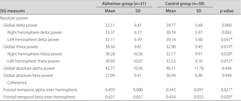
Table 2 shows the global means for the absolute powers in the 4 frequency bands, as well as in the left and right hemispheres for delta and theta bands. It should be noted that, in the theta band, the global amplitude and those of the right and left hemispheres were signiicantly greater in the AD group, whereas in the delta band they were greater in the AD group, but there was only a trend signiicance.
With respect to coherence, there was only a trend sig-niicant diference with respect to the frontal-temporal coherence (means for coherence for F3-F4 and F7-F8) in the alpha and beta bands (Table 2).
EEG relationships with the results of the CERAD neuropsychological battery
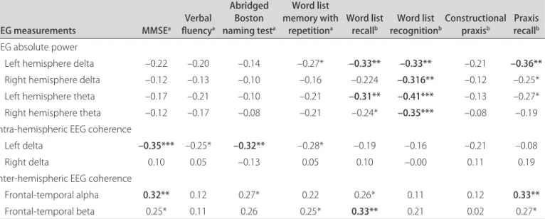
powers of the delta and theta activities can be observed in the results of the CERAD neuropsychological battery.
Signiicance was found for the left hemisphere abso-lute delta and theta powers with the word list recognition.
For MMSE there was a negative correlation with the left delta intra-hemispheric coherence and positive with frontal-temporal alpha inter-hemispheric coherence. Negative left delta intra-hemispheric coherence also oc-curred for abridged Boston naming test and word list recognition.
Positive correlations were observed for word list re-call and praxis rere-call, respectively, with frontal-temporal beta inter-hemispheric coherence and frontal-temporal alpha inter-hemispheric coherence.
here were no signiicant correlations between the
EEG measurements and those of CERAD for the verbal luency, word list memory with repetition, and construc-tional praxis.
AD group – In this group there were no signiicant correlations between EEG power measures and CERAD results.
It should be noted that the correlations were negative with the coherences for the slow activities delta and theta of the left hemisphere, and that the correlation coei-cients for these same coherences in the right hemisphere, were low. With respect to the inter-hemispheric coher-ences, it can be seen that the correlations were diferenti-ated according to the CERAD item under study, and that the negative correlations referred to delta activity and positive correlations to alpha or theta activities (Table 4). Table 1. Clinical characteristics of the study sample.
Clinical characteristics Alzheimer (n=38) Controls (n=31) p value
Age (years) 73.82 (6.60) 71.39 (4.36) 0.083a
Male/female 14/24 8/23 0.328b
Education (years) 3.22 (3.18) 5.66 (4.77) 0.018c
Verbal luency 8.31 (3.45) 16.58 (5.23) 0.000a
Abridged Boston naming test 8.37 (2.68) 13.35 ( 2.0) 0.000c
Mini-Mental State Examination 17.28 (3.48) 26.74 (2.08) 0.000b
Word list memory with repetition 7.19 (3.99) 16.71 (3.52) 0.000a
Constructional praxis 3.28 (1.25) 4.58 (0.56) 0.000c
Word list recall 1.00 (1.37) 5.16 (1.77) 0.000c
Word list recognition 3.54 (4.04) 8.48 (1.91) 0.000c
Constructional praxis recall 1.14 (1.46) 3.30 (1.32) 0.000c
When mean values are shown, the standard deviations (SD) are given in parentheses. at test; bχ2 test; c
Mann-Whitney test.
Table 2. EEG measures: comparison between patients with Alzheimer’s disease and the controls.
EEG measures
Alzheimer group (n=31) Control group (n=38)
p value
Mean SD Mean SD
Absolute power
Global delta power 32.51 6.41 29.77 5.48 0.060
Right hemisphere delta power 33.37 6.37 30.74 5.97 0.082
Left hemisphere delta power 32.11 6.59 29.14 5.60 0.047*
Global theta power 38.50 9.81 32.90 9.45 0.019*
Right hemisphere theta power 38.28 10.36 32.77 9.91 0.028*
Left hemisphere theta power 38.49 10.07 32.53 9.70 0.015*
Global absolute alpha power 42,77 10.56 40.71 11.78 0.446
Global absolute beta power 27.09 9.41 26.94 6.96 0.944
Coherence
Frontal-temporal alpha inter-hemispheric 0.493 0.080 0.542 0.091 0.021*
Frontal-temporal beta inter-hemispheric 0.421 0.057 0.454 0.055 0.020*
Table 3. EEG measures: corrreleation coeicients for the global group constituted of Alzheimer’s disease and controls.
EEG measurements MMSEa luencyVerbal a
Abridged Boston naming testa
Word list memory with
repetitiona Word list recallb recognitionWord list bConstructional praxisb recallPraxis b EEG absolute power
Left hemisphere delta –0.22 –0.20 –0.14 –0.27* –0.33** –0.33** –0.21 –0.36**
Right hemisphere delta –0.12 –0.13 –0.10 –0.16 –0.224 –0.316** –0.12 –0.25*
Left hemisphere theta –0.17 –0.21 –0.10 –0.21 –0.31** –0.41*** –0.13 –0.27*
Right hemisphere theta –0.12 –0.17 –0.08 –0.21 –0.24* –0.35*** –0.08 –0.19
Intra-hemispheric EEG coherence
Left delta –0.35*** –0.25* –0.32** –0.28* –0.19 –0.16 –0.21 –0.08
Right delta 0.10 0.05 –0.13 0.05 0.10 –0.00 0.11 0.19
Inter-hemispheric EEG coherence
Frontal-temporal alpha 0.32** 0.12 0.27* 0.22 0.26* 0.11 0.12 0.33**
Frontal-temporal beta 0.25* 0.11 0.26 0.25* 0.33** 0.21 0.02 0.27*
aPartial correlation coeicients; bSpearman’s rank correlation coeicients; *p<0.05; **p<0.01, ***p<0.005. Signiicant correlations are marked in bold; MMSE:
Mini-Mental State Examination.
Table 4. EEG measurements: correlation coeicients for the group with Alzheimer’s disease.
EEG measurements MMSEa luencyVerbal a
Abridged Boston naming testa
Word list memory with
repetitiona Word list recallb recognitionWord list b Constructional praxisb Intra-hemispheric EEG coherence
Left delta –0.62*** –0.41* –0.60*** –0.47* –0.22 –0.53** –0.39*
Right delta –0.24 –0.10 –0.42* –0.01 0.11 –0.19 –0.17
Left theta –0.44** –0.49*** –0.49*** –0.49*** –0.13 –0.48** –27
Right theta 0.045 –0.22 –0.30 –0.11 0.17 –0.04 –0.05
Inter-hemispheric EEG coherence
FTCP delta –0.36* –0.13 –0.46** 0.04 0.22 –0.20 –0.47**
O1-O2 theta 0.21 –0.07 –0.12 0.32 0.42** 0.26 –0.07
O1-O2 alpha 0.30 0.03 0.01 0.37* 0.38* 0.38* 0.07
aPartial correlation coeicients; bSpearman’s rank correlation; *p <0.05; **p<0.01; MMSE: Mini-Mental State Examination.
Control group (data not shown) – For the control group signiicant correlations between EEG measures and CERAD results were not observed.
DISCUSSION
In the present casuistic, the inding of an increase in the theta power and to a lesser degree in the delta band in the AD group is in agreement with the literature4,21.
In this survey the alterations in coherence were lim-ited to the alpha and beta bands and the frontal and tem-poral regions (F3-F4 and F7-F8), similarly to that de-scribed in the literature5-7. he present casuistic can be
considered typical of mild and moderate AD from the point of view of the qEEG.
Relationships between absolute power and cognitive aspects
With respect to MMSE, elevated negative correla-tion between delta and theta activities and the MMSE score was reported for patients with dementia9, whereas
in other studies they were not found21. On analyzing
to-gether normal elderly subjects and those with cognitive compromise, Onishi et al.12 found a signiicant
regres-sion model to forecast the MMSE score using theta and alpha 1 powers.
In the present study, correlations were found for the slow activities delta and theta in the left hemisphere with MMSE, but they did not reach statistical signiicance.
slow activities and MMSE in the various surveys were probably due to the composition of the series with re-spect to the proportion and severity of the cognitive dys-functions, and to the diferent EEG measures used.
Studies evaluating the relationship between EEG power measures and specific cognitive aspects are scarce15. In agreement with those studies, trend
signif-icant relationships were found in the present study be-tween slow band powers and activities linked to memory, such as word list recall and recognition and praxis recall. Also, similarly to this study, the relationship between as-pects of the EEG and the Boston naming test was not signiicant15.
he association of an increase in slow activity with cognitive deicit could be explained by the hypothesis that basal forebrain neurons are severely afected in AD, re-sulting in a cerebral cholinergic deicit which, for its part, is involved in the genesis of memory loss and of other cognitive symptoms22, as also in slowing of the EEG.
Relationships between coherence and cognitive aspects
Various studies have failed to ind any relationship be-tween EEG coherence and MMSE7,14,15, but those studies
used global coherence measurements that did not in-clude the delta band or did not have any normal controls. In the present study, in addition to the short dis-tance intra-hemispheric measurements (for example C3-P3, T3-T5 for the left hemisphere) as used in other studies14,15, long distance measures such as F3-O1 or
F4-O2 were also used. Thus C3-P3, T3-T5 and also F3-O1 entered the mean for left coherence, resulting in both long and short distance measurements being rep-resented, which could contribute to more ample knowl-edge of the functional relationships between areas of the brain. It is known that distance is a factor that interferes with coherence, and direct comparisons were not made between the long and short coherence measurements.
In the joint group of AD plus the controls, a negative correlation was found between MMSE and the delta co-herence of the left hemisphere, and positive correlation with the inter-hemispheric frontal-temporal alpha co-herence. he inter-hemispheric coherences also showed positive correlation with word list recall and praxis recall. Adler et al.7 also observed a decreased alpha
coher-ence associated with immediate verbal recall perfor-mance, which could suggest that in the organization of the electrical activity of the brain, the sub-cortical con-trol of alpha activity has a relationship with working memory mechanisms.
Both in the global group and in AD group theta, and particularly delta intra-hemispheric coherences on the left, were inversely and strongly correlated with various
types of cognitive aspects. Also negative was the corre-lation between delta frontal-temporal-central-parietal coherence with the abridged Boston naming test in the AD group. On the other hand, the occipital inter-hemi-spheric coherences in alpha and theta bands showed pos-itive correlation with word list recall.
With respect to the physiopathological interpreta-tion, the association of less coherence in the alpha band with worse cognitive performance in AD would be a re-sult of reduced functional connections between areas of the brain beneath the electrodes, or reduced control of two areas by a third area, supporting the hypothesis that Alzheimer’s disease is, in part, a disconnection syn-drome. his could be the result of a loss of the cortico-cortical association ibers necessary for functional inter-actions, or to reduced cholinergic coupling interactions between cortical neurons23.
With respect to coherence in the slow bands, a de-crease in acetylcholine can cause an inverse efect with respect to alpha and beta bands, that is, an increase in coherence. In fact, in healthy subjects anti-cholinergic drugs induce an increase in coherence in slow bands24
and there are reports of an increase in slow coherence in AD25. It is also possible that the association of greater
co-herence in the slower bands with a fall in cognition could also be due to a decrease in acetylcholine in AD subjects.
he predomination of correlations between the slow coherences in the left hemisphere and cognitive aspects could be related to the preponderant representation of language, whose compromise would afect performance in the tests7. In patients with AD there would also be
pre-ponderant metabolic and morphological alterations in the left hemisphere26.
EEG proiles and cognition
The MMSE showed correlations with left delta and theta intra-hemipheric EEG in common with other more speciic items from CERAD. In patients with AD, verbal luency showed correlations that were restricted to the theta coherence of the left hemisphere that in-volved short (C3-P3 and T3-T5) and long (F3-O1) dis-tance coherences, with participation of the frontal and temporal areas, in accordance, respectively, with their executive functions and connected to the semantic lu-ency for animals27.
Positive correlations with the coherence of the alpha and beta activities appeared in the tests involving memory, speciically the word list recall, and praxis recall, for both the general casuistry and in the Alzheimer group. Con-nections between alpha rhythm and memory have al-ready been pointed out in the literature28, especially for
immediate memory7,16. These relationships would be
rhythm is mainly modulated by thalamo-cortical and cortico-cortical interactions, facilitating or inhibiting the transmission of information amongst the sub-cor-tical and corsub-cor-tical pathways and the retrieval of semantic information from brain storage29.
he abridged Boston naming test showed correla-tions with both inter- and intra-hemispheric coherences in the delta band, which could be related to two categor-ical stages of object recognition: a perceptual stage in the right hemisphere and a semantic stage that depends on the left hemisphere30.
Although the analysis carried out in this research was for resting EEG, the diferences in the aspects of the EEG for the diferent cognitive aspects indicated the existence of varied aspects of organization and control of the com-position of the electrical activity related to the various cognitive aspects. he original aspect of the present work was to evaluate the relationships among speciic cogni-tive aspects in AD subjects using simple EEG coherence and power measures involving delta, theta, alpha and beta bands, available in the majority of equipments. With this information one can arrive at the value of the study with the delta coherences of the cerebral hemispheres to-gether with other measures of coherence and power, in the relationships with various cognitive aspects.
A practical and relevant aspect of this study is being a basis for the use of speciic variables of the EEG for the study of the registers made during speciic functional ac-tivations, which could eventually provide a greater diag-nostic value to the EEG in clinical practice, in assessing cognitive dysfunctions.
REFERENCES
1. Robinson DJ, Merskey H, Blume WT, Fry R, Williamson PC, Hachinski VC. Electroencephalography as an aid in the exclusion of Alzheimer’s. Arch Neurol 1994;51:280-284.
2. Claus JJ, Strijers RL, Jonkman EJ, et al. The diagnostic value of electro-encephalography in mild senile Alzheimer’s disease. Clin Neurophysiol 1999;110:825-832.
3. Hogan MJ, Swanwick GRJ, Kaiser J, Rowan M, Lawlor B. Memory-related EEG power and coherence reduction in mild Alzheimer’s disease. Int J Psychophysiol 2003;49:147-163.
4. Miyauchi T, Hagimoto H, Ishii M, Tanaka K, Kajwara A, Kosaka K. Quantita-tive EEG in patients with presenile and senile dementia of the Alzheimer type. Acta Neurol Scand 1994;89:56-64.
5. Besthorn C, Förstl H, Geiger-Kabisch C, Sattel H, Gasser T, Schreiter-Gasser U. EEG coherence in Alzheimer disease. Electroencephalogr Clin Neuro-physiol 1994;90:242-245.
6. Knott V, Mohr E, Mahoney C, Ilivitsky V. Electroencephalographic coher-ence in Alzheimer’s disease: comparisons with a control group and pop-ulation norms. J Geriatr Psychiatry Neurol 2000;13:1-8.
7. Adler G, Brassen S, Jajcevic J. EEG coherence in Alzheimer’s dementia. J Neural Transm 2003;110:1051-1058.
8. Jelic V, Kowalski J. Evidence-based evaluation of diagnostic accuracy of resting EEG in dementia and mild cognitive impairment. Clin EEG Neurosci 2009;40:129-142.
9. Brenner RP, Ulrico RF, Spiker DG, et al. Computerized EEG spectral analisis in elderly normal, demented and depressed subjects. Electroencephalogr Clin Neurophysiol 1986;64:483-492.
10. Schreiter-Gasser U, Gasser T, Ziegler P. Quantitative analysis in early onset Alzheimer’s disease; correlations with severity, clinical charateristics, vi-sual EEG and CCT. Electroencephalogr Clin Neurophysiol 1994;90:267-272. 11. Strijers RL, Scheltens P, Jonkman EJ, de Rijke W, Hooijer C, Jonker C.
Diagnosing Alzheimer’s disease in community-dwelling elderly; a com-parison of EEG and MRI. Dementia 1997;8:198-202.
12. Onishi J, Suzuki Y, Yoshiko K, Hibino S, Iguchi A. Predictive model for assessing cognitive impairment by quantitative electroencephalography. Cog Behav Neurol 2005;18:179-184.
13. Gianotti LRR, Künig G, Lehmann D, et al. Correlation between disease severity and brain electric LORETA tomography in Alzheimer’s disease. Clin Neurophysiol 2007;118:16-196.
14. Jiang ZY. Study on EEG power and coherence in patients with mild cog-nitive impairment during working memory task. J Zhejiang Univ Sci B 2005;6:1213-1219.
15. van der Hiele K, Vein AA, Reijntjes RH, et al. EEG correlates in the spectrum of cognitive decline. Clin Neurophysiol 2007;118:1931-1939.
16. Babiloni C, Cassetta E, Binetti G, et al. Resting EEG sources correlate with attentional span in mild cognitive impairment and Alzheimer’s disease. Eur J Neurosci 2007;25:3742-3757.
17. American Psychiatric Association. Diagnostic and Statistical Manual of Mental Disorders. 4th ed. Washington.D.C. American Psychiatric Asso-ciation, 1994.
18. McKhann G, Drachman D, Folstein M, et al. Clinical diagnosis of Alzheimer’s disease: report of the NINCDS-ADRDA Work Group under the auspices of Department of Healthy and Human Services Task force on Alzheimer’s disease. Neurology 1984;34:939-944.
19. Nitrini R, Caramelli P, Bottino CMC, Damasceno BP, Anghinah R. Diag-nóstico de doença de Alzheimer no Brasil. Avaliação cognitiva e funcional. Recomendações do Departamento Científico de Neurologia cognitiva e do Envelhecimento da Academia Brasileira de Neurologia. Arq Neurop-siquiatr 2005;63:720-727.
20. Bertolucci PH, Okamoto IH, Brucki SN, Siviero MO, Tonioto J Neto, Ramos LR. Applicability of the CERAD neuropsychological battery to Brazilian elderly. Arq Neuropsiquiatr 2001;59:532-536.
21. Chiaramonti R, Muscas GC, Paganini M, et al. Correlations of topograph-ical EEG features with clintopograph-ical severity in mild and moderate dementia of Alzheimer type. Neuropsycobiology 1997;36:153-158.
22. Bartus RT, Dean III RL, Beer B, Lippa AS. The cholinergic hypothesis of geriatric memory dysfunction. Science 1982;217:408-414.
23. Jeong J. EEG dynamics in patients with Alzheimer disease. Clin Neuro-physiol 2004;115:1490-1505.
24. Sloan EP, Fenton GW, Standage KP. Anticholinergic drug effects on quantitative electroencephalogram, visual evoked potential, amd verbal memory. Biol Psychiatry 1992;31:600-606.
25. Locatelli T, Cursi M, Liberati D, Franceschi M, Comi G. EEG coherence in Alzheimer’s disease. Electroencephhalogr Clin Neurophysiol 1998;106: 229-237.
26. Loewenstein DA, Barker WW, Chang JY, et al. Predominant left hemisphere metabolic dysfunction in dementia. Arch Neurol 1989;46:146-152. 27. Damásio H, Grabowski TJ, Tranel D, Hichwa RD, Damásio AR. A neural basis
for lexical retrieval. Nature 1996;380:499-505.
28. Klimesch W. EEG alpha and theta oscillations reflect cognitive and memory performance: are view and analysis. Brain Res Rev 1999;29:169-195. 29. Pfurtscheller G, Lopes da Silva F. Event-related EEG/MEG synchroniza-tion and desynchronizasynchroniza-tion: basics principles. Clin Neurophysiol 1999;110: 1842-1857.