Faculdade de Medicina de Ribeirão Preto – USP
Mailing Address: Luiz Tadeu M. Figueiredo – Depto. Clínica Médica – Faculdade de Medicina de Ribeirão Preto-USP - 14049-900 - Ribeirão Preto, SP - Brazil
Objective - To analyze the epidemiology, diagnosis, cli-nical aspects causes and evolution of infectious endocarditis.
Methods - The patients analyzed were treated at the University Hospital of the Faculdade de Medicina of Ribei-rão Preto-USP and had a diagnosis of infectious endocar-ditis defined by Duke’s criteria, which classifies infectious endocarditis as native, prosthetic valve or that occurring in intravenous drug users.
Results - One hundred and eighty episodes of infectious endocarditis in 168 patients were observed. Echocardio-grams in 132 (73.3%) provided a diagnosis of infectious endocarditis in 111 (84%) patients; mitral valves were affected in 55 (30.5%), tricuspid valves in 30 (16.6%) and the aortic valve in 28 (15.5%) patients. Hemocultures were performed in 148 (93.8%) episodes of IE. The most commonly isolated infectious organisms were Staphylococcus aureus in 46 (27.2%) patients and Streptococcus viridans in 27 (15.9%). Complications occurred in 116 (64.4%) patients and 73 (40.5%) of the patients died.
Conclusion – The general profile of the observed in-fectious endocarditis was similar to that reported in studi-es performed in other countristudi-es and included users of in-travenous drugs. The high degree of mortality observed is not compatible with progress in diagnosis and treatment of infectious endocarditis and is probably due to the ab-sence of diagnostic suspicion. The high frequency of fatal cases of septicemia (45.1% of deaths) in the patients studi-ed indicates that unnoticstudi-ed cases of infectious endocar-ditis had only been diagnosed at necropsy.
Key words: infectious endocarditis, endocarditis in Brazil, bacterial endocarditis.
Arq Bras Cardiol, volume 74 (nº 3), 225-231, 2000
Everaldo Ruiz Jr, Tarciso Schirmbeck, Luiz Tadeu Moraes Figueiredo
Ribeirão Preto, SP - Brazil
A Study of Infectious Endocarditis in Ribeirão Preto, SP
-Brazil. Analysis of Cases Occurring Between 1992 and 1997
present situation of the disease as it currently exists. To achieve this aim, epidemiological factors, risk factors, clinical presentation, etiology, complicating factors, diag-nostic methods, treatment and evolution of cases diagno-sed as IE at the Clinics Hospital of the Medical School of Ribeirão Preto of the University of São Paulo (HCFMRP-USP) between 1992 and 1997 were analyzed.
Methods
The files of 198 individuals diagnosed as having in-fectious endocarditis, collected between 1992 and 1997 at the HCFMRP-USP, a general hospital belonging to the SUS chain, serving the entire Ribeirão Preto region, part of the State of São Paulo and the southern part of the State of Mi-nas Gerais, were retrospectively analyzed.
IE cases were defined according to Duke’s criteria, res-pectively, as definitive, possible or rejected 5. Possible cases had clinical and complementary examination data insuffi-cient for a definitive diagnosis, but clearly did not qualify for rejection. Eighteen cases were rejected and not inclu-ded in the study.
Episodes of IE were classified into four groups as follows: (I) patients without a history of intravenous use of drugs presenting with attacked native valves; (II) users of intravenously applied drugs; (III) carriers of a recently pla-ced prosthetic valve (in which IE had developed within 60 days after valve replacement); (IV) carriers of pre-existing (old) prosthetic valves (in which endocarditis had develo-ped 60 days or more after valve replacement).
Data were compiled and analyzed using the computer program Epi-Info 6.0 (CDC, USA). Statistical calculations were made by the bicaudal Fisher’s exact test, and conside-red significant when P<0.05, using the Graph Pad program in Stat (Graph Pad Software Inc., San Diego, USA)
Results
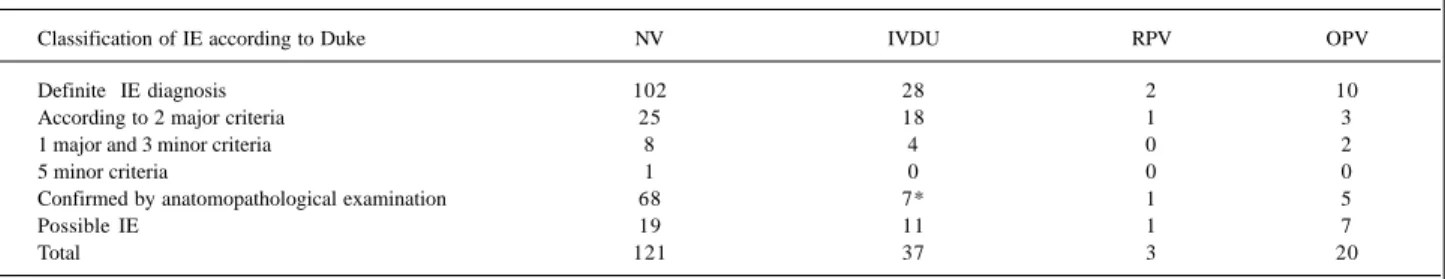
One hundred and eighty episodes of infectious endo-carditis occurred in 168 patients over a period of six years. As shown in table I, according to Duke’s criteria, 142 (79%) were definitive cases and 38 (21%) were classified as possible cases. Among the 180 occurrences, 12 individuals had more than one episode. Table I also shows the
occur-rence of 121 episodes of IE in the native valve of patients wi-thout a history of the use of intravenous drugs, in 37 users of such drugs, in 3 cases of recent and in 20 cases of old prosthetic valves.
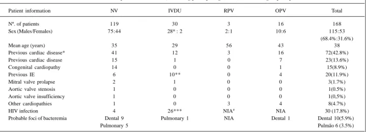
Table II shows sex, age and predisposing factors of the 168 IE patients. Male individuals (68.4%) predominated over females, especially in the group of intravenous drug users. Patient ages fell between 2 months and 106 years (mean age, 38 years). Cardiac diseases predisposing to IE were observed in 41 patients with affected native valves who did not use intravenous drugs, and in 12 intravenous drug users. Twenty-six (86.6%) of the patients using intravenous drugs were HIV-infected. Conditions predisposing towards bacteremia were found in 14 nonusers of intravenous drugs presenting with endocarditis in normal valves, and in one patient with an old prosthetic valve. Among patients presen-ting with endocarditis in native valves and nonusers of intravenous drugs, 9 (7.4%) had undergone dental proce-dures, and 5 (4.1%) had an infectious focus in the respiratory tract prior to the appearance of IE. In the group of intrave-nous drug users, 1 (2.5%) case of pulmonary infection as a risk factor over and above the risk inherent to drug addiction, was noted. Among patients with an old prosthetic valve, 1 (5.8%), related the existence of a predisposing dental focus.
Based on the duration of signs and symptoms, the time of infection preceding the IE diagnosis in the 180 episodes varied between one and 150 days (table III). Regarding the clinical picture the majority (87.2%) of the patients was febrile and prostrated (78.8%). Other frequent symptoms were loss of weight (44.4%) and myalgia (20.5%). Cardiac murmurs were detected in 73.3%; hemorrhagic phenomena either in the skin the mucosa, or both were found in 21.6%. Hepatomegaly was observed in 39.4% and splenomegaly in 27.7%. Complementary examinations showed anemia in 47.7% and leukocyte levels over 11,000/mm3 in 47.2% of the patients. Proteinuria (31.6%), hematuria (27.7%) and increa-sed levels of serum creatinine (25.5%) were also observed. Echocardiograms were performed in 73.3% of the patients. The preferred site of attack of IE in native valves of nonusers of intravenous drugs was the mitral and aortic valves; this was also found in carriers of old prosthetic valves. The tricuspid valve was more frequently attacked (48.3%) in intravenous drug users. The site of IE was not found in 53 (29.4%) episodes (table III).
Table I – Episodes of infectious endocarditis (IE), identified by Duke's criteria in the four groups of patients studied.
Classification of IE according to Duke NV IVDU RPV OPV
Definite IE diagnosis 102 28 2 10
According to 2 major criteria 25 18 1 3
1 major and 3 minor criteria 8 4 0 2
5 minor criteria 1 0 0 0
Confirmed by anatomopathological examination 68 7* 1 5
Possible IE 19 11 1 7
Total 121 37 3 20
Table II – Information about the 168 patients who had IE, including predisposing factors, in the four groups of patients studie d.
Patient information NV IVDU RPV OPV Total
Nº. of patients 119 30 3 16 168
Sex (Males/Females) 75:44 28*: 2 2:1 10:6 115:53
(68.4%:31.6%)
Mean age (years) 35 29 56 43 38
Previous cardiac disease* 41 12 3 16 72(42.8%)
Previous cardiac disease 15 1 0 7 23(13.6%)
Congenital cardiopathy 14 0 0 1 15(8.9%)
Previous IE 6 10** 0 4 20(11.9%)
Mitral valve prolapse 2 1 0 0 3(1.7%)
Aortic valve stenosis 1 0 0 0 1(0.5%)
Aortic valve insufficiency 1 0 0 0 1(0,5%)
Other cardiopathies 1 0 3 4 8(4.7%)
HIV infection 4 26*** NIA# NIA 30 (17.8%)
Probable foci of bacteremia Dental 9 Pulmonary 1 NIA Dental 1 Dental 10(5.9%)
Pulmonary 5 Pulmão 6 (3.5%)
IE- infectious endocarditis; NV- native valve; RPV- recent prosthetic valve; OPV- old prosthetic valve; IVDU- intravenous drug users. *Significantly higher proportion (P=0.0009) of male individuals among IVDU compared with the sum of the other groups. ** Significantly higher proportion (P=0.0003) of patients with a history of previous IE in IVDU compared with the sum of the other groups. *** Significantly higher proportion (P<0.0001) of HIV + in IVDU compared with the sum of the other groups. # NIA – no information available.
Table III– Clinical, laboratory and diagnostic aspects of the 180 episodes of Infectious endocarditis of the four groups of patients studied.
Clinical, laboratory and diagnostic IE NV IVDU RPV Total
Beginning of symptoms/diagnosis of IE* 21,2 25,5 7 20,3
General condition 56 11 1 10 78(43.3%)
Regular 51 20 1 6 78(43.3%)
Poor 13 7 0 0 20(11.1%)
Fever 111 30 1 15 157(87.2%)
Prostration 97 30 2 13 142(78.8%)
Cardiac murmurs 87 29 3 13 132(73.3%)
Weight loss 56 22 0 2 80(44.4%)
Hepatomegaly 39 25 0 7 71(39.4%)
Splenomegaly 32 13 0 5 50(27.7%)
Hemorrhagic phenomena 29 8 0 2 39(21.5%)
Myalgia 22 13 0 2 37(20.5%)
Artralgia 18 8 0 0 26(14.4%)
Serum hemoglobin <10mg/100ml 59 20 20 7 86(47.7%)
Leukocytes >11000/ mm3 59 20 2 4 85(47.2%)
Proteinuria >3,5g/dia 36 14 0 7 57(31.6%)
Hematuria 27 14 0 9 50(27.7%)
Serum creatinine >1,5mg/100ml 32 9 1 4 46(25.5%)
Serum AST >40mUI/ml 16 10 0 3 29(16.1%)
Echocardiogram/IE diagnosis 84 / 71 31/ 26 2 / 2 15 / 12 132/111
(73.3%/84%) Endocardic attack at:
Mitral valve 41 6 2 6 55(30.5%)
Aortic valve 19 3 1 5 28(15.5%)
Tricuspid valve 15 15*** 0 0 30(16.6%)
Mitral and aortic valves 5 0 0 1 6(3%)
Mitral and tricuspid valves 1 1 0 0 2(1.1%)
Aortic and tricuspid valves 3 0 0 0 3(1.5%)
Pulmonary valve 1 0 0 0 1(0.5%)
Pulmonary artery 1 0 0 0 1(0.5%)
Membranous septum 1 0 0 0 1(0.5%)
Unknown 36 14 0 5 53(29.4%)
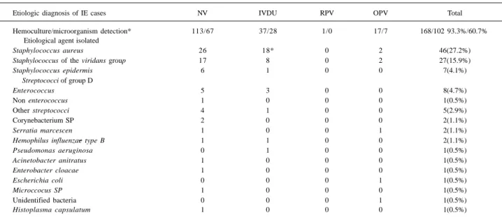
Hemocultures were performed in 168 episodes of IE (table IV). Microorganisms were isolated in 102 cases; in five of them, more than one bacterial species was isolated. The most frequent causal agent was Staphylococcus
aureus in 27.2% of the cases, followed by Streptococcus viridans in 15.9%. IE caused by gram-negative rods
occurred in 5.9% of the episodes.
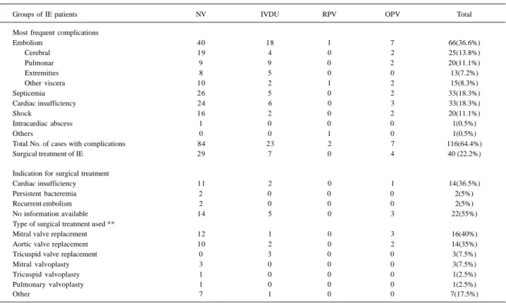
Complications observed in the 180 episodes of IE were: embolisms in 36.1%, septicemia in 18.3% and serious heart failure in 18.3%, respectively. Surgical replacement or repair of valves was performed in 40 (22.2%) patients. The most frequent indication for surgical treatment was serious heart failure in 36.5% of the cases; in 40% surgery was aimed at the replacement of the mitral valve, and in 35.5 % of the aortic valve. Replacement of the tricuspid valve was performed only in users of intravenous drugs (table V).
The mortality observed in the 180 episodes of IE was 40.5% (73 deaths). Deaths occurred in 42.9% of cases of IE in native valves in the absence of intravenous drug ad-diction, in 37% of users of intravenous drugs, in 66.5% of those with recent prosthetic valves and in 25% of those with old prosthetic valves. The main cause of death among the patients was septicemia, followed respectively by, embolic phenomena and serious cardiac insufficiency (table VI).
Discussion
Cases of infectious endocarditis (IE) were divided into four groups: those in which native valves were affected in patients without a history of the use of intravenous drugs; those in users of such drugs; those affecting patients’ recent prosthetic valves and those affecting patients’ old prosthetic valves. This division was based on clinical and
etiological differences observed among these patients 3,6. However, because only three cases of IE in recent prosthe-ses were observed, the analysis of this group was rendered imprecise. In our study, 2/3 of the patients with IE were males, in agreement with an evaluation of 300 cases of IE during the 1980s in the city of São Paulo. In the United Kingdom, an analysis of 118 cases of IE 6 and in Canada a study of 135 cases 3 also showed a predominance of male patients.
The average age of our IE patients was 38 years. Man-sur et al. 11 observed that the majority of their patients were between 21 and 30 years old. Cecchi et al. 12 in Italy noted an average age of 40 years of their IE patients; in Sweden, Hogevik et al. 2 found it to be 69 years. Our results, compa-red with those of the earlier Brazilian study in the 1980s, shows the average age of IE patients to have risen; this was also noted in other countries according to the European so-urces cited.
Cardiopathies prior to the onset of IE were observed in 42.8% of the cases, rheumatic causes being the most preva-lent among them (table II). A higher percentage of cardiopa-thies occurring prior to IE have been reported 11; Sandre et al.3 reported 35% prior cardiopathies. Hogevik et al. 2 observed rheumatic cardiopathies prior to IE in 18% of their cases.
Foci of possible bacteremia, of dental or pulmonary origin, which could have caused IE, were reported by 9.5% of the patients. Hogevik et al. 2 described dental and gum diseases capable of producing bacteremia due to S.
viri-dans in 45.4% of IE patients. In our study, bacterial foci and
procedures capable of evoking bacteremia had not been properly identified.
The average time periods between beginning symp-toms of IE and diagnosis were of 20-25 days (table III); this period is shorter than the one previously reported in the city
Table IV - Positivity of hemoculture and cause of the 180 cases of IE in the four groups of patients studied.
Etiologic diagnosis of IE cases NV IVDU RPV OPV Total
Hemoculture/microorganism detection* 113/67 37/28 1/0 17/7 168/102 93.3%/60.7% Etiological agent isolated
Staphylococcus aureus 26 18* 0 2 46(27.2%)
Staphylococcus of the viridans group 17 8 0 2 27(15.9%)
Staphylococcus epidermis 6 1 0 0 7(4.1%)
Streptococci of group D
Enterococcus 5 3 0 0 8(4.7%)
Non enterococcus 1 0 0 0 1(0.5%)
Other streptococci 4 1 0 0 5(2.9%)
Corynebacterium SP 2 0 0 0 2(1.1%)
Serratia marcescen 1 0 0 1 2(1.1%)
Hemophilus influenzae type B 1 1 0 0 2(1.1%)
Pseudomonas aeruginosa 0 1 0 0 1(0.5%)
Acinetobacter anitratus 1 0 0 0 1(0.5%)
Enterobacter cloacae 1 0 0 0 1(0.5%)
Escherichia coli 0 0 0 1 1(0.5%)
Microccocus SP 1 0 0 0 1(0.5%)
Unidentified bacteria 0 0 0 1 1(0.5%)
Histoplasma capsulatum 1 0 0 0 1(0.5%)
of São Paulo 11, which was 30 days or more in half of the cases. Because our results refer to cases of IE obtained 10 years later than these, it appears probable that the shorter time periods noted by us are due to progress in diagnostic methods (echocardiogram) and greater access to medical facilities. Hogenik et al. 2 in 1995 in Sweden reported an average period between onset of symptoms and IE diagnosis of 13.5 days.
Only 11% of our patients were in poor general clinical condition. This observation, although of a subjective natu-re, suggests that duration of the disease and degree of toxe-mia caused by the infection were not important determining factors in most cases. Nevertheless, this finding is not cor-related with the high degree of mortality observed in these
patients. Fever, prostration and the presence of cardiac mur-murs were noted in over of the cases, as has also been obser-ved by others 1,2,4,6. However, splenomegaly, which is related to the presence of prolonged bacteremia leading to a hyper-trophied reticulo-endothelial system, occurred in 27.7% of the cases studied. Due to the increased frequency of acute infections by S. aureus and the reduction of the period bet-ween onset and diagnosis of IE, splenomegaly is being obser-ved less and less. Earlier studies 11 showed splenomegaly in 47% of cases. However, studies in the 1990s in other countries relate the occurrence of splenomegaly in only 0 to 12% of the cases; such low values have been related to the acute form of the illness in patients in a higher age group 2, 6, 10. Table V – Complicating factors, surgical treatment and mortality in the 180 cases of IE in the four groups of patients studied.
Groups of IE patients NV IVDU RPV OPV Total
Most frequent complications
Embolism 40 18 1 7 66(36.6%)
Cerebral 19 4 0 2 25(13.8%)
Pulmonar 9 9 0 2 20(11.1%)
Extremities 8 5 0 0 13(7.2%)
Other viscera 10 2 1 2 15(8.3%)
Septicemia 26 5 0 2 33(18.3%)
Cardiac insufficiency 24 6 0 3 33(18.3%)
Shock 16 2 0 2 20(11.1%)
Intracardiac abscess 1 0 0 0 1(0.5%)
Others 0 0 1 0 1(0.5%)
Total No. of cases with complications 84 23 2 7 116(64.4%)
Surgical treatment of IE 29 7 0 4 40 (22.2%)
Indication for surgical treatment
Cardiac insufficiency 11 2 0 1 14(36.5%)
Persistent bacteremia 2 0 0 0 2(5%)
Recurrent embolism 2 0 0 0 2(5%)
No information available 14 5 0 3 22(55%)
Type of surgical treatment used **
Mitral valve replacement 12 1 0 3 16(40%)
Aortic valve replacement 10 2 0 2 14(35%)
Tricuspid valve replacement 0 3 0 0 3(7.5%)
Mitral valvoplasty 3 0 0 0 3(7.5%)
Tricuspid valvoplasty 1 0 0 0 1(2.5%)
Pulmonary valvoplasty 1 0 0 0 1(2.5%)
Other 7 1 0 0 7(17.5%)
IE- infectious endocarditis; NV- native valve; RPV recent prosthetic valve; OPV- old prosthetic valve; IVDU- intravenous drug users.* Total cases with complications, some more than one. **Some patients underwent more than one surgical treatment.
Table VI – Mortality due to IE of the 180 patients, according to the four groups studied
Groups of patients with IE NV IVDU RPV OPV Total
(mortality)
Nº of deaths 52 14 2 5 73 (40.5%)
Causes of death
Septicemia 19 7 1 1 28 (38.3%
Embolism 10 0 0 0 10 (13.6%)
Cardiac insufficiency 6 1 0 1 8 (10.9%)
Cardiac insufficiency plus septicemia 4 0 0 1 5 (6.8%)
Other causes 8 4 0 1 13 (17.8%)
Not determined 5 2 1 1 9 (12.3%)
Petechiae and other hemorrhagic phenomena in skin and mucosa were observed in 21.6% of the patients. Others 7,6 reported similar frequencies (22% and 33.3%, respectively).
Approximately half of our patients presented with leukocytosis and anemia (Table III). Leukocytosis is a frequent finding in acute IE; normochromic and normocytic anemia are described in up to 70% of the patients 13. In the Canadian study 3, anemia was observed in 30% of the cases. Hematuria, described as having a high incidence in IE, was observed in of our patients.
Transthoracic echocardiography, an important me-thod for the diagnosis of IE, was performed in 73.3% of our patients; its results suggested episodes of IE with, res-pectively, definitive, probable or possible vegetation in 84% (table III). In some cases, in which the transthoracic echo-cardiogram gave negative results, examination by the transesophageal approach showed vegetations. The per-centage of patients who underwent echocardiography by us can be considered low in comparison with that in other countries, 93-100% 2,3,6,12. The diagnostic efficiency of the echocardiogram was shown to be adequate, being equal to that observed by others 11,3 (83% and 83.9%, respectively). The site of the endocarditic attack in our patients was predominantly the mitral valve (30.5%), followed by the tri-cuspid and the aortic valves in 16.6% and 15.5% of the ca-ses, respectively. Our results show an increased effect on the tricuspid valve compared with those of others 14, who observed the mitral valve to be affected in 40% of the cases, aortic in 18% and tricuspid in 10.7%. The increased attack of the tricuspid valve is due to acute IE by S. aureus in pati-ents using intravenous drugs, corresponding to 19.8% of all individuals studied.
Hemoculture is a fundamental examination for the dif-ferential diagnosis of IE. In our study, hemocultures were made in samples obtained from 93.8% of the IE cases, leading to the isolation of the microorganism in 60.7% (table IV). The percent positivity was slightly lower than that observed by others 12, (65%). In another study 2, hemocul-ture positivity was 75%. We believe that a large part of our negative hemoculture results were due to the concomitant or previous use of antimicrobial agents by the patients.
The causal agents most frequently found in our patients were S. aureus in 27.2% and S. viridans in 15.9%.
Strepto-coccus faecalis (group D StreptoStrepto-coccus, EnteroStrepto-coccus) was
the third cause of IE, affecting 4.7% of the cases. Gram-negative rods caused 5.9% of the IE cases, while fungal endocarditis caused by Histoplasma capsulatum occurred in one case. These results differ from those of others 14 in which S. viridans predominated in about 31% of the epi-sodes, S. aureus staying in second place with about 20%. IE by gram-negative rods and fungi was of low frequency 6, simi-lar to that observed in our study. In the study by Hogevik et al. 2 IE by Streptococci predominated; in other studies, mainly those in which intravenous drug users participated, the predominant microorganism was S. aureus 3,6,12.
Establishing treatment for IE patients and determining
their sensitivity to antimicrobial agents are aims of our further work, and are presently under development.
Complications occurred in 64.4% of the cases, most fre-quently embolisms (36.1%), mainly in the brain and lungs (table V). Heart failure and septicemia occurred in 18% of the cases, each. Other authors 3, observed embolism in 38.5% and heart failure in 37.7% of the cases.
Surgical treatment was used in 22.2% of the patients following indication of heart failure refractory to clinical treatment in 36.5% of the patients presenting with persistent bacteremia not relieved by antimicrobial therapy, and in 5% of patients with recurrent embolism (table V). Surgical treatment was more frequently used by us than by the Swedish workers (15%) 2. It was however less frequent than in the Brazilian study of the 1980s (34%) 14 and the Canadian study 12 with 33% of the patients, respectively. In both the Brazilian and Canadian studies, the predominant indication for surgery was serious heart failure. Mitral valve and aortic valve replacement were employed in our study in 40% and 35% of the cases, respectively.
High mortality (40.5%) was observed in our patients. Its most frequent cause was septicemia (38.3%) accompa-nied by heart failure in 6.8% or embolism in 13.6% (Table VI). This mortality rate was higher than that observed in the São Paulo study 11 (27.1%) and much higher than that reported in recent studies from other countries (19-23%) 2,3,14. Septi-cemia was the predominant cause of death, corroborating other observations 11, but differing from those 3 in which the major cause of death was heart failure.
Our 180 cases of IE had differences in the four groups of patients. In IE attacking native valves of patients without a history of drug use, a predominance of men with an ave-rage age of 35 years was observed. One third of the patients had a history of previous cardiac disease, echocardiograms made in 69.4% of the patients being positive in 84.5 of them, mitral and aortic valves being affected. Hemocultures in 93.3% of the cases, showed a 62.8% degree of positivity and the predominant isolation of S. aureus in 23% of the cases and of S. viridans in 15%.
Complicating factors, especially embolic episodes oc-curred in 69.4% of the cases; due to serious cardiac insuf-ficiency, 23.9% of these patients underwent valve, mainly mitral, replacement. Mortality of IE cases affecting native valves of nondrug users was 43.6%, the second highest among the four groups studied; the main cause of death in these patients was septicemia.
comparison with that found in other patients. It was also noted that 86.6% were infected with HIV, a proportion signi-ficantly higher (P<0.0001), than that in other patients with IE. It must be pointed out that these drug users, although infec-ted with HIV, probably did not have AIDS because mortality in the group was lower than that of the average of the other patients, which in their majority were immunocompetent. Echocardiograms performed in 83.7% of the drug users demonstrated IE in 83.8% of patients, the tricuspid being the most affected valve (48.3%, P=0.0002) in these cases.
Association of IE in the right heart with the use of illicit in-travenous drugs is known 3,6,12,13. In 83.7% of our cases of IE in drug users, hemocultures were made; bacterial isolation was obtained in 83.8% of them. S. aureus predominated in 64.2% (P=0.0139) of the cases. A similar finding has been reported by others 3, S. aureus having been isolated in 40% of the drug users. In our study, embolism mainly at the pulmonary level occurred in 48% of cases of drug users. Surgery, predominan-tly replacement of the tricuspid valve, was performed in 18.9%. A 37.8% mortality rate observed in this group was mainly due to septicemia. It is interesting to note that although high and caused by S. aureus in patients infected with the HIV virus, this mortality ratio was lower than the average mortality ratio in all other cases. It is known that IE of the right heart causes a more efficient local immune response and has a better prognosis.
IE cases affecting old prosthetic valves were more fre-quent in older patients, of average age above 43 years: 25.5% of them had had previous episodes of IE. Echocardio-graphy performed in 75% of the cases confirmed the diag-nosis in 80%, in which mitral and aortic prostheses were most affected. Hemocultures made in 85% of these cases permitted bacterial isolation of predominantly S. aureus and S. viridans (28.6% each) in 41.4%. Gram-negative rods provided for 28.6% of the infections, demonstrating the sensitivity of valve prostheses to these germs. Embolism in 35% and heart failure in 15% were the most frequent compli-cations in these cases. Surgical treatment was effected in 20%, with a mortality ratio of 25%, the lowest observed
among the four groups, caused by septicemia or serious heart failure. Other authors 3 reported a mortality of 18% in these cases, having heart failure as their major cause.
Three cases of IE in recent valve prostheses occurred in older individuals, mean age of 56 years. These patients had the highest mortality rate observed in this study, 66.6%. Others 3 reported a mortality of only 14% in patients of mean age of 59 years, in such cases.
The analysis of the 180 episodes of IE of this study showed differences in the cause and site of cardiac attack, compared with results of studies performed in the 1980s 11, 14 in Brazil. Such differences also appeared in results publi-shed in recent years in other countries, especially regarding intravenous drug users 3,11. However, the high mortality ra-tios observed in our study, (40.5%) do not fit into the picture of recent progress in diagnosis and treatment of IE. Taking into consideration that IE is usually fatal when not adequa-tely controlled by antimicrobial therapy, we believe that high mortality ratios result in part from the absence of correct diagnosis and adequate therapy. This is shown by the fact that in 26.7% of the cases in our files not even echocardio-graphy had been performed 1,13. The low mortality ratio observed in cases of affected old prosthetic valves indica-tes that in this group of patients known to be of high risk, IE diagnosis was always checked for. The low degree of posi-tive responses of hemocultures in our studies is also worth pointing out. In 39.7% of the cases, no microorganism could be isolated. Either previous or inadequate use of anti-microbial agents or both or technical laboratory problems may explain this finding. Furthermore, the high frequency of septicemia as the cause of death (45.1%) renders it likely that in many cases endocarditis may have passed unnoti-ced, evolving into systemic infection.
Acknowledgment
We are thankful to CNPq for financing the project by the Institutional Program of Scientific Initiation.
1. Scheld WN, Sande MA. Endocarditis and intravascular infections. In: Mandell GL, Bennett JE, Dolin R (eds). Mandell, Douglas and Bennett’s Principles and Practice of Infectious Diseases. New York: Churchill Livingstone, 1995: 740. 2. Hogevik H, Olaison L, Andersson R, Lindberg J, Alestig K. Epidemiologic
as-pects of endocarditis in an urban population. Medicine 1995; 74: 324-38. 3. Sandre RM, Shafran SD. Infective endocarditis: review of 135 cases over 9 years.
Clin Infect Dis 1996; 22: 276-86.
4. Levison ME. Endocardite Infecciosa. In: Bennett JC, Plumm MD (eds). Tratado de Medicina Interna, 6ª Ed. Rio de Janeiro: Guanabara Koogan, 1996: 1763. 5. Durack DT, Lukes AS, Bright DK. Duke endocarditis service. New criteria for
ac-tive infecac-tive endocarditis: utilization of specific echocardiographic findings. Am J Med 1994; 96: 2000-9.
6. Lamas, CC, Eykyn SJ. Suggested modifications to the Duke Criteria for the clini-cal diagnosis of native and prosthetic valve endocarditis: analysis of 118 patho-logically proven cases. Clin Infect Dis 1997; 25: 713-9.
7. Nettles RE, McCarty DE, Corey GR, Li J, Sexton DJ. An evaluation of the Duke criteria in 25 pathologically confirmed cases of prosthetic valve endocarditis. Clin Infect Dis 1997; 25: 1401-3.
References
8. Olaison L, Hogevik H. Comparison of the von reyn and Duke criteria for diagno-sis of infective endocarditis: A critical analydiagno-sis of 161 episodes. Scand J Infect Dis 1996; 28: 399-406.
9. Rios-Gonçalves AJ, Cunha RQ, Rozembaum R, et al. Endocardite infecciosa, aneurisma (s) micótico (s), hemorragia subaracnóidea e outras lesões neurológi-cas. Arq Bras Med 1991; 65: 559-71.
10. Tiossi CLD, Franken RA, Rivetti LA, Brasil SAB. Endocardite infecciosa. Aná-lise de 20 casos de óbito na Santa Casa de São Paulo. Arq Bras Cardiol 1994; 62: 403-6.
11. Mansur AJ, Grinberg M, Gallucci SDD, Bellotti G, Jatene A, Pileggi. Endocardi-te infecciosa: análise de 300 episódios. Arq Bras Cardiol 1990; 54: 13-21. 12. Cecchi E, Parrini I, Chinaglia A, et al. New diagnostic criteria for infective
endo-carditis. A study of sensitivity and specificity. European Heart Journal 1997; 18: 1149-56.
13. Sohsten RV, Kaye D. Endocardite infecciosa. In: Veronesi R, Focaccia R (eds.). Tratado de Infectologia. São Paulo: Atheneu, 1996: 633.