Effects of ischemic preconditioning in a pig model of
large-for-size liver transplantation
Antonio Jose´ Gonc¸alves Leal,IAna Cristina Aoun Tannuri,I,IIAlessandro Rodrigo Belon,IIRaimundo Renato Nunes Guimara˜es,IMaria Cecı´lia Mendonc¸a Coelho,IJosiane de Oliveira Gonc¸alves,ISuellen Serafini,I Evandro Sobroza de Melo,III Uenis TannuriI*
IFaculdade de Medicina da Universidade de Sa˜o Paulo, Pediatric Surgery Division, Pediatric Liver Transplantation Unit and Laboratory of Research in
Pediatric Surgery (LIM 30), Sa˜o Paulo/SP, Brazil. IIFaculdade de Medicina da Universidade de Sa˜o Paulo, Department of Surgical Technique and Experimental Surgery, Sa˜o Paulo/SP, Brazil.IIIFaculdade de Medicina da Universidade de Sa˜o Paulo, Liver Function Research Laboratory (LIM-14), Sa˜o Paulo/SP, Brazil.
OBJECTIVE:In most cases of pediatric liver transplantation, the clinical scenario of large-for-size transplants can lead to hepatic dysfunction and a decreased blood supply to the liver graft. The objective of the present experimental investigation was to evaluate the effects of ischemic preconditioning on this clinical entity. METHODS:Eighteen pigs were divided into three groups and underwent liver transplantation: a control group, in which the weights of the donors were similar to those of the recipients, a for-size group, and a large-for-size+ischemic preconditioning group. Blood samples were collected from the recipients to evaluate the pH
and the sodium, potassium, aspartate aminotransferase and alanine aminotransferase levels. In addition, hepatic tissue was sampled from the recipients for histological evaluation, immunohistochemical analyses to detect hepatocyte apoptosis and proliferation and molecular analyses to evaluate the gene expression ofBax
(pro-apoptotic),Bcl-XL(anti-apoptotic),c-Fosandc-Jun(immediate-early genes), ischemia-reperfusion-related inflammatory cytokines (IL-1, TNF-alpha and IL-6, which is also a stimulator of hepatocyte regeneration), intracellular adhesion molecule, endothelial nitric oxide synthase (a mediator of the protective effect of ischemic preconditioning) andTGF-beta(a pro-fibrogenic cytokine).
RESULTS:All animals developed acidosis. At 1 hour and 3 hours after reperfusion, the animals in the large-for-size and large-for-large-for-size+ischemic preconditioning groups had decreased serum levels of Na and increased serum
levels of K and aspartate aminotransferase compared with the control group. The molecular analysis revealed higher expression of theBax, TNF-alpha, I-CAMandTGF-betagenes in the large-for-size group compared with the control and large-for-size+ischemic preconditioning groups. Ischemic preconditioning was responsible for
an increase inc-Fos,IL-1, IL-6ande-NOSgene expression.
CONCLUSION:Ischemia-reperfusion injury in this model of large-for-size liver transplantation could be partially attenuated by ischemic preconditioning.
KEYWORDS: Liver Transplantation; Models; Animal; Reperfusion; Apoptosis; Ischemic Preconditioning.
Leal AJ, Tannuri AC, Belon AR, Guimara˜es RR, Coelho MC, Gonc¸alves JO, et al. Effects of ischemic preconditioning in a pig model of large-for-size liver transplantation. Clinics. 2015;70(2):126-135.
Received for publication onOctober 13, 2014;First review completed onDecember 5, 2014;Accepted for publication onDecember 5, 2014
E-mail: [email protected]
*corresponding author
Tel.: 55 11 3061-7246
& INTRODUCTION
With the improvement in the management of pediatric patients with terminal liver diseases and the technical refinement and advances in intensive care and
immuno-suppression, liver transplantation has been performed in increasingly young children (1); a large series of liver transplantations in recipients that were less than 10 or even 5 kg have been reported with satisfactory results (1,2). However, a problem encountered by surgeons in such cases is the graft-to-body weight ratio (GBWR). Classically, the ideal ratio varies from 1 to 3%. However, in small children, the ratio is higher than 5% (3). This situation is known as large-for-size (LFS) and is characterized by the discrepancy between the small size of the abdominal cavity and the large size of the graft, which can lead to hepatic dysfunction and a diminished blood supply to the liver graft (3). Higher incidences of graft dysfunction, vascular complications and
Copyrightß2015CLINICS– This is an Open Access article distributed under the terms of the Creative Commons Attribution Non-Commercial License (http:// creativecommons.org/licenses/by-nc/3.0/) which permits unrestricted non-commercial use, distribution, and reproduction in any medium, provided the original work is properly cited.
No potential conflict of interest was reported.
acute rejection have been reported in the literature in cases involving LFS grafts (3). The mechanisms of injury involved in this scenario are not yet well established.
Among the main mechanisms of graft injury in trans-plantation is ischemia-reperfusion injury (IRI) (4), which has been related to complications such as poor graft function. Immediate-early genes (IEGs) encode transcription factors that regulate the expression of cellular response genes after injury and are associated with tissue repair and cell apoptosis (5,6). Many studies have shown thatc-Fosand c-Junare the IEGs that are activated during the early phase after IR and that they play an important role in the regulation of the tissue response to injury stimulation (7,8). Tissue inflammation triggered by IRI occurs immedi-ately after reperfusion and is related to the production of cytokines (TNF-alpha, IL-1, IL-6) and increased expression of adhesion molecules (such as ICAM) (9). The inflamma-tory cascade that culminates in apoptotic cell death and the cellular mechanisms of regeneration are the elements involved in IRI (10,11,12,13,14). The apoptosis phenomenon results from alterations in the balance between the expres-sion of pro-apoptotic (BaxandBak) and anti-apoptotic (Bcl-2
and Bcl-XL) genes. The activation of liver regeneration, which is essential for graft survival, is mainly mediated by IL-6 (15,16,17). After liver transplantation, IRI is responsible for the induction of pro-fibrogenic cytokines, represented mainly by TGF-beta (18).
Some techniques that attenuate IRI, such as ischemic preconditioning (IPC), have been reported and the nitric oxide (NO) produced by endothelial nitric oxide synthase (eNOS) is an important mediator of the effects of IPC. eNOS produces a protective action mediated by vasodilation and platelet aggregation inhibition, thereby improving the microcirculation in addition to inhibiting neutrophil activa-tion and neutralizing reactive oxygen species (18,19,20). Furthermore, it has been demonstrated that IPC stimulates liver regeneration (21). This effect has been demonstrated in rats that underwent warm ischemia and hepatectomy and interleukin-6 (IL-6) has been shown to be a likely mediator (22). No experimental studies have evaluated early hepatic injury in the LFS transplant scenario or the effects of IPC in this situation. Therefore, this was the objective of the present experimental investigation, which utilized a LFS pig experimental model of liver transplantation.
& MATERIALS AND METHODS
The institution’s animal welfare regulatory committee approved all study protocols and all protocols were in compliance with the Guide for the Care and Use of Laboratory Animals published by the National Institutes of Health 86-23, which was revised in 1985. Our study was performed using 36 Landrace pigs, with body weights between 20 and 50 kg. All of the experiments were performed under general anesthesia and all of the animals were killed after the experiments. The operations were performed by the same team of surgeons (the first five authors).
Eighteen large, white Landrace pigs (weighing 17 to 20 kg) underwent OLT with whole liver grafts and were divided randomly into three experimental groups, as follows:
1- Control group (n = 6): the weights of the donors were similar to the recipients (17-20 kg);
2- Large-for-size group (LFS, n = 6): the weights of the donors were nearly 2 times the weights of the recipients (40-50 kg);
3- Large-for-size+ ischemic preconditioning (LFS+IPC,
n = 6): the weight distributions were similar to the LFS group and IPC was performed in the donors.
Anesthetic procedures and intraoperative monitoring
After fasting for a period of 12 hours, the donors were anesthetized and maintained with a continuous intravenous infusion of 0.005 mg/kg/h fentanyl and 3.5 mg/kg/h propofol.
After an open cut was made down the right carotid sheath, a double lumen catheter was placed into the internal jugular vein for invasive venous pressure monitoring and fluid administration. In addition, a single lumen catheter was inserted into the carotid artery for arterial pressure monitoring. A total volume of approximately 300 ml of blood was collected from the donors before liver perfusion for infusion into the recipient pigs if necessary.
Donor procedure
In the donor operation, a long, longitudinal, midline thoracic and abdominal incision was performed to widely expose all organs. The portal vein, infrahepatic inferior vena cava and suprahepatic inferior vena cava were dissected. The hepatic artery was maintained in continuity with the celiac trunk and abdominal aorta up to the iliac bifurcation. In situ cold liver perfusion (with Euro CollinsHand Ringer’s lactate solutions) was then performed through both the portal vein and the aorta. The harvested graft was weighed, immersed in a plastic bag filled with Euro CollinsH
perfusion solution at 4
˚
C and placed in a basin for the subsequent back-table procedure.In the donors of the LFS+IPC group, after opening the
abdominal cavity, the hepatic hilum was clamped with a vascular clamp. Blood flow was stopped for 15 minutes, followed by reperfusion for 15 minutes.
Recipient procedure
A total hepatectomy was performed via a longitudinal incision and the supraceliac aorta was dissected close to the diaphragmatic crura and clamped during the anhepatic phase. The graft was put in place and the suprahepatic inferior vena cava, infrahepatic inferior vena cava and portal vein were anastomosed. Before the completion of infrahepa-tic inferior vena cava anastomosis, the graft was flushed with 1 L of saline solution at 4
˚
C though the portal vein.All anastomoses were performed using continuous Prolene 5-0 or 6-0 sutures. After completion of the venous anastomoses, the liver graft was reperfused. The recipient hepatic artery was anastomosed to the donor celiac trunk patch. The biliary reconstruction consisted of a choledo-chus-choledochus stented anastomosis.
The abdominal wall was kept open and only a few stitches were placed in the skin to retain the bowels and to avoid hypothermia. After 3 hours, the animal was euthanized.
Blood and serum analyses
pH and the sodium and potassium levels. Aspartate aminotransferase (AST) and alanine aminotransferase (ALT) levels were measured in the blood samples collected 1 and 3 hours after reperfusion.
Tissue analyses
Hepatic tissue was sampled from the recipient at 1 and 3 hours after portal reperfusion. Each biopsy sample was divided into two sections. One was preserved in 10% formaldehyde for subsequent embedding in paraffin and the other was snap-frozen in liquid nitrogen at2170
˚
C for molecular analyses.Histological examination was performed via hematox-ylin-eosin staining and light microscopy for the presence of centrilobular necrosis, sinusoidal neutrophils, steatosis, apoptotic bodies and sinusoidal dilatation. For each of these parameters, the following scoring system was used:
0 - absent; 1 - mild; 2 - moderate, 3 - intense; 4 - very intense.
& IMMUNOHISTOCHEMICAL METHODS
Detection of hepatocyte apoptosis
For the detection of early apoptosis, M30 CytoDEATH, a mouse monoclonal antibody (Clone M30, Roche, Mannheim, Germany), was used to detect a specific epitope of cytokeratin 18 (cleaved cytokeratin 18), which is produced after cleavage by caspase.
The paraffin-embedded liver tissue specimens were sectioned into 5-mm-thick sections. These sections were dewaxed in xylene and hydrated in graded concentrations of alcohol. Endogenous peroxidase activity was blocked with 3% H2O2in methanol for 15 minutes.
The M30 CytoDEATH (Roche) antibody was used at a dilution of 15100 for 1 hour at room temperature. The
sections were then incubated with anti-mouse-Ig Biotin (Chemicon AP302B) at 37
˚
C for 30 minutes, followed by Streptavidin-POD (Roche) for 30 minutes. The location of the antibody was detected using diaminobenzidine, (DAB, SigmaH) before the sections were counterstained with hematoxylin, dehydrated with graded alcohol concentra-tions (70%, 90%, 100%) and Histoclear, and mounted with Entellan (Merck).Detection of hepatocyte proliferation
To quantify cellular proliferation, immunohistochemical staining for Ki-67 was performed on the liver tissue samples using a rabbit anti-human Ki-67 monoclonal antibody (Clone SP6, Spring Bioscience, Pleasanton, California, USA). Ki-67 is a nuclear protein that is detected in all active stages of the cell cycle except for G0 (quiescent) and is therefore a marker of cellular proliferation.
After fixation with 10% formaldehyde, the liver tissues were embedded in paraffin, cut into 5-mm-thick sections, depar-affinized and hydrated. The sections were then processed for antigen recovery, endogenous peroxidase blocking and immunohistochemical detection of Ki-67. For that purpose, the sections were incubated with the rabbit anti-human Ki-67 monoclonal antibody (Clone SP6, Spring Bioscience, Pleasanton, California, USA) at a 15100 dilution. Staining
was performed using DAB, followed by counterstaining with Harris hematoxylin, dehydration, and mounting with Entellan (Merck).
The stained tissue sections were examined at magnifica-tions of 1506, 6006and 15006(under oil immersion) by a
researcher who was blinded to the experimental groups. The stained hepatocyte nuclei were counted at a magnifica-tion of 6006using an ocular lens containing a 1-cm2grid
(Nikon). Among all of the nuclei included in the grid area (500 – 800 nuclei), all nuclei stained for Ki-67 and cleaved cytokeratin 18 were counted and expressed as percentages of the total number of nuclei.
Molecular analysis
The expression of the pro-apoptotic Bax, anti-apoptotic
Bcl-XL, IEGsc-Fosandc-Jun,IL-1,IL-6,TNF-alpha,TGF-beta,
ICAMand eNOSgenes were evaluated using quantitative reverse-transcriptase polymerase chain reaction (qRT-PCR). In all experiments, theb-actin gene was used as a house-keeping control gene.
Total RNA was isolated from all liver samples using the TRIzol reagent (Invitrogen, Carlsbad, CA, USA). Approximately 100.0 mg of tissue was fragmented (Mikro-Dismembrator U; Sartorius AG, Goettingen, Germany) after the addition of liquid nitrogen and homogenization in 1 mL of TRIzol reagent. The RNA was then isolated following standard procedures. Total RNA was quantified by spectro-photometry via a Biophotometer (Eppendorf AG, Hamburg, Germany) at an absorbance of 260 nm and the purity was assessed by measuring the 260/280 nm ratio. This ratio ranged from 1.8 to 2.0 for all samples.
The integrity of the isolated RNA sample was assessed by denaturing agarose gel electrophoresis through the visuali-zation of the 18S and 28S ribosomal RNA bands after ethidium bromide staining. Complementary DNA (cDNA) was prepared from 2.0mg of total RNA by reverse transcription using 200.0 U of SuperScript III RNase H-RT (Invitrogen) and oligo (dT)s as primers. The resulting cDNA solution was stored at220
˚
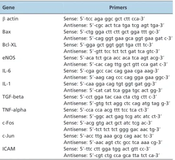
C.Quantitative real-time PCR was performed in a 15.0-mL reaction mix using 7.5mL of Platinum SYBER Green qPCR SuperMix-UDG (Invitrogen Carlsbad, CA-USA), 0.3mL of gene-specific forward and reverse primers (10mM), 1.0mL of cDNA, and 5.9mL of nuclease-free water. The primers used are shown in Table 1.
The cycling conditions were as follows: initial template denaturation at 95
˚
C for 1 minute, followed by 40 cycles of denaturation at 95˚
C for 20 seconds, annealing at 60˚
C for 30 seconds, and extension at 72˚
C for 30 seconds. The reactions were prepared separately for each of the genes and were amplified in a Rotor-Gene RG-3000 (Corbett Research, Australia). The reactions were run in triplicate. Blank controls were included in parallel for each master mix. Amplification was followed by a melting curve analysis to check the PCR product specificity. The data were analyzed by relative quantification and the 22DDCtvalue was calculated (23).
Statistical analysis
groups were compared using a nonparametric Kruskal-Wallis test followed by Dunn’s multiple comparisons test. Differences with a p value less than 0.05 were considered significant.
& RESULTS
The mortality rate was 16.5% (1/6) in all groups. The death in the control group was related to bleeding from a laceration on the graft surface. In the LFS and LFS+ IPC groups, the deaths were associated with hemodynamic instability immediately after aortic unclamping.
The anthropometric data and transplant characteristics of the groups are shown in Table 2. The recipient weights, total ischemia times and warm ischemia times were similar between the groups. The donor weights and GBWRs were significantly higher in the LFS and LFS + PCI groups
(p= 0.005).
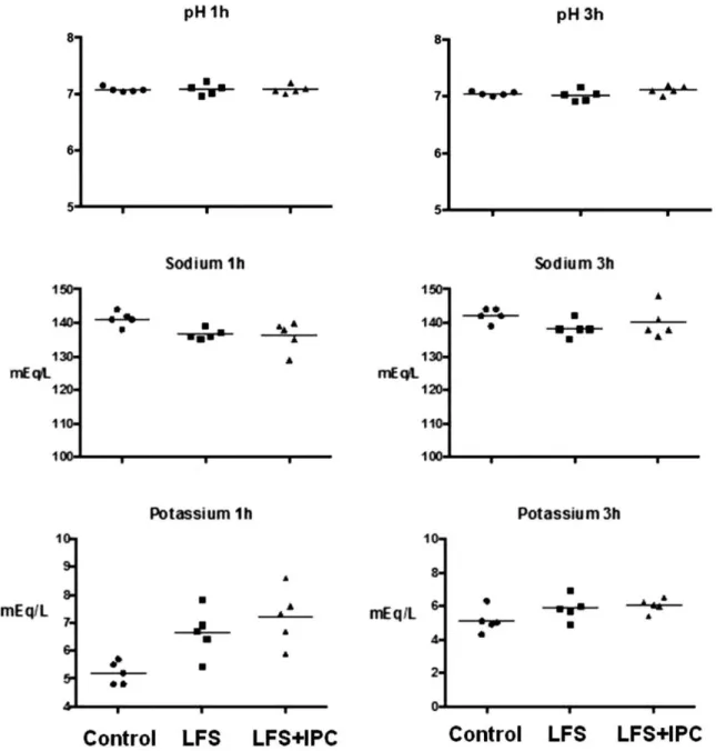
Blood analysis: All animals developed acidosis, but there was no difference in the pH values among the groups during the procedure. At 1 hour and 3 hours after reperfu-sion, the animals in the LFS and LFS + IPC groups had
decreased serum levels of Na and increased serum levels of K compared with the control group (Figure 1). The AST levels were markedly higher in the LFS and LFS + IPC
groups at both 1 and 3 hours after portal reperfusion
compared with the control group. No alterations were observed in the levels of ALT (Figure 2).
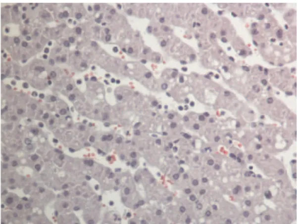
Tissue analysis: Histological alterations were observed in all groups, including marked sinusoidal dilatation and the presence of sinusoidal neutrophils and steatosis (Figure 3). However, there was no difference between the experimental groups regarding these findings.
Immunohistochemical analysis: The numbers of hepato-cytes that stained positive for Ki-67, expressed as the percentage of the total number of counted cells, are shown in Table 3. The comparisons between the groups demon-strated that there were no significant differences. Regarding the apoptotic index represented by cleaved cytokeratin-18-positive cells, the statistical analysis showed that there were no differences between the groups.
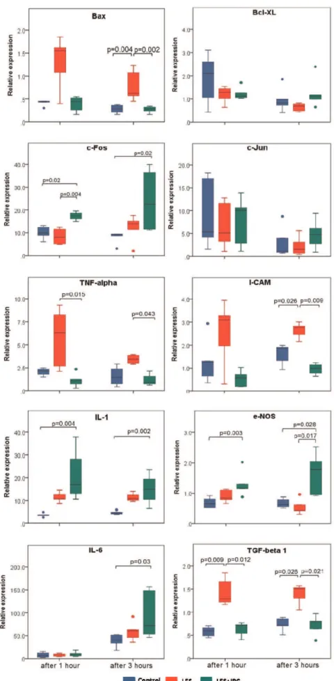
Molecular analysis: The results of the molecular analyses are shown in Figure 4. Higher Bax gene expression was observed at 3 hours after reperfusion in the LFS group compared with the control and LFS+IPC groups (p= 0.004
andp= 0.002, respectively).Bcl-XLgene expression did not differ among the groups.c-Fosgene expression was higher in the LFS+IPC group compared with the control and LFS
groups (p= 0.02 and p= 0.004, respectively). This elevated gene expression persisted after 3 hours of reperfusion (p= 0.02 compared with the control group). c-Jun gene expression did not differ among the groups and the times after reperfusion.
Regarding cytokines, adhesion molecules and e-NOS, elevated TNF-alpha gene expression was observed in the LFS group compared with the LFS + IPC group (p= 0.015
andp= 0.043 1 hour and 3 hours after reperfusion, respec-tively). I-CAM expression was elevated at 3 hours after reperfusion in the LFS group compared with the control and LFS + IPC groups (p= 0.026 and p= 0.009, respectively).
After 1 hour, no differences were observed.IL-1ande-NOS
gene expression was significantly elevated in the LFS+IPC group compared with the control, at both time points after reperfusion (p= 0.004 and p= 0.002; p= 0.003 and p= 0.028, respectively). Additionally, e-NOS gene expression was elevated in the LFS + IPC group compared with the LFS group, 3 hours after reperfusion (p= 0.017). IL-6 gene expression in the LFS+IPC group was increased 3 hours
after reperfusion compared to the control group (p= 0.03). Finally, TGF-beta gene expression was elevated in the LFS group compared with the control and LFS + IPC groups at both time points after reperfusion (p= 0.009 and
p= 0.012 after 1 hour; p= 0.026 and p= 0.021 after 3 hours, respectively).
& DISCUSSION
Given the complexity of liver transplantation in children, the difference between the recipient weight and the size of liver grafts that are most commonly harvested from adults may lead to an LFS clinical scenario, which can involve multiple complications.
We previously reported an original LFS experimental model of liver transplantation in large animals with aortic clamping (23). One technical difficulty in porcine liver transplantation is the occasional lethal hemodynamic instability caused by simultaneous portal and caval clamp-ing; to compensate for this, venovenous bypass (VVB) is often used during the anhepatic phase (24). However, VVB substantially increases the morbidity and complexity of the
Table 1 -Primer sequences.
Gene Primers
bactin Sense: 59-tcc aga ggc gct ctt cca-39 Antisense: 59-cgc act tca tga tcg agt tga-39 Bax Sense: 59-ctg gga ctt ctt gct gga ttt gc-39
Antisense: 59-cag ggt gaa gca ggt gaa gat c-39 Bcl-XL Sense: 59-gga gct ggt ggt tga ctt tc-39
Antisense: 59-gtt tcc tct tct gat tca gtc-39 eNOS Sense: 59-aca tct gca acc aca tca agt acg-39
Antisense: 59-cac cag ttg gct gtt cca gat c-39 IL-6 Sense: 59-cga gcc cac cag gaa cga aag-39
Antisense: 59-aag cag ccc cag gga gaa ggc-39 IL-1 Sense: 59-caa gga cag tgt ggt gat gg-39
Antisense: 59-cat cat tca gga tgc act gg-39 TGF-beta Sense: 59-cct gga tac caa cta ctg ctt c-39
Antisense: 59-gtg tct agg ctc cag atg tag g-39 TNF-alpha Sense: 59-cca cca acg ttt tcc tca ct-39
Antisense: 59-ggc act gag tcg atc atc ct-39 c-Fos Sense: 59-acg gtg act gct atc tcg ac-39
Antisense: 59-tct tct tct ggg gac aac tg-39 c-Jun Sense: 59-acc ttg aaa gcg cag aac tc-39
Antisense: 59-aac agt ctc gcc tca aaa cg-39 ICAM Sense: 59-ttc ctt gga tgg act gtt cc-39
Antisense: 59-cgt ctg cca gca tta tct ca-39
Table 2 -Anthropometric data and transplant characteristics of the groups (mean¡sd).
Control LFS LFS+IPC
Donor weight (kg) 20.3¡0.58 44.8¡6.21* 48.24¡2.89** Recipient weight (kg) 20.86¡1.08 22.2¡4.76 20.6¡2.79 GBWR 2.9¡0.22 6.3¡0.73* 6.4¡0.98*** Total ischemia time (min.) 138.8¡17.8 147.6¡8.3 152.0¡14.5 Warm ischemia time (min.) 44.8¡3.7 46.8¡2.7 47.0¡3.4
procedure (25,26). Aortic clamping during the anhepatic phase is well tolerated, results in excellent hemodynamic stability and considerably reduces the operative time because VVB and mesocaval anastomosis construction become unnecessary (27). Although there are potential harmful consequences of aortic clamping, these complica-tions are dependent on the level of aortic cross clamping (clamping of the thoracic aorta produces more complica-tions than clamping of the infrarenal aorta), the duration and patient comorbidities (28). In our experiments using young, healthy pigs and an anhepatic phase lasting no more than 45 minutes, we believe that the complication rate should be low.
Up to 60% of the indications for liver transplantation in children are due to biliary atresia (1), a condition that progresses with hypoplasia or even thrombosis of the portal
vein (29) and these complications lead to an early indication for liver transplantation. The low flow in the portal vein results in difficulty with graft perfusion and this difficulty can be even more pronounced when the graft is relatively large. Using a flowmetry study, we compared portal flow in control and LFS pig liver transplantations. We observed a significant decrease in portal flow from the donor to the recipient in the LFS group, and this decrease was correlated with the higher expression of inflammatory genes related to IRI and progressive elevation of AST levels (30). To simulate the clinical situation in humans, we initially attempted to use pigs weighing approximately 10 kg, but they were still suckling and did not tolerate the procedure; deaths occurred during the anhepatic phase. Therefore, we began to use weaning pigs (weighing approximately 20 kg), which produced better survival indices.
Figure 1 -The results of serum analyses. There were no differences in the pH values among the groups. Decreased serum levels of Na and increased serum levels of K were observed at 1 hour and 3 hours after reperfusion in the LFS and LFS+IPC groups compared with
In the present study, we aimed to assess the early phase of IRI in LFS transplantation cases. The abdominal cavity was kept open after the experiment in an attempt to avoid the deleterious effects of increased abdominal pressure and inferior vena cava obstruction.
The GBWR was significantly higher in the LFS and LFS+
IPC groups (mean 6.3% and 6.4%, respectively). This ratio is consistent with liver transplantation in children. Although the numbers of transplantations and recipients per group were not high, they were similar to many pig experimental
studies in the literature. The complexity of the procedure and the difficulties associated with the maintenance of large animals in urban laboratories justify the low number of animals per group in the current study. However, this number was sufficient to detect many significant differences among the groups. Another point of discussion is the cold ischemia time, which was relatively short compared with liver transplantations with cadaveric donors but prolonged compared with liver transplantations with living donors, which represent the majority of pediatric transplantations in many centers. Although the period of three hours after reperfusion is a relatively short follow-up, we focused our study on the early repercussions of IRI and the expression of genes that are triggered immediately in this process. Thus, the present model is suitable for assessing injury in the LFS
Figure 2 -The AST levels were markedly higher in the LFS and LFS+IPC groups at 1 and 3 hours after portal reperfusion compared with
the control group (p= 0.013 andp= 0.002, respectively). No alterations were observed in the ALT levels.
Figure 3 - The histological aspects of a transplanted liver at 3 hours after reperfusion. Note the presence of sinusoidal dilatation, steatosis and sinusoidal neutrophils.
Table 3 -Results of immunohistochemical analyses (median, minimum and maximum values). No differences were observed among the groups.
Control LFS* LFS+IPC*
1 h 3 h 1 h 3 h 1 h 3 h
Ki-67 10 (5-10) 15 (8-25) 7.5 (3-15) 20 (6-25) 15 (6-25) 10 (3-15) Cytokeratin 1 (0-3) 2 (0-4) 2 (1-5) 1 (0-3) 1 (0-4) 2 (0-4)
*p= 0.7 and p = 0.8 relative to the control group at 1 h and 3 h, respectively, for Ki-67.
scenario and we aimed to evaluate acute graft lesions in this context.
The standardization of the IPC interval (15 minutes of portal clamping followed by 15 minutes of reperfusion) was based on a literature review. Schulz et al. demonstrated in a study conducted in pigs that IPC with 10-minute intervals had no protective effect on IRI (31), whereas the utilization of longer IPC times (15 minutes) produced significant effects (32). Similar to the clinical scenario, all animals developed acidosis that was not related to the size of the graft because there were no differences in the pH values between the groups. The acidosis was likely related to conditions of hypoperfusion caused by the clamping of the inferior vena cava and the portal vein during the anhepatic phase in addition to the clamping of the aorta.
Hyperkalemia was observed in all groups, especially in the first hour after reperfusion. This condition is also similar to that observed in clinical transplantation. The liver graft, which is stored in a cold preservation solution, is the main source of potassium in this situation. During reperfusion, a large quantity of potassium is released from the graft into the bloodstream (33,34). Higher values were observed in the LFS and LFS+IPC groups and as described above, this can
be attributed to the higher mass of the transplanted liver graft. After 3 hours, the serum potassium values in the LFS and LFS+ IPC groups decreased and were comparable to
the control group values.
Intraoperative natremia disturbances are less often discussed in the literature. The sodium values were lower in the LFS and LFS+IPS groups one hour after reperfusion.
During the period of ischemia, cellular swelling occurs because of the influx of sodium and water into the intracellular space (35). As the liver mass was higher in the LFS and LFS+IPC groups, this result may have been
caused by a greater accumulation of intracellular sodium in these groups.
The pathophysiology of hepatic ischemia/reperfusion injury (IRI) includes a number of complex and diverse mechanisms involving the interactions between hepato-cytes, Kupffer cells, neutrophils, macrophages, sinusoidal endothelial cells and platelets. IRI is one of the main causes of graft dysfunction after transplantation and contributes to decreased graft and patient survival (14-16). Therefore, the identification of the factors that contribute to increased tissue damage, as well as measures to attenuate this damage, is critical to improving survival. In the current investigation, we aimed to study whether IPC has positive effects on the graft in the experimental LFS liver transplan-tation model. IPC consists of an adaptive mechanism in which brief episodes of ischemia protect an organ from injury caused by prolonged ischemia. IPC contributes to improving hepatic microcirculation and regeneration, thereby attenuating IRI. Biochemical, histological and molecular biology analyses were used in this study to evaluate the effects of IRI and IPC in the LFS liver transplantation model.
The serum AST levels were higher in the LFS and LFS+
IPC groups during both time periods studied. This change highlights the potential deleterious effect of implanting a disproportionately large graft into a recipient. The AST levels in the LFS group increased from the first to the third hour after reperfusion; however, in the LFS+IPC group, the AST levels tended to decrease from the first to the third hour. Similarly, immunohistochemical analyses of cellular
proliferation revealed that IPC was responsible for a tendency towards an increased peak in Ki-67 expression. These facts may suggest that IPC provokes an earlier but less harmful response to IRI.
The histological analysis revealed no differences between the groups. This result is most likely explained by the fact that the study only analyzed the acute phase of the experiment. Nevertheless, it is important to note that some typical IRI-related changes, such as the presence of sinusoidal dilatation, sinusoidal neutrophils and hepatic steatosis, were observed.
Apoptosis was evaluated based on Bax (pro-apoptotic) and Bcl-XL (anti-apoptotic) gene expression. Bax gene expression was significantly higher in the LFS group compared with the control and LFS+IPC groups at 3 hours
after reperfusion. This finding supports the hypothesis that apoptosis due to IRI is exacerbated in LFS scenarios. In addition, IPC plays a protective role by decreasingBaxgene expression.
Regarding the anti-apoptotic effects of IPC,Bcl-XL gene expression did not differ between the studied groups, but there was a tendency for higher Bcl-XL expression in the LFS+IPC group at 3 hours after reperfusion. The difference between the LFS and LFS + IPC groups almost reached statistical significance (p= 0.05), demonstrating that IPC likely plays an anti-apoptotic role in IRI.
The co-existence of apoptosis and hepatocyte regenera-tion in IRI shows that liver tissue remodeling is regulated by both processes. IEGs are important factors in the cellular proliferation induced by IRI as a part of tissue repair (9,10). Cells lose the ability to respond to proliferation signals by the blockade of Fos protein; thus, c-Fos is considered an important factor in the cell regeneration signal transduction pathway (36). Some studies suggest that the apoptosis/ proliferation processes are doubly regulated by different c-Fosandc-JunmRNA expression levels. c-Fos/c-Jun hetero-dimers are thought to promote cell regeneration and c-Jun
homodimers may lead to cell apoptosis (10,11). In fact, we observed that c-Jun levels were similar in all groups; however, IPC was responsible for increasing c-Fos levels. This result may be one explanation for the protective effect of IPC against hepatocyte apoptosis.
as decreased ICAM expression in the LFS situation, thereby attenuating IRI.
Interestingly, in our study, IPC led to an increase in IL-1 expression. Shin et al., analyzing IPC-induced neuroprotec-tion, observed that IPC was responsible for a parallel increase in IL-1band IL-1 receptor antagonist expression in the ischemic cortex, resulting in a significant reduction in the brain cortex infarct area (40). Unfortunately, we did not measure IL-1 receptor antagonist expression, but it is reasonable to assume that in the LFS+IPC group, higher
IL-1 receptor antagonist expression would also be observed. Along these lines,eNOSgene expression was significantly higher in the LFS+IPC group than in the other groups at 3 hours after reperfusion. IPC resulted in increased eNOS
expression. This enzyme is responsible for NO production, which in turn contributes to improving microcirculation, thereby reducing tissue injury (41). Liver regeneration is closely related to the expression of IL-6, which is released in IRI by Kupffer cells. Previous studies have demonstrated the role of IL-6 in liver regeneration and the increased release of IL-6 promoted by IPC (20,42). At 1 hour after reperfusion, there was no difference between the groups. However, after 3 hours, IL-6 gene expression was higher in the LFS+IPC group. This finding suggests that IL-6, along
with other factors, may play an important role in the protective mechanisms of IPC in attenuating IRI.
A relationship between IRI and the stimulation of fibrogenesis has been suggested and studied in the literature. TGF-beta is one of the main cytokines related to liver fibrogenesis and has been related to the transdiffer-entiation of stellate cells into myofibroblasts and to the deposition of portal collagen. Yu et al., using a model of selective portal vein ligation, aimed to determine whether IR in clamped lobes facilitated liver regeneration of non-clamped lobes (43). In fact, the authors observed that IR pretreatment of the clamped lobes facilitated the liver regeneration of non-clamped lobes after selective portal vein ligation, which may have resulted from down-regulated TGF-beta 1 expression in the non-clamped lobes. Our results suggest that the more intense IRI in the large graft stimulated TGF-beta production and that IPC is responsible for the attenuation of these liver fibrogenesis stimuli.
In summary, IRI is more pronounced in this model of LFS liver transplantation and is partially attenuated by IPC, mainly through the improvement of the gene expression of early molecular markers. The long-term implications and the graft and animal survival were not examined in this study because this research was conducted to assess the acute phase of IRI. Future studies to evaluate the long-term outcomes, liver perfusion and techniques to modify liver perfusion are important to establish a better understanding of the biological processes involved in this scenario.
& ACKNOWLEDGMENTS
The authors are grateful to the Fundac¸a˜o de Amparo a Pesquisa do Estado de Sa˜o Paulo - FAPESP, which financed the project (grant number 2011/ 12550-0).
& AUTHOR CONTRIBUTIONS
Leal AJ, Tannuri AC, Belon AR and Guimara˜es RR performed the experiments. Coelho MC, Gonc¸alves JO, Serafini S and Melo ES
performed the laboratory analyses. Tannuri U performed the final revision of the manuscript.
& REFERENCES
1. Arnon R, Annunziato R, Miloh T, Sogawa H, Nostrand KV, Florman S. Liver transplantation in children weighing 5 kg or less: analysis of the UNOS database. Pediatr Transplant. 2011;15(6):650-8.
2. Mekeel KL, Langham MR Jr, Gonzalez-Peralta RP, Hemming AW. Liver transplantation in very small infants. Pediatr Transplant. 2007; 11(1):66-72, doi: 10.1111/j.1399-3046.2006.00610.x.
3. Fukazawa K, Nishida S, Volsky A, Tzakis AG, Pretto EA Jr. Body surface area index predicts outcome in orthotopic liver transplantation. J Hepatobiliary Pancreat Sci. 2011;18(2):216-25, doi: 10.1007/s00534-010-0334-9.
4. Teoh NC, Farrell GC. Hepatic ischemia reperfusion injury: pathogenic mechanisms and basis for hepatoprotection. J Gastroenterol Hepatol. 2003;18(8):891-902, doi: 10.1046/j.1440-1746.2003.03056.x.
5. Itoh H, Yagi M, Fushida S, Tani T, Hashimoto T, Shimizu K, et al. Activation of immediate early gene, c-fos, and c-jun in the rat small intestine after ischemia/reperfusion. Transplantation. 2000;69(4):598-604, doi: 10.1097/00007890-200002270-00022.
6. Taguchi T, Shima Y, Nakao M, Fujii Y, Tajiri T, Ogita K, et al. Activation of immediate early genes in relation to proliferation and apoptosis of enterocytes after ischemia-reperfusion injury of small intestine. Transplant Proc. 2002;34(3):983, doi: 10.1016/S0041-1345(02)02728-8. 7. Xiao JS, Cai FG, Niu Y, Zhang Y, Xu XL, Ye QF. Preconditioning effects
on expression of proto-oncogenes c-fos and c-jun after hepatic ischemia-reperfusion in rats. Hepatobiliary Pancreat Dis Int. 2005;4(2):197-202. 8. Shima Y, Tajiri T, Taguchi T, Suita S. Increased expression of fos and
c-jun in the rat small intestinal epithelium after ischemia-reperfusion injury: a possible correlation with the proliferation or apoptosis of intestinal epithelial cells. J Pediatr Surg. 2006;41(4):830-6, doi: 10.1016/j. jpedsurg.2005.12.025.
9. Datta G, Fuller BJ, Davidson BR. Molecular mechanisms of liver ischemia reperfusion injury: insights from transgenic knockout models. World J Gastroenterol. 2013;19(11):1683-98.
10. Clavien PA, Harvey PR, Strasberg SM. Preservation and reperfusion injuries in liver allografts. An overview and synthesis of current studies. Transplantation. 1992;53(5):957-78.
11. Varotti G, Grazi GL, Vetrone G, Ercolani G, Cescon M, Del Gaudio M, et al. Causes of early acute graft failure after liver transplantation: analysis of a 17-year-single-centre experience. Clin Transplant. 2005; 19(4):492-500, doi: 10.1111/j.1399-0012.2005.00373.x.
12. Kupiec-Weglinski JW, Busuttil RW. Ischemia and reperfusion in liver transplantation. Transplant Proc. 2005;37(4):1653-6, doi: 10.1016/j.trans-proceed.2005.03.134.
13. Klune JR, Tsung A. Molecular biology of liver ischemia/reperfusion injury: established mechanisms and recent advancements. Surg Clin North Am. 2010;90(4):665-77, doi: 10.1016/j.suc.2010.04.003.
14. de Rougemont O, Dutkowski P, Clavien PA. Biological modulation of liver ischemia-reperfusion injury. Curr Opin Organ Transplant. 2010;15(2):183-9, doi: 10.1097/MOT.0b013e3283373ced.
15. Taki-Eldin A, Zhou L, Xie HY, Zheng SS. Liver regeneration after liver transplantation. Eur Surg Res. 2012;48(3):139-53, doi: 10.1159/000337865. 16. Camargo CA Jr, Madden JF, Gao W, Selvan RS, Clavien PA. Interleukin-6 protects liver against warm ischemia/reperfusion injury and promotes hepatocyte proliferation in the rodent. Hepatology. 1997;26(6):1513-20, doi: 10.1002/hep.510260619.
17. Debonera F, Aldeguer X, Shen X, Gelman AE, Gao F, Que X, et al. Activation of interleukin-6/STAT3 and liver regeneration following transplantation. J Surg Res. 2001;96(2):289-95, doi: 10.1006/jsre.2001.6086. 18. Serracino-Inglott F, Habib NA, Mathie RT. Hepatic ischemia-reperfusion
injury. Am J Surg. 2001;181(2):160-6.
19. Shah V, Kamath PS. Nitric oxide in liver transplantation: pathobiology and clinical implications. Liver Transpl. 2003;9(1):1-11, doi: 10.1053/jlts. 2003.36244.
20. Koti RS, Tsui J, Lobos E, Yang W, Seifalian AM, Davidson BR. Nitric oxide synthase distribution and expression with ischemic precondition-ing of the rat liver. FASEB J. 2005;19(9):1155-7.
21. Koti RS, Seifalian AM, Davidson BR. Protection of the liver by ischemic preconditioning: a review of mechanisms and clinical applications. Dig Surg. 2003;20(5):383-96, doi: 10.1159/000072064.
22. Bedirli A, Kerem M, Pasaoglu H, Erdem O, Ofluoglu E, Sakrak O. Effects of ischemic preconditioning on regenerative capacity of hepatocyte in the ischemically damaged rat livers. J Surg Res. 2005;125(1):42-8, doi: 10. 1016/j.jss.2004.11.028.
23. Leal AJ, Tannuri AC, Belon AR, Guimara˜es RR, Coelho MC, Oliveira Gonc¸alves Jd, et al. A simplified experimental model of large-for-size liver transplantation in pigs. Clinics. 2013;68(8):1152-6, doi: 10.6061/ clinics/2013(08)15.
alterations and the estimation of appropriate bypass flow during the period of the anhepatic state. Hokkaido Igaku Zasshi. 1987;62(4):616-28. 25. Eason J, Tan K, Howard E, Bras P, Pastellopoulos A, Patrapinyokul S, et al. Comparative hemodynamics of venovenous and venoarterial bypass during liver transplantation in the pig. Transplant Proc. 1989;21(3):3525. 26. Tojimbara T, Fuchinoue S, Sannomiya A, Murakami T, Sawada T, Tsuji
K, et al. Porcine liver transplantation from non-heart-beating cadaver donors: effect of passive/active venovenous bypass on graft function. Transplant Proc. 2000;32(7):1626-7, doi: 10.1016/S0041-1345(00)01444-5. 27. Lo´pez-Santamaria M, Migliazza L, Gamez M, Murcia J, Paz Cruz JA,
Mun˜oz J, et al. Supraceliac aortic clamping during the anhepatic phase of experimental orthotopic liver transplantation. J Pediatr Surg. 1999;34(9):1374-7, doi: 10.1016/S0022-3468(99)90014-3.
28. Gelman S. The pathophysiology of aortic cross-clamping and unclamping. Anesthesiology. 1995;82(4):1026-60, doi: 10.1097/00000542-199504000-00027. 29. Gibelli NE, Tannuri AC, Tannuri U, Santos MM, Pinho-Apezzato ML, Maksoud-Filho JG, et al. Rex shunt for acute portal vein thrombosis after pediatric liver transplantation in children with biliary atresia. Transplant Proc. 2011;43(1):194-5, doi: 10.1016/j.transproceed.2010.11.011. 30. Rangel Moreira DA, Aoun Tannuri AC, Belon AR, Mendonc¸a Coelho
MC, Oliveira Gonc¸alves J, Serafini S, et al. Large-for-size liver transplantation: a flowmetry study in pigs. J Surg Res. 2014;189(2):313-20, doi: 10.1016/j.jss.2014.03.018.
31. Schulz R, Walz MK, Behrends M, Neumann T, Gerken G, Heusch G. Minimal protection of the liver by ischemic preconditioning in pigs. Am J Physiol Heart Circ Physiol. 2001;280(1):198-207.
32. Ricciardi R, Schaffer BK, Kim RD, Shah SA, Donohue SE, Wheeler SM, et al. Protective effects of ischemic preconditioning on the cold-preserved liver are tyrosine kinase dependent. Transplantation. 2001;72(3):406-12, doi: 10.1097/00007890-200108150-00008.
33. Borland LM, Roule M, Cook DR. Anesthesia for Pediatric Orthotopic Liver Transplantation. Anesth Analg. 1985;64(2):117-24.
34. Carmichael FJ, Lindop MJ, Farman JV. Anesthesia for Hepatic Transplantation: Cardiovascular and Metabolic Alterations and their Management. Anesth Analg. 1985;64(2):108-16.
35. Belzer FO, Southard JH. Principles of solid-organ preservation by cold storage. Transplantation. 1988;45(4):673-6, doi: 10.1097/00007890-198804000-00001.
36. Michalopoulos GK, Defrance MC. Liver regeneration. Science. 1997;276(2):60-6, doi: 10.1126/science.276.5309.60.
37. Perry BC, Soltys D, Toledo AH, Toledo-Pereyra LH. Tumor necrosis factor-alpha in liver ischemia/reperfusion injury. J Invest Surg. 2011; 24(4):178-88, doi: 10.3109/08941939.2011.568594.
38. Zhang A, Chi X, Luo G, Hei Z, Xia H, Luo C, et al. Mast cell stabilization alleviates acute lung injury after orthotopic autologous liver transplanta-tion in rats by downregulating inflammatransplanta-tion. PLos One. 2013;8(10): e75262, doi: 10.1371/journal.pone.0075262.
39. Cziga´ny Z, Turo´czi Z, o`nody P, Harsa´nyi L, Lotz G, Hegedus V, et al. Remote ischemic preconditioning protects the liver from ischemia-reperfusion injury. J Surg Res. 2013;185(2):605-13, doi: 10.1016/j.jss.2013. 07.018.
40. Shin JA, Park EM, Choi JS, Seo SM, Kang JL, Lee Ke, et al. Ischemic preconditioning-induced neuroprotection is associated with differential expression of IL-1beta and IL-1 receptor antagonist in the ischemic cortex. J Neuroimmunol. 2009;217(1):14-9, doi: 10.1016/j.jneuroim.2009. 06.001.
41. Alfieri A, Ong AC, Kammerer RA, Solanky T, Bate S, Tasab M, et al. Angiopoietin-1 regulates microvascular reactivity and protects the microcirculation during acute endothelial dysfunction: role of eNOS and VE-cadherin. Pharmacol Res. 2014;80(1):43-51, doi: 10.1016/j.phrs. 2013.12.008.
42. Kannerup AS, Gronbaek H, Funch-Jensen P, Tonnesen E, Jorgensen RL, Mortensen FV. Cytokine changes during warm ischemia and reperfusion of the pig liver with or without preconditioning. Eur Surg Res. 2009;42(4):216-22, doi: 10.1159/000208520.