Case Report
Case Report
Arquivos Brasileiros de Cardiologia - Volume 85, Nº 6, Dezembro 2005
Restrictive Cardiomyopathy due to Myocardial
Cysticercosis
Rodrigo Morel Vieira de Melo, Almiro Vieira de Melo Neto, Luís Cláudio L. Corrêa,
Almiro Vieira de Melo Filho
Universidade Federal da Bahia - UFBA – Salvador, BA - Brazil
Mailing Address: Rodrigo Morel Vieira de Melo • Rua Martagão Gesteira, 274/501 - 40150-390 – Salvador, BA - Brazil E-mail: [email protected] Received on 01/14/05 • Accepted on 07/01/05
There is no description of cysticercosis affecting heart function. In the present report, the authors describe the case of a 46-year-old woman with cardiac cysticercosis and heart failure, presenting with echocardiographic
fi ndings suggestive of restrictive cardiomyopahy and myocardial microcalcifi cations suggestive of cardiac infi ltration by the disease.
Cysticercosis is a disease caused by an infestation of the larval form of Taenia solium. In human cysticercosis, the person is an accidental intermediate host. The infection is caused by the ingestion of eggs containing viable oncospheres that invade the intestine, enter the vasculatory system and lodge in tissues. The global prevalence of cysticercosis caused by cisticerco cellulosae has been estimated at three hundred thousand people1. Although any organ or tissue can house the cysts, the most commonly affected areas are the brain, skeletal musculature and subcutaneous tissue. While the topic of neurocysticercosis has been documented in numerous reports, cardiac cysticercosis and particularly myocardial cysticercosis are rare and consequently few studies have been conducted.
C
ASE
R
EPORT
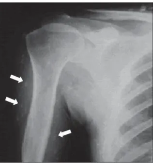
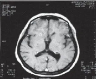
The patient is a 46 year old female born in the city of Jacobina – BA, with a history of seizures since 1970, believed to be caused from epilepsy, using phenobarbital on a regular basis. In 1996, a simple x-ray of soft tissues revealed microcalcifi cations (fi g.1). Examination of the cerebrospinal fl uid revealed a positive hemagglutination reaction of 1:4 for cysticercosis and a positive ELISA reaction for the detection of the antibody Cysticercus cellulosae. A CT scan of the brain revealed cerebral and right cerebellar calcifi cations which are also compatible with neurocysticercosis (fi g.2). As such, the patient was diagnosed with neurocysticercosis.
In 1998 she began to present dyspnea while performing out-of-the-ordinary exertions and palpitations.
Fig. 1 – Simple x-ray of soft tissues, showing microcalcifi cations
An electrocardiogram revealed a sinus rhythm, strain on the left ventricle (LV), secondary alterations of repolarization and supraventricular ectopic activity.
Arquivos Brasileiros de Cardiologia - Volume 85, Nº 6, December 2005 aspect of the diffusion of myocardial microcalcifi cations
in both ventricles called attention, in view of the aspect of other sites attacked by the disease (fi g. 3). Additionally, a concentric increase in the LV wall thickness was observed and slight dilation of the left atrium.
A hemodynamic study and angiocardiography showed normal coronaries. The diagnosis was therefore confi rmed as diastolic heart failure as a result of restrictive cardiomyopathy and due to the pattern of calcifi cation is probably secondary to myocardial cysticercosis.
D
ISCUSSION
The clinical picture presented by the patient starting in 2001 indicated a syndromic diagnosis of heart failure. The Doppler echocardiogram revealed alterations compatible with a restrictive cardiomyopathy and presence of microcalcifi cations suggestive of cysticercosis. In this case a myocardial biopsy was not performed to confi rm the diagnosis, however, the lesions found on
Fig. 2 – Computerized tomography of the brain showing cerebral calcifi cations
Fig. 3 – A) Doppler Echocardiogram showing myocardial microcalcifi cations and slight failure of the mitral and tricuspid valves. B) Doppler of the mitral valve showing diastolic fl ow with a single wave and a deceleration time of 170 ms
A B
the echocardiogram are very suggestive of cysticercosis since they correspond to the calcifi cation pattern in other areas.
There are other possible causes of the diastolic impairment that could be suggested, such as: hypertensive heart disease, constrictive pericarditis, endomyocardial fibrosis and Loeffler’s idiopathic hypereosinophilic syndrome.
Hypertensive heart disease is unlikely since the patient did not present a prior history of systemic hypertension. In reference to constrictive pericarditis, the Doppler echocardiogram did not reveal any indicative signs of this disease, such as mitral valve respiratory fl ow variations, paradoxal movement of the interventricular septum or morphological alterations of the pericardium.
The increased thickness of the LV walls and the absence of signs of endocardial fi brosis on the echocardiogram, such as abnormal echoes in the left ventricle apical region, infl ow pathway or apex of the right ventricle did not substantiate a diagnosis of endomyocardial fi brosis or Loeffl er’s idiopathic hypereosinophilic syndrome2.
The distribution of topographic locations of cysticercosis varies among different papers on the subject. Nevertheless, there is a relative consensus that the encephalic and muscular skeletal forms have a greater prevalence.
Reports in medical literature are controversial regarding the prevalence of cardiac cysticercosis. Gobbi et al3 and Lino et al.4 discovered cysticerci in the hearts of 26% and 22% of cysticercosis autopsies, respectively, while Vianna et al. discovered them in only 8%5.
The cardiac variety can cause functional diastolic alteration which is a minor symptom in the majority of cases3,6-9.
Most cases related in medical literature of myocardial cysticercosis consist of anatomical pathological studies of autopsy8 series and there are few data about the functional importance of these alterations and clinical evolution of these patients’ condition.
Arquivos Brasileiros de Cardiologia - Volume 85, Nº 6, December 2005 The patient’s condition progressed with signifi cant clinical improvement after specifi c treatment for restrictive cardiomyopathy and was referred to a cardiologist for outpatient treatment.
The relevance of this clinical case is the signifi cant clinical manifestation of cardiac disease in the case of myocardial cysticercosis; there are no documented cases in medical literature of restrictive cardiomyopathy associated
with this disease. Additionally, medical literature lacks data about echocardiograms for myocardial cysticercosis and this case illustrates these alterations.
C
ONCLUSION
Cardiac cysticercosis presents an unusual echocar-diograph aspect and can provoke a signifi cant manifestation of diastolic heart failure.
R
EFERENCES
1. Nascimento E. Teníase e cisticercose. In: Neves DP. Parasitologia Humana. 8ª ed. São Paulo: Atheneu, 1991: 230-42.
2. Acquatella H, Schiller NB, Puigbo JJ et al. Value of two-dimensional echocardiography in endomyocardial disease with and without eosinophilia. Circulation 1983; 67:1219-26.
3. Gobbi H, Adad SJ, Neves RR, Almeida HO. Ocorrência de cisticercose (Cysticercus cellulosae) em pacientes necropsiados em Uberaba, MG. Rev Patol Trop 1980; 9: 51-9.
4. Lino RS, Reis MA, Teixeira VPA. Ocorrência de cisticercose (Cysticercus cellulosae) encefálica e cardíaca em necropsias. Rev Saúde Públ 1999; 33: 495-8.
5. Vianna LG, Macedo V, Costa JM. Cisticercose músculo-cutânea e visceral – Doença rara? Rev Inst Med Trop S Paulo 1991; 33: 129-36. 6. García HH, Gonzales AE, Evans CAW et al. Taenia solium cysticercosis.
Lancet 2003; 362:547–56.
7. Agapejev S. Epidemiology of neurocysticercosis in Brazil. Rev Inst Med Trop S Paulo 1996; 38: 207-16.
8. Lino RS, Ribeiro PM, Antonelli EJ. Developmental characteristics of Cysticercus cellulosae in the human brain and heart. Rev Soc Bras Med Trop 2002; 35: 617-22.
9. Garcia HH, Del Brutto OH. Taenia solium cysticercosis. Infect Dis Clin North Am 2000; 14:97-119.