Hospital do Coração de Ribeirão Preto/Fundação Waldemar Pessoa
Mailing address: Rosana G. G. Mendes Rua ibiriçá, 1094/802 14015120 -Ribeirão Preto, SP - Brazil
Rosana G. G. Mendes, Paulo Roberto B. Evora
Ribeirão Preto, SP - Brazil
Atrial Infarction is a Unique and
Often Unrecognized Clinical Entity
A patient with heart failure and acute atrial fibrilla-tion received the final diagnosis of atrial infarcfibrilla-tion asso-ciated with ventricular infarction based on clinical fin-dings of ischemia in association with atrial fibrillation and heart failure (mechanisms probably involved: contractile dysfunction and loss of atrial contribution). Although a transesophageal echocardiography, which could refine the diagnosis of anatomic abnormalities, was not performed, all evidence led to the diagnosis of atrial involvement. Elec-trocardiographic findings were consistent with Liu’s major criterion 3.Therapy with digitalis, quinidine and angioten-sin-converting enzyme inhibitors was chosen, as the patient had acute pulmonary edema. The use of beta-blockers and verapamil was res-tricted. No other complications, such as thrombo-embolism or atrial rupture, were noted.
So far, atrial infarction is a poorly studied condition. This fact is reflected by the lack of published reports in the literature. In contrast with ventricular infarction, it repre-sents a unique clinical entity that, when diagnosed, requires significant changes in therapy. In this report, a patient presenting with heart failure and acute atrial fibrillation, who received a final diagnosis of atrial infarction associated with ventricular infarction, is described.
Case report
A 67-year-old man was hospitalized due to pneumonia. One week later, he developed dyspnea at rest and tachycar-dia. Ten days previously, he had been treated for a dyspeptic disease due to severe epigastric pain. His physical exa-mination showed marked bilateral pulmonary rales on aus-cultation, extending to the medium third of his lungs. He had an irregular heart rate and an apical systolic murmur ++/6+. Inferior limb edema, as well as varicose veins, were not noted. The results of the diagnostic workup included: 1) chest X-ray: an enlarged left ventricle (LV) and a bilateral alveolar infiltrate in pulmonary bases; 2) electrocardiogram
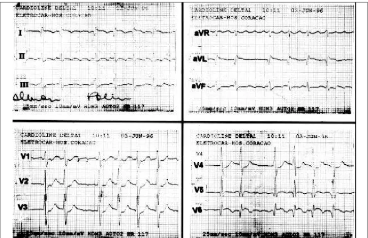
(ECG): atrial fibrillation, necrosis and ST segment elevation in the lateral wall (fig. 1); 3) Transthoracic echocardiogram: lateral hypokinesis, papillary muscle dysfunction with a slight mitral regur-gitation; 4) Routine laboratory test results within the normal range, with slightly increased cardiac enzymes.
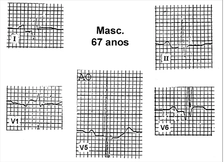
After treatment for pulmonary edema and reversion of the atrial fibrillation (with the use of digitalis, diuretics, angiotensin-converting enzyme inhibitors, oxygen and quinidine), the clinical condition was controlled and the sinus rhythm was restored. A new control ECG showed sinus rhythm, Q waves and ST segment elevation in the la-teral wall, PR-segment depression in leads DI, DII, V4, V5 e V6, as well as clear PR-segment elevation in leads V1 and V2 (figs. 2 and 3). At this moment, the diagnosis of atrial infarction associated with myocardial infarction was consi-dered in view of the ischemic abnor-malities associated with the atrial arrhythmia.
During clinical investigation, cardiac catheterization with coronary angiography showed: a 30% obstruction of the dominant right coronary artery in its medium third, a normal left main coronary artery, a 90% obstruction of the anterior descending coronary artery next to the first septal branch, a 70% obstruction of the circumflex artery at the origin of posterior ventricular and left marginal branches, a subocclusion in the emergence of the ramus medianus, apical akinesia and a slight mitral regurgitation.
As the patient was clinically stable, he underwent a coronary artery bypass graft surgery with in situ anasto-mosis of the left mammary artery to the anterior descending coronary artery and saphenous vein bypass grafting to the ramus medianus and circumflex artery. In the days following surgery, he developed pleuropericarditis that improved only after the use of corticosteroids and required a few pleural drainages due to fluid accumulation.
The last transthoracic echocardiogram performed showed normally functioning LV, slight dilation of the left atrium (LA), slight mitral regurgitation and no pericardial effusion.
Discussion
Fig. 1 – Initial electrocardiogram showing atrial fibrillation, necrosis and subepicardial current of injury in the lateral and upper lateral areas.
Fig. 3 - Amplification of leads showing in detail PR-segment abnormalities consistent with the diagnosis of atrial infarction in leads DI, DII, V1, V5 and V6.
disease, atrial infarctions have been less often related to: 1) chronic obstructive pulmonary disease with cor pulmonale and normal coronary arteries; 2) primary pulmonary hyper-tension; 3) muscular dystrophy; 4) Friedreich’s ataxia and 5) aluminum phosphide intoxication. A comprehensive report was published by Lazar et al1, with an excellent review of
the history, incidence, anatomy, pathology and etiological, clinical and diagnostic aspects of acute atrial infarction.
This case report was motivated by the following aspects of atrial infarction, based on the selected biblio-graphic references: 1) so far, atrial infarction has been a poorly studied condition. This fact is reflected by the scar-city of published reports in the literature; 2) atrial infarction represents a unique clinical entity apart from ventricular in-farction and, when diagnosed, requires significant changes in therapy; 3) in routine medical practice, it is felt that the diagnosis of atrial infarction has little clinical relevance; 4) the prognosis of atrial infarction, more than its occurrence per se, is related to the associated ventricular infarction; 5) in addition to the associated supraventricular arrhythmias, atrial infarction may present with lethal complications, such as thromboembolism and atrial rupture; 6) in order to make the diagnosis, with the patient alive or postmortem, the physician must be aware of this condition and pay
atten-tion to the possible electrocardiographic abnormalities of the PR-segment and to the higher incidence of supraventri-cular arrhythmias compared with ventrisupraventri-cular infarctions and 7) diagnosis in daily routine practice is based on electrocar-diographic findings and on the transthoracic echocardio-gram. However, these simpler tests are limited. More appro-priate imaging techniques, such as transesophageal echo-cardiography, are required, as well as the acquisition of new diagnostic resources, that is, the use of graphic resources with increased resolution.
In most cases, atrial infarction involves the right side of the heart. Its characteristics may be divided into two groups: 1) those cases where the effects of ventricular infarction predominate and 2) those that occur as a direct result of atrial involvement. Many times it is difficult to make this distinction. In addition, it is of note that a series of complications resulting from atrial infarction may occur, including arrhythmias, rupture, thromboembolic pheno-mena and reduced heart output as a result of the loss of atrial contribution 1,2.
infarc-tion abnormalities may often be absent or hidden by in-creased ventricular depolarization 1,3. However, some
elec-trical criteria for the diagnosis of atrial infarction have been proposed and include: abnormalities of the PTa-segment and P wave morphology (fig. 4), as well as the development of supraventricular arrhythmias 3. A large review of a series
of reports concludes that there are no specific data for the electrocardiographic diagnosis of atrial infarction4. The
most commonly used electrocardiographic diagnostic criteria have been those proposed by Liu et al, who catego-rized them into major and minor criteria (Table I) 5. More
recently, Mayuga & Singer6 suggested the addition of the
following electrocardiographic criteria to those established by Liu: 1) follow-up abnormalities of P wave shape , J-point, and PR-segment in serial ECG tracings and 2) simultaneous occurrence of atrial arrhythmias and abnormalities of P wave shape and PR-segment. These authors emphasize that preexistent P wave abnormalities must be taken into consi-deration when interpreting Liu’s criteria. Maybe the use of specialized electrical tracings with high resolution (atrio-grams or transesophageal recordings), as well as intraatrial tracings during cardiac catheterization, may be more spe-cific for the diagnosis of atrial infarction.
of cardiac atrial chambers and may be an obligatory test to confirm the presence of atrial infarction and thrombi 7.
Some aspects deserve special mention in the routine management of acute myocardial infarction (AMI): 1) the treatment of supraventricular arrhythmias; 2) the use of anticoagulation due to the frequency of thromboembolism and 3) the treatment of heart failure, when this condition is present. The use of beta-blockers requires special attention. This group of drugs has been proved useful in decreasing the frequency of tachyarrhythmias and atrial fibrillation/ flutter during hospitalization. It is not clear, however, whether this effect occurs as a result of an antiarrhythmic effect or due to the decrease in the size of the infarction. One can assume, although without a definite conclusion, that patients with atrial infarction may benefit from the early use of beta-blockers. Atrial infarction alone does not seem to cause heart failure. Thus, poor responses with the use of beta-blockers are not expected 1.
In conclusion, this case report has some aspects that deserve special mention: 1) the presence of ischemia asso-ciated with atrial fibrillation and heart failure (mecha-nisms probably involved: contractile dysfunction and loss of atrial contribution) which led to the diagnosis of atrial infarction associated with AMI; 2) transesophageal echocardiogra-phy was not performed; this test could have refined the diagnosis of anatomic abnormalities, but all evidence led to the diagnosis of atrial involvement; 3) electrocardiographic findings were consistent with Liu’s major criterion 3; 4) the therapy, which included digitalis, quinidine and angio-tensin-converting enzyme inhibitors, was chosen because the patient had acute pulmonary edema and the use of beta-blockers and verapamil was restricted; 5) the patient did not have other complications such as thromboembolism or atrial rupture.
Table I - Electrocardiographic diagnostic criteria according to Liu et al5.
Major criteria:
1) PTa-segment elevation >0.5 mm in leads V3 and V6 with reciprocal depression of PTa segments in V
1 and V2 leads.
2) PTa-segment elevation >0.5 mm in lead I with reciprocal depressions in leads II and III.
3) PTa-segment depression >1.5mm in precordial leads and 1.2mm in leads I, II and III, associated with any atrial arrhythmia. Minor criteria:
Abormal P-waves, flattening of P-wave in M, flattening of P-wave in W, irregular or notched P wave.
Fig. 4 – P wave morphology and PR-segment abnormalities in atrial infarction. A) Normal P wave and PR-segment; B and C) P wave abnormalities and elevation and depression of the PR-segment, respectively (Adapted 7).
1. Lazar EJ, Goldberger J, Peled H, Sherman M, Frishman WH. Atrial infarction: diagnosis and management. Am Heart J 1988; 116: 1058-63.
2. Alonso-Orcajo N, Izquierdo-Garcia F, Simarro E. Atrial rupture and sudden death following atrial infarction. Int J Cardiol 1994; 46: 82-4.
3. Liu CK, Greenspan G, Piccirilio RT. Atrial infarction of the heart. Circulation 1961; 23: 331-8.
4. Christensen JH, Nielsen FE, Falstie-Jensen N, Schmidt EB. Interobserver variation in interpretation of electrocardiographic signs of atrial infarction. Clin Cardiol 1993; 16: 603-6.
References
5. Mayuga RD, Singer DH. Atrial infarction: clinical significance and diagnostic criteria. Pratical Cardiol 1985; 11: 142-60.
6. Vargas-Barron J, Romero-Cardenas A, Keirns C, et al. Transesophageal echocardiography and right atrial infarction. J Am Soc Echocardiogr 1993; 6: 543-7.