ABSTRACT
Apparent mineralocorticoid excess (AME) syndrome results from defec-tive 11-hydroxysteroid dehydrogenase type 2 (11-HSD2). This enzyme is co-expressed with the mineralocorticoid receptor (MR) in the kidney and converts cortisol (F) to its inactive metabolite cortisone (E). Its defi-ciency allows the unmetabolized cortisol to bind to the MR inducing sodium retention, hypokalemia, suppression of PRA and hypertension. Mutations in the gene encoding 11-HSD2 account for the inherited form, but a similar clinical picture to AME occurs following the ingestion of bioflavonoids, licorice and carbenoxolone, which are competitive inhibitors of 11-HSD2. Reduced 11-HSD2 activity may explain the increased sodium retention in preeclampsia, renal disease and liver cir-rhosis. Relative deficiency of 11-HSD2 activity can occur in Cushing’s syndrome due to saturation of the enzyme and explains the mineralo-corticoid excess state that characterizes ectopic ACTH syndrome. Reduced placental 11-HSD2 expression might explain the link between reduced birth weight and adult hypertension. Polymorphic variability in the HSD11B2 gene in part determines salt sensitivity, a forerunner for adult hypertension onset. AME represents a spectrum of mineralocorti-coid hypertension with severity reflecting the underlying genetic defect in the 11-HSD2; although AME is a genetic disorder, several exogenous compounds can bring about the symptoms by inhibiting 11-HSD2 enzyme. Substrate excess as seen in Cushing’s syndrome and ACTH ectopic production can overwhelm the capacity of 11-HSD2 to convert F to E, leading up to an acquired form of AME.(Arq Bras Endocrinol Metab 2004;48/5:687-696)
Keywords: Hypertension; 11-HSD2; AME syndrome; Cortisol; Cortisone
RESUMO
Síndrome do Excesso Aparente de Mineralocorticóides: Uma Revisão.
A síndrome do excesso aparente de mineralocorticóides (SEAM) resul-ta de defeito na 11-hidroxisteróide desidrogenase tipo 2 (11-HSD2). Esta enzima é co-expressa com o receptor mineralocorticóide (RM) nos rins e converte cortisol (F) em cortisona (E), seu metabólito inativo. Defi-ciência desta enzima permite que o cortisol não metabolizado se ligue ao RM, induzindo retenção de sódio, hipocalemia, supressão da APR e hipertensão. Mutações no gene que codifica a 11-HSD2 são respon-sáveis pela forma herdada, mas um quadro clínico semelhante de SEAM ocorre durante ingestão dos bioflavonóides, alcaçuz e car-benoxolona, que são inibidores competitivos da 11-HSD2. Redução na atividade da 11-HSD2 pode explicar o aumento da retenção de sódio na pré-eclâmpsia, na doença renal e na cirrose hepática. Deficiência relativa de atividade da 11-HSD2 pode ocorrer na síndrome de Cush-ing devido à saturação da enzima e explicar o estado de excesso min-eralocorticóide que caracteriza a síndrome do ACTH ectópico. Redução da expressão placentária da 11-HSD2 poderia justificar a lig-ação entre baixo peso ao nascer e hipertensão no adulto.
Variabili-Mario Palermo
Marcus Quinkler
Paul M. Stewart
Institute of Endocrinology, University of Sassari (MP), Sassari, Italy; and Division of Medical Sciences, University of Birmingham, Queen Elizabeth Hospital (MQ, PMS), Edgbaston, Birmingham, UK.
dade polimórfica no gene HSD11B2 determina, em parte, a sensibilidade ao sódio, um preditor do surg-imento da hipertensão no adulto. A SEAM represen-ta um espectro de hipertensão mineralocorticóide cuja severidade reflete o defeito genético de base na 11-HSD2; embora a SEAM seja uma doença genética, vários compostos exógenos podem provocar os sintomas pela inibição da 11-HSD2. O excesso de substrato, visto na síndrome de Cushing e na produção ectópica de ACTH, pode sobrepujar a capacidade da 11-HSD2 de converter F em E, levando a uma forma adquirida de SEAM. (Arq Bras Endocrinol Metab 2004;48/5:687-696)
Descritores: Hipertensão; 11-HSD2; Síndrome do EAM; Cortisol; Cortisona
A
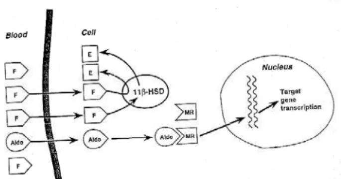
PPARENT MINERALOCORTICOID EXCESS SYNDROME(AME) is characterized by clinical features sug-gesting excessive production of a mineralocorticoid-like substance with hypertension, plasma volume expansion, hypokalemic alkalosis and a suppressed renin-angiotensin-aldosterone system (1). It can be classified on the basis of whether it is congenital or acquired, but the two forms share the same patho-physiology: AME is the outcome of defective 11 -hydroxysteroid dehydrogenase type 2 (11-HSD2) (2,3). This enzyme is predominantly expressed, together with the mineralocorticoid receptor (MR), in the renal distal tubules and collecting ducts (4), in the distal colon, in the salivary glands and also in the pla-centa where it protects the fetus from an excessive amount of maternal cortisol (F) (5,6) (figure 1). 11 -HSD2 converts F to its inactive metabolite cortisone (E). Since F, but not E, is a potent agonist of epithe-lial type 1 mineralocorticoid receptors, reduced activi-ty or total deficiency of the enzyme exposes the kidney to an excess of F, which can then act as a potent min-eralocorticoid (7,8). Minmin-eralocorticoid receptor (MR) has the same affinity for F and aldosterone in vitro(9), and the inactivation of cortisol to cortisone by 11 -HSD2 at the site of the MR enables aldosterone to bind to this receptor in vivo (figure 2) (10).
Aldos-terone is not metabolized by 11-HSD2 because it forms a C11-C18hemi-ketal group in aqueous solution.
Circulating levels of adrenal corticosteroids and 11-HSD2 activity are then involved in blood pressure regulation. Their importance is highlighted by patho-physiological situations such as Cushing’s syndrome or ectopic production of ACTH, but even in essential hypertension decreased activity of 11-HSD2 has been described.
A distinct isozyme of 11-hydroxysteroid dehy-drogenase exists (11-HSD1). It is widely distributed, but most abundant in liver and adipose tissue. It func-tions mainly as an oxoreductase, converting cortisone to cortisol, and plays a crucial role in the organ-specif-ic modulation of F effect (11) (figure 1).
This review discusses the consequence of con-genital or acquired deficiency of 11-HSD2 activity, in humans.
CONGENITAL DEFICIENCY OF 11-HSD2
Apparent Mineralocorticoid Excess Syn-drome
Cortisol metabolism
To understand the metabolic consequence of defective 11-HSD2 activity, it is important to know the nor-mal metabolism of F.
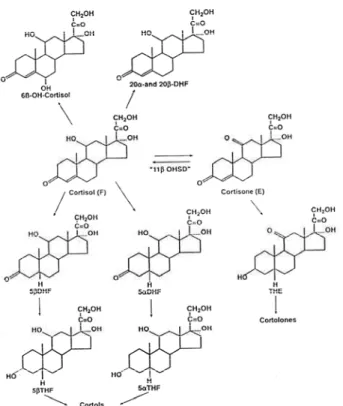
Cortisol is interconverted with cortisone by 11-HSD2 and the principal site of conversion is the kidney (12), whilst the liver is the place where
corti-11-HSD type I 11-HSD type II
Location Liver, adipose tissue Kidney, colon, placenta Cofactor NADP+ NAD+
Substrate affinity Low High
Bi-directional? Yes, mainly reductase No, only dehydrogenase DNA 1287 bp 1840 bp
Aminoacids 292 405 Molecular mass 34 kDa 45 kDa Chromosome 1 16
Figure 1. 1 1-Hydroxysteroid dehydrogenase (11- H S D ) isozymes.
sone is mainly converted to cortisol by 11-HSD1. Both are substrates for a series of enzymatic activities in the liver, including the reduction of 4double bond
(yields 5- and 5-dihydrocortisol and 5- and 5 -dihydrocortisone), reduction of 3-keto group (yields 5- and 5-tetrahydrocortisol and 5- and 5 -tetrahy-drocortisone), reduction of 20-keto group (yields to 20- and 20-DHF, cortols and cortolones) (figure 3). Most of the products are excreted in the urine as glucuronides. Only a small part of cortisol metabolites is excreted unconjugated mainly as 3-oxo-4-ene steroids (36).
In the case of AME (partial or complete defi-ciency of 11-HSD2), urinary steroid metabolite pro-files indicate that the majority of cortisol metabolites are excreted as A-ring reduced metabolites of cortisol itself (5-tetrahydrocortisol (THF) and 5-THF or allo-THF) with very low or absent levels of tetrahydro-cortisone (THE) in the urine. The excretion of 5- c o r-tisol metabolites exceeds that of 5-cortisol metabolites and results in a high urinary allo-THF/THF ratio
sug-gesting an additional defect in 5-reductase activity (13,14). The incremental increase in the THF+allo-THF/THE compared to the allo-THF/THF ratio, however, is much larger, with typical THF+allo-THF/THE ratios ranging from 3 to over 70 in AME (normal ratio is approximately 1). The THF+allo-THF/THE ratio has been used in the past in the diag-nosis of AME (13,14), but probably provides an index of “global” 11-HSD activity within the body, i.e. principally 11-HSD1 in the liver and 11-HSD2 in the kidney. The conversion of cortisone to cortisol mediated by 11-HSD1 is normal in AME (67). The plasma half-life of [11-3H]-cortisol (which when
metabolized by 11-HSD yields tritiated water and cortisone) may more accurately reflect renal 11 -HSD2 activity (10), as may the ratio of urinary free cor-tisol/urinary free cortisone (UFF/UFE) (15). Normal subjects excrete 2-3-fold more UFE than UFF, reflect-ing the significant activity of renal 11-HSD2. In AME, however, UFE excretion is virtually undetectable (16) resulting in a high UFF/UFE ratio. Plasma corti-sol half-life is prolonged (120-190min vs. 70-90min in controls), but patients with AME are not cushingoid; the cortisol secretion rate falls often to very low levels due to a normal intact negative feedback mechanism. This maintains normal circulating concentrations in the face of impaired cortisol metabolism.
A variant of AME, so-called “type II AME” has been documented in several patients (17,18). This variant is characterized by a milder phenotype, with onset in late adolescence or early adulthood and only a mildly deranged urinary THF+allo-THF/THE ratio. However, the UFF/UFE excretion is high in the type II variant, and the metabolism of 11-triated cortisol
Figure 3.Cortisol metabolism. 11-HSD2 deficiency reduces the production of THE; as a consequence, (THF+allo-THF)/THE ratio increases. Despite normal circulating cortisol levels, patients with AME show a decrease in the total uri-nary excretion of cortisol metabolites reflecting a reduction secretion rate consequent upon a prolonged plasma half-life. In addition, 5-reduced cortisol metabolites predomi-nate over 5-reduced cortisol metabolites consistent with a reduction of 5-reductase activity in patients with AME.
(directly reflecting 11-HSD2 dehydrogenase activity) is grossly deranged, confirming deficiency of 11 -HSD2 (16) (figure 5A).
Pathophysiology
The pathophysiology of AME has now been satisfac-torily explained in terms of its clinical, biochemical and genetic basis. An inability of the renal 11-HSD2 enzyme to inactivate F to E is the cause of sodium retention, PRA and aldosterone suppression and hypertension.
Firstly, in 1974 Werder et al. (19) described a child with features similar to primary hyperaldostero-nism, but presenting suppressed plasma aldosterone. Afterwards, New et al. (20) and Ulick et al. (21) described other children presenting similar clinical pic-tures. The distinctive feature of the patients was the high excretion of 11-hydroxycortisol metabolite (THF and cortols) to the extremely low excretion of 11-oxo-metabolites (THE and cortolones). Since hypertension, low renin and hypokalemia, but low lev-els of aldosterone and deoxycorticosterone were pre-sent, the term “Apparent Mineralocorticoid Excess” (AME) was coined. In 1983, Oberfield et al. (22) doc-umented the mineralocorticoid effect of hydrocortisol and the marked hypotensive effect of spironolactone and metyrapone. For these reasons, they suggested the presence of a defective conversion of F to E and a min-eralocorticoid-like action of cortisol on MR (22). In 1985, the first adult case of AME was reported. It was described the beneficial effect of dexamethasone and the deleterious action of hydrocortisone on blood pressure and hypokalemia in this patient, confirming the involvement of a deranged cortisol metabolism in the pathogenesis of the syndrome (23). The physio-logical explanation of this theory was given by the demonstration that the MR has the same affinity for cortisol and aldosteronein vitro, but 11-HSD pro-tects the MR in vivoby the action of F hundreds of times higher in concentration compared to aldos-terone (9,24). This enzyme was then proposed as the one responsible for the syndrome. In 1989, Ulick et al. (25) described the case of 4 Italian children with the same clinical presentation of classical AME, but less severe biochemical features. They called this syndrome AME type II (25). On the basis of the markedly decreased ring-A reduction constant (THF+allo-THF/F), they indicated the impaired ring-A reduc-tion and/or defective interconversion of F and E in both directions (F to E and E to F, leaving the ratio between 11-and 11-oxo steroid unchanged) as the principal abnormalities of AME (25,26). In 1995-1996, information on the structure and sequence of
the HSD11B2 gene has enabled the identification of mutations in AME patients. HSD11B2 is 6.2kb in length containing five exons and is located on chro-mosome 16q22 (27,28). At present, more than 30 dif-ferent mutations have been defined within the HSD11B2 gene in approximately 60 affected kindreds (figure 3) (3,28-30). Genetics entirely explain the clin-ical and biochemclin-ical features of AME.
The congenital form of AME is thus attribut-able to deficiency of 11-HSD2. Cortisol and aldos-terone have similar affinities in vitrofor the type I MR and 11-HSD2 confers aldosterone specificity on the intrinsically non-specific MR by converting cortisol to its inactive metabolite cortisone. This way, 11-HSD2
in vivoprotects MR from the hundreds of times high-er circulating levels of cortisol.
AME is an autosomal recessive inherited form of hypertension. Most type I AME patients are homozygous for HSD11B2 mutations causing full, or partial loss of activity. It is most commonly found in consanguineous families (3,28,30,31).
Type II AME is also explained on the basis of mutations in the HSD11B2 gene (32,33). In an extensive Sardinian kindred, a novel homozygous mutation (R279C) was found in all 4 affected cases. In keeping with the mild phenotype the mutation result-ed in a mutant enzyme with only minor disturbances in activity. Classification of AME into distinct variants is therefore inappropriate (figure 5A). In keeping with this, a close correlation is reported between disease phenotype (as measured by the THF+allo-THF/THE ratio, serum potassium and blood pressure) and geno-type (34). Patients with mutant 11-HSD2 cDNAs that demonstrate little or no activityin vitro, present in early life with severe, often life-threatening, hyper-tension and hypokalemia. In contrast patients present-ing in late adolescence or early adulthood with so-called “mild” forms of AME have been found to have mutations that result in an 11-HSD2 protein with only attenuated activity.
Clinical Picture
car-diomegaly and hypertensive retinopathy. The mortali-ty is more than 10%, due to stroke, cerebral hemor-rhage and infarction. Less severe forms in adults have been described. These patients were in the past includ-ed in the type II AME. The less severe biochemical and clinical features in type II patients compared to type I appear to be explained on the basis of muta-tions, which result in some residual functional
enzy-matic activity. The decision to assign the individual patients to AME type I or II group is therefore rather arbitrary (35,18) (figure 5A).
Diagnosis
Biochemical abnormalities comprise suppressed PRA, undetectable serum aldosterone levels and hypokalemia. Traditionally, the THF+alloTHF/THE ratio has been used in the diagnosis of AME. A very high ratio can be found (normal ratio ranges from 1 to 3) together with evidence of a more general defect in steroid ring-A reduction (i.e. a higher allo-THF/THF ratio and a lower ring-A reduction constant THF+allo-THF/F).
The “net”in vivoconversion of F to E involves both isoforms of 11-HSD in tissue expressing these enzymes. As AME is a disorder of the renal 11 -HSD2, a direct measure of the ratio of urinary free cortisol/free cortisone fractions (UFF/UFE) should better reflect 11-HSD2 isozyme activity with respect to the ratio of liver-reduced metabolites (THF+allo-THF)/THE (15,16,36). As a consequence, UFF/UFE ratio proves extremely sensitive and accurate when used as an index of clinical disorder. In 24 patients suf-fering from AME syndrome, where urinary E is virtu-ally absent, THE was always detectable although 12 subjects had undetectable UFE (16). This suggests that UFE may be more sensitive than THE in the diagnosis of AME. Moreover, if used in monitoring the enzymatic activity in heterozygotes, we often found a significant increase in UFF/UFE ratio, but not in the (THF+allo-THF)/THE ratio (37). A com-parison of the UFF/UFE to (THF+allo-THF)/THE ratios in patients with AME after licorice ingestion or in patients suffering from ectopic ACTH syndrome, shows that any deviation from normal in the (THF+allo-THF)/THE ratio resulted in a much more marked change in UFF/UFE (15). The higher sensi-tivity of UFF/UFE probably occurs because it derives from the activity of the renal isozyme 11-HSD2, expressed together with the MR in the distal tubule and collecting duct, whereas the reduced fraction THF, allo-THF and THE are products of the hepatic metabolism of F (38).
AME patients are not cushingoid because they have a normal intact negative feedback mechanism. This maintains normal circulating concentration in the face of impaired cortisol metabolism.
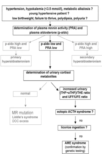
Figure 6 illustrates how AME might be diag-nosed in a patient presenting with mineralocorticoid excess.
Therapy
Therapy is directed at correcting life-threatening
Figure 5. A)(THF+allo-THF)/THE and UFF/UFE are significant-ly increased both in AME type I and type II.B)11 -Hydrox-ysteroid dehydrogenase is saturated by substrate excess. Increase in UFF/UFE ratio depends by the amount of circu-lating cortisol.
n o r m a i s AME Type 2 AME Type 1 normais vs AME type 1 p < 0.04
normais vs AME type 2 p < 0.5 THF+aTHF/THE IN AME SYNDROME
FREE F/E IN AME SYNDROME
normais vs AME type 1 p < 0.05
normais vs AME type 2 p < 0.05
n o r m a i s AME Type 1 AME Type 2
F/E In CUSHING’S DISEASE
normais vs ectopic p < 0.001 normais vs pituitary p < 0.01 normais vs adrenal p < 0.5 pituitary vs ectopic p < 0.002 adrenal vs pituitary p < 0.06
hypokalemia and hypertension (18,35). Dexametha-sone is the treatment of choice. Doses ranging from 1.5 to 2mg/day brought serum potassium levels to normal in 7-10 days in approximately 60% of cases by suppressing cortisol and progressively decreasing blood pressure. Additional antihypertensive medica-tion may be required. Patients have been successfully treated with the potassium sparing diuretics tri-amterene and/or amiloride. Thiazide diuretics are indicated when hypercalciuria and/or nephrocalci-nosis are present. Spironolactone, a MR antagonist, has been of variable benefit, presumably because very high doses are required to block the mineralocorticoid effects of cortisol on the MR. Its side effects include menstrual disturbances in women, gynecomastia, impotence and decreased libido in men and are main-ly due to its inhibition of steroid biosynthetic P-450 enzyme and its action as an antiandrogen. Sometimes it is important to reduce dietary sodium. AME was
reported “cured” in one patient following kidney transplantation due to the normal 11-HSD2 activity of the transplanted kidney (39). The case suggests a new strategy in a selected cohort of patients such as drug-unresponsive children and in patients with end-stage kidney failure.
11-HSD2 and “Essential” Hypertension
Although patients with essential hypertension do not have overt signs of mineralocorticoid excess, some positive correlations between blood pressure and plas-ma sodium levels or a negative correlation with serum potassium levels have been described.
Regarding 11-HSD2, studies have demon-strated variations in 11-HSD activity in hypertensive subjects with either increases in the plasma [11˜-3
H]-cortisol half-life or the THF+allo-THF/THE ratio (40,41), but mineralocorticoid excess in patients with impaired 11-HSD2 activity could not be demon-strated.
Recently, association and linkage studies have been performed. One study has reported an associa-tion between a microsatellite marker close to the HSD11B2 gene and hypertension in African Ameri-cans with hypertensive end stage renal disease (42). These data were confirmed using a polymorphic restriction site in exon 3 of the HSD11B2 gene. In terms of hypertension per se, however, linkage and/or association studies have been negative (43,44).
Increased sensitivity to salt is a forerunner to “essential” hypertension. Salt sensitive individuals appear to have impaired 11-HSD2 activity as mea-sured by increased urinary cortisol/cortisone ratios. Studies have evaluated a microsatellite within intron 1 of the HSD11B2 gene, and documented association with salt sensitivity in both normal subjects and patients with hypertension (45,46). Short microsatel-lite alleles were more common in salt sensitive com-pared to salt resistant subjects. The same phenomenon was observed in Blacks compared to Caucasians (47), in keeping with the predisposition to low-renin, salt-sensitive hypertension in this ethnic group.
In addition to enhanced renal sodium reten-tion, the modulation of active glucocorticoid concen-tration by 11-HSD in vascular smooth muscle cells could be an additional factor underlying hypertension (48).In vitro and in vivo studies indicate that 11 -HSDs regulate vascular tone at an autocrine level through the amplification of responses to vasocon-strictors (49). Inhibition of 11-HSD2 in vascular smooth muscle cells resulted in increased responses to angiotensin-II (50) and phenylephrine (51). 11
HSD2 knockout mice demonstrate increased arterial reactivity to norepinephrine and decreased endotheli-um-derived nitric oxide synthase activity (52).
ACQUIRED DEFICIENCY OF 11-HSD2
Licorice
Licorice roots and their extract have been used for over one thousand years as a medical herb product and as sweeteners and mouth fresheners (53). The active ingredient of licorice is glycyrrhizic acid, which is hydrolyzed into its aglicone glycyrrhetinic acid in vivo. Licorice products are made from peeled and unpeeled dried root. There are powdered and finely cut root preparations; the most important are the liquid and the dry extracts. These formulations have different concentrations of the active ingredient, glycyrrhizic acid, and can vary from 20% to trace amount, based on the extraction process. In addition, a number of com-mercial preparations containing licorice are available such as herboristic and cosmetic; moreover some preparations are used as a cough remedy and are usu-ally mixed with Arabic gum, sugar, alcohol and tobac-co. A preparation of the root of the licorice plant was successfully used to treat patients with peptic ulcera-tion. Such observations were the basis for the develop-ment of the effective anti-ulcer drug, carbenoxolone, which is a hemisuccinate derivative of 18- g l y-cyrrhetinic acid. Licorice possesses some endocrino-logical effects such as glucocorticoid activity, antian-drogen effect, and estrogenic activity. Whorwood (54) described an inhibitory effect of licorice on prolactin gene expression in vivo. Its mineralocorticoid effect was first documented in the 1940’s. Patients consum-ing excessive quantities of licorice present with hyper-tension and hypokalemia, which may be severe enough to cause myopathy and cardiac arrhythmia. Both PRA and aldosterone levels are suppressed and exchange-able sodium levels are increased. The condition responds to spironolactone and is reversible upon stopping licorice ingestion (55). Glycyrrhizic and gly-cyrrhetinic acids have a very low affinity for the MR, but are very potent competitive inhibitors of 11 -HSD2 (Ki of approx. 5-10nM) (56). Licorice admin-istration to normal volunteers results in a mineralocor-ticoid excess state, an increase in the urinary THF+allo-THF/THE ratio, an increase in plasma cor-tisol half-life, and a decrease in circulating cortisone values, indicative of inhibition of 11-HSD2in vivo. Thus it is now established that licorice induces an acquired and milder form of AME, causing its miner-alocorticoid effects through inhibition of 11-HSD2.
Flavonoids Consumption
The flavonoids naringin and its aglycone naringenin present in some kind of fruits, such as grapefruit, seem to have an inhibitory effect on 11-HSD2 similar to licorice. In sensitive individuals, 250mg/day of grape-fruit juice for 7 days causes significant inhibition of the enzyme causing an increase in the UFF/UFE ratio, reduction of PRA and mild hypokalemia (57).
OTHER DISEASES
Ectopic ACTH Syndrome
Eighty per cent of patients with Cushing’s syndrome have hypertension, and in the subgroup of patients with ectopic ACTH syndrome this increases to over 95%. The severity of hypertension is a key factor in predicting morbidity and mortality from the disease, yet its pathogenesis has been poorly understood. The ectopic ACTH syndrome is characterized by mineralo-corticoid excess, with hypokalemic alkalosis found in 95-100% of cases, in contrast to < 10% in other forms of Cushing’s syndrome. Although elevated plasma lev-els of deoxycorticosterone have been postulated to play a role, it is the level of cortisol secretion, which corre-lates best with the degree of mineralocorticoid excess.
ACTH has no direct effect on 11-HSD2, but the enzyme is saturated in ectopic ACTH syndrome by very high concentrations of ACTH-dependent 11 -HSD substrates such as cortisol and corticosterone. Both the urinary ratio of THF+allo-THF/THE and UFF/UFE are elevated, not because of impaired 11 -HSD2 activity, but because of substrate saturation (58) (figure 5B). In severe hypercortisolism all avail-able cortisol cannot be inactivated to cortisone and “spills over” onto the MR to cause mineralocorticoid hypertension (15).
Renal Disease
unchanged. Impaired 11-HSD2 activity in patients with renal disease might underpin the increased sodi-um retention observed in some pathologies, notably nephrotic syndrome. ACE inhibitors are known to increase renal 11-HSD2 activity and this, in part, may explain their natriuretic effect (61).
Liver Disease
Activation of MR in patients with liver cirrhosis leads to renal sodium retention and hypokalemia. The same is described during alcoholic and non-alcoholic chron-ic liver disease or bile duct obstruction where an increase in (THF+allo-THF)/THE ratio is present as a consequence of an inhibitory effect of bile acid on 11-HSD2 activity (62).
Fetal Growth
Glucocorticoid excess in uterus decreases fetal growth and the high levels of placental 11-HSD2 may pro-tect the fetus from maternal glucocorticoid excess. Impaired enzymatic activity causing an excess of glu-cocorticoid in uterus can lead to the poor growth rate seen in many children with AME (63). Impaired pla-cental 11-HSD2 activity has been associated with intrauterine growth restriction and with programming of hypertension in adult life (64).
Preeclampsia
Sodium retention is a feature in preeclampsia and pregnancy-induced hypertension caused probably by activation of MR. Progesterone and its metabolites can favor this by inhibiting 11-HSD2 (65). Reduced 11-HSD2 expression has been reported in placentas of women with preeclampsia and pregnancy induced hypertension (66).
CONCLUSION
1 1-HSD is a key enzyme for cortisol metabolism. Its activity in converting F to its inactive metabolite E regu-lates at “pre-receptor” site the action of glucocorticoid steroids in the body. The isozyme type II is involved in sodium and potassium homeostasis giving specificity to aldosterone for the mineralocorticoid receptor. AME syn-drome is caused by total or partial 11-HSD2 deficiency and is characterized by hypertension, suppressed PRA and aldosterone and hypokalaemia. It represents a spectrum of mineralocorticoid hypertension with severity reflecting the underlying genetic defect in the 11-HSD2, from very severe and life threatening to mild. Several acquired forms of AME exist. Licorice, carbenoxolone and flavonoids may
cause sufficient 11-HSD2 inhibition to produce meta-bolic and clinical disorders. Mineralocorticoid excess is also a feature of the ectopic ACTH syndrome, because 1 1-HSD2 is overwhelmed by its substrate cortisol. Poly-morphic variability in the HSD11B2 gene determines salt sensitivity and might play a role in patients with “essential” hypertension. Impaired 11-HSD2 activity in patients with renal or hepatic disease or in preeclampsia might be involved in sodium retention in these diseases.
REFERENCES
1. Stewart PM. Mineralocorticoid hypertension. L a n c e t 1999;353(9161):1341-7.
2. White PC, Mune T, Agarwal AK. 11- H y d r o x y s t e r o i d dehydrogenase and the syndrome of apparent miner-alocorticoid excess.Endocr Rev 1997;18(1):135-56.
3. Wilson RC, Nimkarn S, New MI. Apparent mineralocorticoid excess.Trends Endocrinol Metab 2001; 1 2 ( 3 ) : 1 0 4 - 1 1 .
4. Albiston AL, Obeyesekere VR, Smith RE, Krozowski ZS. Cloning and tissue distribution of the human 11 -hydroxysteroid dehydrogenase type 2 enzyme. Mol Cell Endocrinol 1994;105:R11-R17.
5. Vogt B, Frey BM, Frey FJ. 11-Hydroxysteroid dehydro-genase: pathophysiology.Adv Nephrol Necker Hosp 1999;29:127-48.
6. Kitanaka S, Tanae A, Hibi I. Apparent mineralocorticoid excess due to 11-hydroxysteroid dehydrogenase defi-ciency: a possible cause of intrauterine growth retarda-tion.Clin Endocrinol (Oxf) 1996;44:353-9.
7. Edwards CR, Stewart PM, Burt D, Brett L, McIntyre MA, Sutanto WS, et al. Localization of 11-hydroxysteroid dehydrogenase - tissue specific protector of the miner-alocorticoid receptor.Lancet 1988;2(8618):986-9.
8. Funder JW, Pearce PT, Smith R, Smith AI. Mineralocorti-coid action: target tissue specificity is enzyme, not receptor, mediated.Science 1988;242(4878):583-5.
9. Arriza JL, Weinberger C, Cerelli G, Glaser TM, Handelin BL, Housman DE, et al. Cloning of human mineralocorti-coid receptor complementary DANN: structural and functional kinship with the glucocorticoid receptor. Sci-ence 1987;237(4812):268-75.
10. Stewart PM, Corrie JE, Shackleton CH, Edwards CR. Syn-drome of apparent mineralocorticoid excess. A defect in the cortisol-cortisone shuttle. J Clin Invest 1988;82(1):340-9.
11. Stewart PM, Krozowski ZS. 11-Hydroxysteroid dehydro-genase.Vitam Horm 1999;57:249-324.
12. Whitworth JA, Stewart PM, Burt D, Atherden SM, Edwards CR. The kidney is the major site of cortisone production in man.Clin Endocrinol Oxf 1989;31(3):355-61.
Corti-costeroid metabolite profiles of four patients and their families.Clin Endocrinol Oxf 1985;22(6):701-12.
14. Monder C, Shackleton CH, Bradlow HL, New MI, Stoner E, Iohan F, et al. The syndrome of apparent mineralo-corticoid excess: its association with 11 -dehydroge-nase and 5-reductase deficiency and some conse-quences for corticosteroid metabolism. J Clin Endocrinol Metab 1986;63(3):550-7.
15. Palermo M, Shackleton CHL, Mantero F, Stewart PM. Uri-nary free cortisone and the assessment of 11 -hydrox-ysteroid dehydrogenase activity in man. C l i n Endocrinol (Oxf) 1996;45:605-11.
16. Palermo M, Delitala G, Mantero F, Stewart PM, Shackle-ton CHL. Congenital deficiency of 11-hydroxysteroid dehydrogenase (apparent mineralocorticoid excess syndrome): diagnostic value of urinary free cortisol and cortisone.J Endocrinol Invest 2001;24:17-23.
17. Ulick S, Tedde R, Mantero F. Pathogenesis of the type 2 variant of the syndrome of apparent mineralocorticoid excess.J Clin Endocrinol Metab 1990;70(1):200-6.
18. Mantero F, Palermo M, Petrelli MD, Tedde R, Stewart PM, Shackleton CHL. Apparent mineralocorticoid excess: Type I and type II.Steroids 1996;61:193-6.
19. Werder E, Zachmann M, Vollmin JA, Veyrat R, Prader A. Unusual steroid excretion in a child with low renin hyper-tension.Res Steroids 1974;6:385-9.
20. New MI, Levine LS, Biglieri EG, Pareira J, Ulick S. Evidence for an unidentified steroid in a child with apparent min-eralocorticoid hypertension.J Clin Endocrinol Metab 1977;44(5):924-33.
21. Ulick S, Levine LS, Gunczler P, Zanconato G, Ramirez LC, Rauh W, et al. A syndrome of apparent mineralocorti-coid excess associated with defects in the peripheral metabolism of cortisol. J Clin Endocrinol Metab 1979;49(5):757-64.
22. Oberfield SE, Levine LS, Carey RM, Greig F, Ulick S, New MI. Metabolic and blood pressure responses to hydro-cortisone in the syndrome of mineralocorticoid excess. J Clin Endocrinol Metab 1983;56:332-7.
23. Edwards CRW, Stewart PM, Nairn IM, Grieve J, Shackle-ton CHL. Cushing’s disease of the kidney.J Endocrinol 1985;104(S):53 (abstr).
24. Krozowski Z, Funder JW. Renal mineralocorticoid recep-tors and hippocampal corticosterone-binding site have identical intrinsic steroid specificity.Proc Nat Acad Sci USA 1983;80:6056-60.
25. Ulick S, Chan CK, Rao KN, Edassery J, Mantero F. A new form of the syndrome of apparent mineralocorticoid excess.J Steroid Biochem 1989;32:209-12.
26. Monder C, Shackleton CHL, Bradlow HL, New MI, Stoner E, Iohan F, et al. The syndrome of apparent mineralo-corticoid excess: its association with 11-hydroxysteroid dehydrogenase and 5-reductase deficiency and some consequences for corticosteroid metabolism.J Clin Endocrinol Metab 1986;63(3):550-7.
27. Agarwal AK, Rogerson FM, Mune T, White PC. Gene struc-ture and chromosomal localization of the human HSD11K
gene encoding the kidney (type 2) isozyme of 11- h y d r o x-ysteroid dehydrogenase.Genomics 1995;29: 195-9.
28. Mune T, Rogerson FM, Nikkilä H, Agarwal AK, White PC. Human hypertension caused by mutations in the kid-ney isozyme of 11-hydroxysteroid dehydrogenase. Nat Genet 1995;10:394-9.
29. Wilson RC, Harbison MD, Krozowski ZS, Funder JW, Shackleton CHL, Hanauske-Abel HM, et al. Several homozygous mutations in the gene for 11 -hydroxys-teroid dehydrogenase type 2 in patients with apparent mineralocorticoid excess. J Clin Endocrinol Metab 1995;80:3145-50.
30. Stewart PM, Krozowski ZS, Gupta A, Milford DV, Howie AJ, Sheppard MC, et al. Hypertension in the syndrome of apparent mineralocorticoid excess due to mutation of the 11-hydroxysteroid dehydrogenase type 2 gene. Lancet 1996;347:88-91.
31. Kitanaka S, Katsumata N, Tanae A, Hibi I, Takeyama KI, Fuse H, et al. A new compound heterozygous mutation in the 11-hydroxysteroid dehydrogenase type 2 gene in a case of apparent mineralocorticoid excess.J Clin Endocrinol Metab 1997;82(12):4054-8.
32. Li AR, Li KXZ, Marui S, Krozowski ZS, Batista MC, Whor-wood CB, et al. Apparent mineralocorticoid excess in a Brazilian kindred: hypertension in the heterozygote state.J Hypertension 1997;15(12):1397-402.
33. Wilson RC, Dave-Sharma S, Wei JQ, Obeyesekere VR, Li K, Ferrari P, et al. A genetic defect resulting in mild low-renin hypertension. Proc Natl Acad Sci USA 1998;95(17):10200-5.
34. Nunez BS, Rogerson FM, Mune T, Igarashi Y, Nakagawa Y, Phillipov G, et al. Mutants of 11-hydroxysteroid dehy-drogenase (11-HSD2) with partial activity – Improved correlations between genotype and biochemical phe-notype in apparent mineralocorticoid excess. J Biol Chem 1999;34:638-42.
35. Shimojo M, Stewart PM. Apparent mineralocorticoid excess syndromes.J Endocrinol Invest 1995;18:518-32.
36. Palermo M, Gomez-Sanchez C, Roitman E, Shackleton CHL. Quantitation of cortisol and related 3-oxo-4ene steroids in urine using gas chromatography/mass spec-trometry with stable isotope-labeled internal standards. Steroids 1996;61:583-9.
37. Li A, Krozowsky ZS, Pala A, Li KXZ, Shackleton CHL, Man-tero F, et al. Molecular basis for hypertension in the “type II variant” of apparent mineralocorticoid excess. Am J Hum Genet 1998;63:370-9.
38. Tannin GM, Agarwal AK, Monder C, New MI, White PC. The human gene for 11-hydroxysteroid dehydroge-nase. Structure, tissue distribution, and chromosomal localization.J Biol Chem 1991;266:16653-8.
39. Palermo M, Cossu M, Shackleton CHL. Cure of apparent mineralocorticoid excess by kidney transplantation.N Engl J Med 1998;329(24):1787-8.
40. Walker BR, Stewart PM, Shackleton CH, Padfield PL, Edwards CR. Deficient inactivation of cortisol by 11 -hydroxysteroid dehydrogenase in essential hyperten-sion.Clin Endocrinol Oxf 1993;39(2):221-7.
dehydroge-nase and 5-reductase activity in subjects with untreated essential hypertension.Hypertension 1995; 2 5 : 6 7 - 7 0 .
42. Watson B Jr, Bergman SM, Myracle A, Callen DF, Acton RT, Warnock DG. Genetic association of 11 -hydroxys-teroid dehydrogenase type 2 (HSD11B2) flanking microsatellites with essential hypertension in blacks. Hypertension 1996;28(3):478-82.
43. Smolenicka Z, Bach E, Schaer A, Liechti-Gallati S, Frey BM, Frey FJ, et al. A new polymorphic restriction site in the human 11-hydroxysteroid dehydrogenase type 2 gene.J Clin Endocrinol Metab 1998;83(5):1814-7. 44. Brand E, Kato N, Chatelain N, Krozowski ZS, Jeunemaitre
X, Corvol P, et al. Structural analysis and evaluation of the 11-hydroxysteroid dehydrogenase type 2 (11 -HSD2) gene in human essential hypertension.J Hyper-tens 1998;16:1627-33.
45. Agarwal AK, Giacchetti G, Lavery G, Nikkila H, Palermo M, Ricketts M, et al. CA-Repeat polymorphism in intron 1 of HSD11B2: effects on gene expression and salt sensi-tivity.Hypertension 2000;36(2):187-94.
46. Lovati E, Ferrari P, Dick B, Jostarndt K, Frey BM, Frey FJ, et al. Molecular basis of human salt sensitivity: The role of the 11-hydroxysteroid dehydrogenase type 2.J Clin Endocrinol Metab 1999;84:3745-9.
47. White PC, Agarwal AK, Li A, Nikkila H, Pratt JH, Caulfield M, et al. Possible association but no linkage of the HSD11B2 gene encoding the kidney isozyme of 11 -hydroxysteroid dehydrogenase to hypertension in Black people. Clin Endocrinol (Oxf) 2001;55(2):249-52. 48. Smith RE, Little PJ, Maguire JA, Stein-Oakley AN, Kro-zowski ZS. Vascular localization of the 11 -hydroxys-teroid dehydrogenase type II enzyme.Clin Exp Phar-macol Physiol 1996;23(6-7):549-51.
49. Walker BR, Connacher AA, Webb DJ, Edwards CR. Glu-cocorticoids and blood pressure: a role for the corti-sol/cortisone shuttle in the control of vascular tone in man.Clin Sci 1992;83(2):171-8.
50. Hatakeyama H, Inaba S, Takeda R, Miyamori I. 11 -Hydroxysteroid dehydrogenase in human vascular cells.Kidney Int 2000;57:1352-7.
51. Souness GW, Brem AS, Morris DJ. 11-Hydroxysteroid dehydrogenase antisense affects vascular contractile response and glucocorticoid metabolism. Steroids 2002;67(3-4):195-201.
52. Hadoke PW, Christy C, Kotelevtsev YV, Williams BC, Kenyon CJ, Seckl JR, et al. Endothelial cell dysfunction in mice after transgenic knockout of type 2, but not type 1, 11- h y d r o x-ysteroid dehydrogenase.Circulation 2001; 1 0 4 ( 2 3 ) : 2 8 3 2 - 7 . 53. Armanini D, Fiore C, Mattarello MJ, Bielenberg J, Paler-mo M. History of the endocrine effect of licorice.Exp Clin Endocrinol Diabetes 2002;110:257-61.
54. Whorwood CB, Sheppard MC, Stewart PM. Licorice inhibits 11-hydroxysteroid dehydrogenase messenger ribonucleic acid levels and potentiates glucocorticoid hormone action.Endocrinology 1993;132:2287-92. 55. Bernardi M, D’Intino PE, Trevisani F, Cantelli-Forti G,
Raggi MA, Turchetto E, et al. Effect of prolonged inges-tion of graded doses of licorice by healthy volunteers. Life Sci 1994;55:863-72.
56. Stewart PM, Murry BA, Mason JI. Human kidney 11 -hydroxysteroid dehydrogenase is a high affinity nicoti-namide adenine dinucleotide-dependent enzyme and
differs from the cloned type I isoform.J Clin Endocrinol Metab 1994;79(2):480-4.
57. Lee YS, Lorenzo BJ, Koufis T, Reidenberg MM. Grapefruit juice and its flavonoids inhibit 11-hydroxysteroid dehy-drogenase.Clin Pharmacol Therapy 1996;59:62-71.
58. Stewart PM, Walker BR, Holder G, O’Halloran D, Shackle-ton CHL. 11-Hydroxysteroid dehydrogenase activity in Cushing’s syndrome: Explaining the mineralocorticoid excess state of the ectopic adrenocorticotropin syn-drome.J Clin Endocrinol Metab 1995;80:3617-20.
59. Kawai S, Ichikawa Y, Homma M. Differences in meta-bolic properties among cortisol, prednisolone, and dexamethasone in liver and renal diseases: accelerat-ed metabolism of dexamethasone in renal failure.J Clin Endocrinol Metab 1985;60(5):848-54.
60. Srivastava LS, Werk EE Jr, Thrasher K, Sholiton LJ, Kozera R, Nolten W, et al. Plasma cortisone concentration as measured by radioimmunoassay.J Clin Endocrinol Metab 1973;36(5):937-43.
61. Riddle MC, McDaniel PA. Renal 11- h y d r o x y s t e r o i d dehydrogenase activity is enhanced by ramipril and captopril.J Clin Endocrinol Metab 1994;78(4):830-4.
62. Quattropani C, Vogt B, Odermatt A, Dick B, Frey BM, Frey FJ. Reduced activity of 11-hydroxysteroid dehy-drogenase in patients with cholestasis. J Clin Invest 2001;108(9):1299-305.
63. McTernan CL, Draper N, Nicholson H, Chalder SM, Driver P, Hewison M, et al. Reduced placental 11 -hydroxys-teroid dehydrogenase type 2 mRNA levels in human pregnancies complicated by intrauterine growth restriction: an analysis of possible mechanisms.J Clin Endocrinol Metab 2001;86(10):4979-83.
64. Lindsay RS, Lindsay RM, Edwards CRW, Seckl JR. Inhibi-tion of 11-hydroxysteroid dehydrogenase in pregnant rats and the programming of blood pressure in the off-spring.Hypertension 1996;27:1200-4.
65. Quinkler M, Johanssen S, Grossmann C, Bähr V, Müller M, Oelkers W, et al. Progesterone metabolism in the human kidney and inhibition of 11-hydroxysteroid dehydroge-nase type 2 by progesterone and its metabolites.J Clin Endocr Metabol 1999; 8 4 ( 1 1 ) : 4 1 6 5 - 7 1 .
66. Schoof E, Girstl M, Frobenius W, Kirschbaum M, Dörr HG, Rasch-er W, et al. Decreased gene expression of 11- h y d r o x y s t e r o i d dehydrogenase type 2 and 15-hydroxyprostaglandin dehy-drogenase in human placenta of patients with preeclampsia. J Clin Endocrin Metabol 2001; 8 6 ( 3 ) : 1 3 1 3 - 7 .
67. Nikkila H, Tannin GM, New MI, Taylors NF, Kalaitzoglu G, Monder G, et al. Defect in HSD1 gene encoding 11 -hydroxysteroid dehydrogenase are not found in patient with mineralocorticoid excess or 11-oxo reductase defi-ciency.J Clin Endocrinol Metab 1993;77:687-91.
Endereço para correspondência:
Mario Palermo
Institute of Endocrinology, University of Sassari Viale S. Pietro 43