ABSTRACT
Improvements in abdominal imaging techniques have increased the detection of clinically inapparent adrenal masses, or incidentalomas (AI), the appropriate diagnosis and management of which have become a common clinical problem for health care professionals. Once an adrenal mass has been detected, the clinician needs to address two questions: 1) is the tumor hormonally active? and 2) is there any chance of the mass being malignant? The majority of AI is non-hypersecretory cortical adenomas, but an endocrine evaluation can lead to the identification of subtle hormone excess. An overnight low-dose dexamethasone suppression test, fractionated urinary or plasma metanephrine assay and, in hypertensive patients, establishing the upright plasma aldosterone/plasma renin activity ratio are recom-mended as preliminary screening steps. Masses greater than 4cm are at greater risk of malignancy. Morphological imaging features may be helpful in the distinction between benign and malignant forms. Fine-nee-dle aspiration biopsy is an important tool in the evaluation of oncologi-cal patients to establish any metastatic disease. Adrenalectomy is indi-cated by evidence of a functional adrenal mass, or a suspected malig-nant form. We advocate adrenalectomy of subtle hypercortisolism, especially in the presence of hypertension, obesity, diabetes or osteo-porosis potentially aggravated by glucocorticoid excess. A close follow-up is needed, particularly in the first year after diagnosis.(Arq Bras Endocrinol Metab 2004;48/5:583-591)
Keywords:Adrenal incidentaloma; Benign adenoma; Carcinoma
RESUMO
Uma Abordagem Abrangente do Incidentaloma Adrenal.
O aperfeiçoamento das técnicas de imagem abdominal tem aumen-tado a detecção de massas adrenais clinicamente não aparentes, ou incidentalomas (IA), cujo diagnóstico e manuseio apropriados têm se tornado um problema clínico comum para os profissionais da saúde. Uma vez detectada uma massa adrenal, o clínico é obrigado a formu-lar duas questões: 1) o tumor é hormonalmente ativo? e 2) existe algu-ma possibilidade de a algu-massa ser algu-maligna?. A algu-maioria dos IA são ade-nomas corticais não hipersecretores, mas uma avaliação endócrina pode resultar na identificação de um excesso hormonal sutil. Como passos preliminares de screening são recomendados um teste de supressãoovernightcom doses baixas de dexametasona, a dosagem de metanefrinas em urina fracionada ou no plasma e, em pacientes hipertensos, estabelecer a relação da aldosterona plasmática/ativi-dade plasmática de renina na posição ortostática. Massas maiores que 4cm têm risco maior de malignidade. Achados morfológicos de imagem podem ser valiosos na distinção entre formas benignas e malignas. A biópsia de aspiração com agulha fina é um procedimen-to importante na avaliação de pacientes oncológicos para se estab-elecer qualquer doença metastática. Adrenalectomia está indicada na evidência de uma massa adrenal funcionante, ou na suspeita de
Franco Mantero
Nora Albiger
Department of Medical and Surgical Sciences, Endocrinology Unit, University of Padua, Padua, Italy.
uma forma maligna. Nós recomendamos adrena-lectomia para casos de hipercortisolismo sutil, espe-cialmente em presença de hipertensão, obesidade, diabetes ou osteoporose, potencialmente agrava-dos pelo excesso de glicocorticóides. Um acom-panhamento rigoroso é necessário, particularmente no primeiro ano após o diagnóstico. (Arq Bras Endocrinol Metab 2004;48/5:583-591)
D e s c r i t o r e s : Incidentaloma adrenal; Adenoma benigno; Carcinoma
A
D REN AL IN CID EN TALO M A is an adren massdis-covered inadvertently during diagnostic tests or treatment for other clinical conditions unrelated to any suspicion of adrenal disease, though patients are often found retrospectively to have sym ptom s or signs of hormone oversecretion by the tumor. The definition of incidentaloma rules out patients undergoing imag-ing procedures for cancer stagimag-ing and work-up. In the last 15-20 years, the widespread use of noninvasive imaging techniques has led to an increased detection of incidentalomas.
After th e in cid en tal d iagn o sis o f an ad ren al mass, it is important to establish whether the tumor is hormonally active. D ifferentiating between malignant and benign masses is also essential because metastases to adrenal glands are common. Adrenocortical carci-nomas are rare, but they have a high mortality rate.
CAUSES AND PREVALENCE
In normal subjects, the prevalence of adrenal inciden-talom as m ay depend on what im aging is done and why. In large series of patients screened using routine transabdominal ultrasonography (U S) during a gener-al hegener-alth examination, 0.1-0.5% had abnormgener-al adrengener-al findings with a prevalence on the right side (1). The figure increases to 0.6-4.4% in series where computed tomography (C T) was performed and among patients with a previous diagnosis of cancer (2,3). In cases eval-uated by CT , there was no apparent difference between sides. There may be bilateral masses in 10% of cases (4). In autopsy series, the prevalence of previ-ously undiagnosed adrenal masses ranges between 1.4 and 8.7% (4,5).
The frequency of adrenal incidentaloma increas-es with age: it is uncommon under 30 years old and peaks between the fifth and seventh decades.
In more than 1000 cases evaluated by a multi-center study organized by the Adrenal Incidentaloma Study Group of the Italian Endocrinology Society
(AI-SIE), the patients’ median age was 58 years and there was a significant female prevalence (584 women versus 420 men) (6). N o gender differences were found in autopsy series, however.
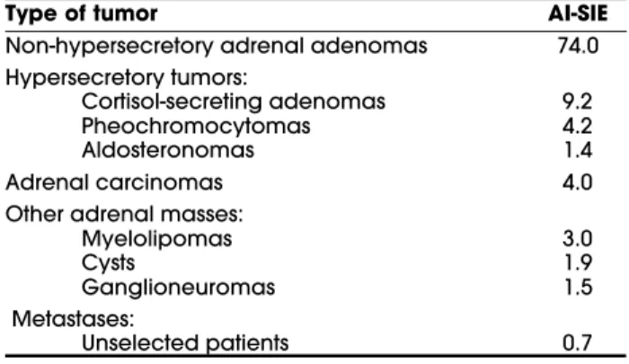
T able 1 summarizes the prevalence of adrenal incidentalomas in the AI-SIE study and the histologi-cal diagnoses are illustrated in figure 1. M ost adrenal incidentalomas are non-hypersecretory adenomas, but man y patients with adrenal in cidentalomas h ave revealed isolated or multiple mild abnormalities of the hypothalamic-pituitary-adrenal axis. M oreover, adren-al insufficiency following the surgicadren-al excision of “ silent” adrenal adenomas has been described in 18-20% of cases, suggesting a condition of mild hypercor-tisolism. This situation of cortisol excess without the classic clin ical sign s m ay b e a co m m o n fin d in g in patients with incidental adrenal adenom as and it is term ed su b clin ical C u sh in g’s syn d ro m e. T h e term subclinical autonomous glucocorticoid hypersecretion (SAGH ) has recently been proposed to define this sit-uation (7). The prevalence of hypercortisolism in clin-ically in apparen t ad ren al m asses repo rt ed ly ran ges from 5 to 47% in various studies using different study protocols and diagnostic criteria (8-12). In our series of 208 adrenal incidentalomas, 29 patients met the
cri-Table 1.Prevalence of causes of adrenal incidentalomas in AI-SIE series.
Type of tumor AI-SIE
Non-hypersecretory adrenal adenomas 74.0 Hypersecretory tumors:
Cortisol-secreting adenomas 9.2
Pheochromocytomas 4.2
Aldosteronomas 1.4
Adrenal carcinomas 4.0
Other adrenal masses:
Myelolipomas 3.0
Cysts 1.9
Ganglioneuromas 1.5
Metastases:
Unselected patients 0.7
teria for subclinical C ushing’s syndrome, correspond-ing to a prevalence of 14%.
Ph eochro m ocytom a is rare (0.01-0.1% of hypertensive patients), and clinical manifestations vary. H ypertension is constant in only about half of the patients, paroxysmal in one third and absent in approximately one fifth. In the M ayo C linic review of a 50-year autopsy series, pheochromocytoma was found in 0.13% of cases and the tumor had not been suspected in 75% of patients while alive, though it con-tributed to death in 55% of cases (13). So, a clinically silent pheochromocytoma is not so rare and its preva-lence in patients with adrenal incidentalomas has been estim ated in 1 .5 -1 3%(4 ). In t he AI -SI E series, pheochrom ocytom a was the second m ost prevalent form of hyperfunctioning tumor, occurring in 4.2% of all m asses. About half of the patients affected were normotensive, while the others had mild-to-moderate hypertension. N one of these patients had paroxysmal symptoms of adrenergic discharge. In the same series, a pheochromocytoma was confirmed by histology in 11% of patients who underwent adrenalectomy (6). A horm one evaluation to exclude pheochrom ocytom a should therefore be done routinely in patients with adrenal incidentalom as. T he m ost frequent form of presentation is a sporadic pheochromocytoma, but up to 30% may be a component of hereditary syndromes. This percentage is now higher than was once reported (10%), due to our increasing knowledge based on gen etic stu d ies. T h e h ered itary syn d ro m es in clu d e m ultiple endocrine neoplasia type 2 (M EN 2), von H ippel-Lindau disease (VH L) and neurofibromatosis type 1 (N F-1), which are associated with an autosomal dominant inheritance pattern and variable penetrance. T h e prevalen ce o f prim ary ald o st ero n ism is m ore frequent nowadays than previously reported. U sing the plasma aldosterone concentration/ plasma renin activity ratio (PAC / PRA ratio) as a screening test for primary aldosteronism and studying patients with normokalemia have improved the recognition of this condition in the hypertensive population, though prevalence rates of 5-13% am ong all hypertensives have been described (14-16). An aldosteronom a is found in 1.6-3.3% of patients with adrenal inciden-taloma (6,17). The low prevalence found in the AI-SIE study (1.6%) is probably explained by the exclusion of cases with severe hypertension and hypokalemia.
Adrenocortical carcinoma is rare, with an inci-dence ranging from 0.6 to 2 cases per million in the general population (18). T he prevalence of prim ary adrenal carcinoma in clinically inapparent adrenal masses is related to the size of the mass. In tumors up
to 4cm, adrenocortical adenomas account for 65% and carcinom as for 2%. T he larger the diam eter of the mass, the greater the risk of it being malignant. M ost pu b lish ed series repo rt a pred o m in an ce o f fem ale patients (up to 90% in som e series). In the AI-SIE series of adrenal incidentalomas, the relative frequency of malignancy was 4.6% (6).
Some incidental adrenal masses are infiltrative disease, fungal and tuberculosis infection, hemorrhage and lesions masquerading as adrenal but arising from adjacent organs (kidney, pancreas, gallbladder, spleen, lymph nodes). Primary adrenal lymphoma is uncom -mon, but it is important to recognize it because it is potentially curable.
EVALUATION OF FUNCTIONAL STATUS
Although most adrenal incidentalomas are non-hyper-secretory adenomas, hormone evaluation is mandatory to identify cases of clinically unsuspected horm one-secreting adrenal tumors.
T he N I H State-of-Science C onference panel (19) recom m ends the 1m g dexam ethasone suppres-sio n test in all patien ts with in cid en tally d etected adrenal masses. After taking 1mg dexamethasone overnight, a serum cortisol of less than 5µg/ d l (< 138nmol/ l) at 8:00 a.m. is considered negative. We believe that cortisol insuppressibility would be better evaluated by lowering the cut-off to 3µg/ dl. M ore recently, this cut-off level has been reduced to less than 1.8µg/ dl (50nmol/ l) (20).
In the authors’ series, the prevalence of cortisol excess in patients with adrenal incidentalom as was considerable. In the AI-SIE multicenter study, other abnormalities were seen in patients with cortisol excess
and the most frequent combinations encountered are shown in figure 2.
Patients with adrenal incidentalomas may have a high prevalence of impaired glucose tolerance, previ-ously undiagnosed diabetes mellitus, high triglyceride levels and arterial hypertension (21).
Fractionated urinary or plasma metanephrines (norm et anephrine an d m etanephrin e) shou ld be assayed in all patien ts with ad ren al in cid en talo m a, including normotensive patients (19). The sensitivity and specificity of 24-hour urine catecholam ines are high, but this test is less sensitive than determ ining free metanephrines. Various medications may produce false-positive results when urinary metanephrines and catecholam ines are m easured (i.e. m ethyldopa, lev-odopa, labetalol, sotalol, tricyclic antidepressants, ben-zod iazepines, drugs containing catecho lam in es, am phetamines, wit hdrawal fro m clon id in e and ethanol) and should be avoided, wherever possible, during evaluation for a suspect pheochromocytoma. D ynam ic tests with clonidine suppression should be reserved for dubious cases. Glucagon testing may trig-ger a hypertensive crisis and is not recommended.
C hromogranin A is not specific for pheochro-mocytoma, since it may be elevated in other neuroen-docrine tumors, but its evaluation may be useful. The level of chromogranin A correlates with tumor mass (7). In patients with hypertension, serum potassium and a PAC/ PRA ratio should be determined to check for prim ary aldosteronism (19). A reference for the PAC / PRA ratio should be ascertained at each center and the m inim um PRA value included in the ratio should never be lower than the detection limit of the assay. The cut-off for the PAC / PRA ratio suggesting the possibility of a primary aldosteronism should be calculated on the basis of normal reference ranges for PAC and PRA at each center. PAC and PRA should be expressed in ng/ dl and ng/ ml/ h, respectively. A ratio higher than 20 with a PAC ≥ 15ng/ dl has been con-sidered suspect for the diagnosis of primary aldostero-nism (22,23). In our experience, a PAC / PRA ratio > 40, with m inim um PRA levels of 0.2ng/ m l/ h, has 100% sensitivity and 84.4% specificity in screening patients with suspected primary aldosteronism. Several drugs can interfere with PAC and PRA measurements. It was classically recommended that patients undergo-ing this evaluation be taken off all antihypertension treatment for at least 2-3 weeks, but such a total wash-out could be unsafe in cases of severe hypertension, for whom calcium channel blockers and -blockers seem to be a valid option. O n the other hand, some authors recommend doing the tests while patients are taking
their antihypertension therapy, except for antialdos-terone drugs and -blo ckers (23). If they have hypokalemia, plasma potassium should be normalized and the ratio measured again 2 weeks later.
D ynam ic tests are usually needed to confirm primary aldosteronism. Sodium-loading tests are usu-ally employed, using fludrocortisone or saline infusion. Fludrocortisone is adm inistered at a dose of 0.1m g four times daily for 4 days during dietary supplemen-tation with 20-30mmol sodium 4 times a day, mea-suring aldosterone at the baseline and on days 3 and 4. Primary aldosteronism is confirmed if upright PAC is
≥ 5ng/ dl. O ral potassium supplementation may be needed (24). The saline infusion test is performed with an i.v. infusion of 2 liters of 0.9% isotonic saline over 4 hours while the patient is in a supine position. Pri-mary aldosteronism is confirmed if PAC levels at the end of the test remain > 10ng/ dl, and it is highly like-ly with levels > 7.5ng/ dl, whereas it is excluded when PAC levels are < 5ng/ dl. T he captopril test has also been considered for screening or as a confirm atory test; various protocols have been proposed. C aptopril can be administrated orally, 25-50mg, measuring the PAC/ PRA ratio 2 hours later (or after 1 hour if 50mg are used) with the patient in the upright position. A ratio > 20 is highly indicative, while a ratio > 30 in a patient whose previous PAC/ PRA ratio was high con-firms the diagnosis of primary aldosteronism (25,26).
There is still controversy regarding the value of measuring dehydroepiandrosterone sulfate (D H EAS). Low D H EAS levels are frequently observed in subclinical Cushing’s syndrome and adrenocortical adeno -mas, but the sensitivity and specificity of this parame-ter are poor (51% and 65%, respectively) (27). Elevat-ed D H EAS levels are frequently seen in patients with adrenocortical carcinoma (28), but other studies have found no convincing data that D H EAS is helpful in discriminating malignant from benign masses (27).
with co rtical tu m o r, with n o sign ifican t d ifferen ce between unilateral and bilateral, or benign and malig-nant lesions. An enhanced 17-O H P response was also observed in the majority of patients with subclinical C ushing’s syndrom e (68%). M oreover, a norm aliza-tion of this endocrine alteraaliza-tion was observed in most of the patients who had unilateral adrenalectomy (6). So, it is sometimes difficult to interpret the ACTH test and an exaggerated response of 17-O H P is not specif-ic for 21-O H defspecif-iciency; it might merely be a sign of disturbed intratumoral steroidogenesis.
EVALUATING MALIGNANCY
Adrenocortical carcinoma can be functional or non-functional as regards horm one synthesis and clinical features. U sing the clin ical definition , fu n ctio n al tumors account for 26-94% of adrenocortical carcino-m as (30,31). H ypercortisoliscarcino-m or a cocarcino-m bination of cortisol and androgen excess, are the most frequent presenting signs of functional tumors. Most androgen-secreting neoplasms are adrenocortical carcinomas rather than benign adenomas and they are more fre-quent in children (32). Estrogen-secreting tumors are rare and can cause feminization.
The lesion’s size is an important parameter of malignancy: the larger the size, the greater the risk. The cut-off for suspected malignancy ranges between 3 and 6cm. Based on the receiver operating characteristic (RO C) curve for the diameter, as calculated from data in the AI-SIE study, 4cm is considered a reasonable cut-off. Among the 47 cases of adrenal carcinoma found at surgery among the 387 patients considered in the AI-SIE study who had adrenalectomy, only 2 lesions were less than 4cm in size. The mean diameter was 7.5cm, range 2.6-25cm (6). M any types of adren-al lesion can present as large masses; so radiologicadren-al fea-tures can facilitate the differential diagnosis (33,34).
IMAGING STUDIES
C omputed Tomography (C T) is an accurate tool for detecting adrenal m asses. Adrenocortical adenom as usually appear on scans as small homogeneous round m asses with sm ooth m argins. M ost lesions sm aller t h an 4 cm appear t o b e b en ign (6 ). C alcificat io n , necrosis and hem orrhage are uncom m on in benign adenomas, but such lesions are not specific. Adenomas frequently contain a large amount of intracytoplasmat-ic lipid, whintracytoplasmat-ich enables a quantitative evaluation by
m easuring the attenuation valu e of the lesio ns (expressed in H ounsfield units, H U ) (35). Adenomas usually have attenuation values below 18H U on unen-hanced CT; when the lesion has an attenuation of less than 10H U , further work-up seems to be unnecessary, since it is probably a lipid-rich adrenal adenoma (36,37). M oreover, adenom as are generally characterized by rapid i.v. contrast washout. Lipid-poor adenomas have much the same enhancement and washout as lipid-rich adenomas, thus enabling their distinction from metas-tases (37,38). A relative enhancement washout of more than 40-50% is highly suggestive of a benign m ass (sensitivity 96%, specificity 100%), whereas lower rela-tive washout percentages strongly suggest a metastasis. By contrast, malignancies exhibit irregular mar-gins and variable density, with strong enhancem ent an d slo w wash o u t . C alcificat io n , h em o rrh age an d necrosis are com m on (39). O n m agnetic resonance imaging (M RI), malignancies usually show high signal in tensit y on T 2-weighted images, whereas m ost benign tumors have isointense or low signal intensity on both T1- and T2-weighted images. Approximately 30% of masses cannot be distinguished reliably on T2-weighted images (40). Both CT and M RI are accurate in assessing tumor spread into tissues such as the liver, lymph nodes, lung and inferior vena cava. In our expe-rience, C T and M RI were equally accurate in estab-lishing the size of adrenal m asses in a group of 67 patients, though the sm allest lesions (< 3cm ) were more accurately evaluated by M RI. The sensitivity of CT and MRI was similar in the differential diagnosis of adrenal tumors, but MRI imaging was more specific in confirming a clinical suspicion of pheochromocytoma and carcinoma. M ost (65%) of the adenomas evaluat-ed with C T, M RI or both, were homogeneous, while both methods showed carcinomas as being non-homogeneous. Carcinomas were variably hyperintense on T 2-weighted M R im ages and the m argins were irregular in all cases. U nfortunately, this was often also true on CT and MRI scans of pheochromocytomas (in 62% and 83% of cases, respectively). For all tumors a positive correlation was found between the size detected on CT or M RI and after surgery (r=0.92; p< 0.0001). Pheochromocytoma is typically isointense with respect to the liver on T1-weighted images and hyper-intense on T2-weighted images, whereas adrenal carci-nomas and metastases may have a similar T2-weighted hyperintensity. In our experience, all pheochromocy-tomas revealed T2 hyperintensity.
been documented at autopsy in as many as 38% of can-cer patients. Morphological CT images are non-specif-ic. The size of the mass varies, but it is less well defined than adenoma. H emorrhage or central necrosis may produce irregular cystic areas. Several studies have demonstrated a significantly slower contrast material washout in metastases than in adenomas (41,42). In the presence of a mass with a high attenuating value (> 20H U ) and when a metastasis is suspected, ultrasound or C T -guided fine-needle aspiration is a useful diag-nostic tool for evaluating adrenal lesions with a sensi-tivity of 81-100%, a specificity of 83-100% and an accuracy of 91% (43,44). Pheochromocytoma should always be ruled out before this procedure is attempted to avoid the risk of a hypertensive crisis (19).
Adrenocortical scintigraphy using 1 3 1I 6 b
-iodo m ethyl-19-n orcho lestero l (N P5 9) or 7 5S e
-methylnorcholesterol assures not only the anatomical localization of the adrenal glands but also theirin vivo
functional characterization (45). A discordant scinti-graphic pattern demonstrating little or no radiocholes-terol uptake by the affected adrenal gland is compati-ble with malignancies (primary or secondary) or destructive adrenal lesions. In our series, 4 out of 5 m alignant tum ors showed this pattern. It has been su ggested th at a co n co rd an t scin tigraph ic pattern , defined as a unilateral adren al visualization or increased radiotracer uptake with virtually no con-tralateral gland, is typical of benign cortical adenomas or nodular hyperplasia. In our series, 54% of benign lesions showed a concordant uptake, while there was only a partially increased uptake on the side of adeno-ma in another 28% of cases. Adrenal adeno-masses less than 2cm in size m ay show a norm al pattern, bilaterally demonstrating a symmetrical uptake, representing the resolution limit of this technique.
Scintigraphy with 131I or 123
I-meta-iodobenzyl-guanidine (M IBG) or111In-octreotide should be
per-form ed to evaluate a suspected pheochrom ocytom a (46).
A more promising technique is positron emis-sion tomography with18F-fluorodeoxyglucose (FD
G-PET). M ost malignant tumors show an enhanced gly-colytic m etabolism with an increased deoxyglucose uptake (47).
MANAGEMENT
After the incidental discovery of an adrenal mass, two m ajor issues should be addressed in form ulating a treatment plan: 1) is the tumor hormonally active even
in the absence of a classic clinical presentation? and 2) is there any chance of the mass being malignant?
If history and physical examination in a patient with a unilateral form suggest a clinical endocrine syn-drome and this is biochemically confirmed, adrenalec-tomy is considered the treatment of choice (figure 3). N owadays, laparoscopic adrenalectomy is preferred by most endocrine surgeons. O pen adrenalectomy is the exception, but it m ay be necessary to convert to an open procedure during laparoscopy due to finding a large, potentially malignant mass. Laparoscopic resec-tion of benign adrenal m asses is a safe and effective m ethod involving a shorter hospital stay (48). T he needle endoscopic approach, the rem ote-controlled ro botic surgical system and gasless laparoscopic adrenalectomy have also been used. Conservative tech-niques such as autotransplantation of adrenocortical tissue or subtotal adrenalectomy have been proposed in some particular cases where bilateral adrenalectomy was indicated. Experience with autotransplantation has been disappointing in m ost studies (49). A variable rate of recurrence has been reported after subtotal adrenalectomy. Recently, unilateral subtotal with con-tralateral total adrenalectomy has been recommended in patients with bilateral familial pheochromocytoma.
I n cases o f co n t rain d icat io n s fo r su rgery o r unresectable lesions, m edical treatm ent m ay be an option. We advocate preoperative medical treatment with an alpha-1 adrenergic antagonist (e.g. prazosin, doxazosin) in patients with incidental pheochromocy-toma, even if they are normotensive. Such treatment enables the expansion of the vascular bed and plasma volum e and reduces the am ount of fluid needed to main tain the blood pressure when the tumor is removed.
In the absence of clinical syndromes, treat-m ent decisions treat-may be treat-m ore difficult. An exceptio n is the “silent” pheochromocytom a, which carries a risk o f hypertensive crisis and should prompt adrena-lectom y. Alt hough the natural hist ory of subclinical C ushin g’s syndrome and it s morbid ity are unclear, we advocat e adrenalectomy for patient s wit h this condition, especially in t he presence o f h ypertension, obesit y, diabetes or osteoporosis potentially aggra-vated by glu cocorticoid excess. Since the hypothala-m us-p itu it ary-ad ren al axis an d t he co nt ralateral adrenal gland m ay b e su ppressed b y prolo nged corti-sol secretion, these patients require glu cocorticoid therapy b oth d uring and after su rgery. While adrena-lectom y has been found to correct biochemical abnormalities, it s effect on lo ng-term outcom e and qualit y of life is unknown, so careful observation has also been suggested as a treatm ent option (1 9).
As m entioned above, the risk of m alignancy increases with the size of the m ass. H om ogeneous lesions with regular margins less than 4cm in size are unlikely to have malignant potential and are generally not resected. According to the N H I State-of-Science Conference panel (19), the general recommendation is to excise lesions larger than 6cm. For lesions between 4 and 6cm, either closer follow-up or adrenalectomy (an option preferred by our group on the strength of our personal experience) are both considered reason-able. In cases of rapid growth, low lipid content and other features described earlier suggestive of adrenal carcinoma, surgery is the treatment of choice. Finally, adrenalectomy does not seem to be beneficial in the case of metastases from a known or unknown primary neoplasm (19).
Long-term follow-up studies suggest that most adrenal lesions remain stable, whereas 5-25% grow and a small percentage may shrink. Follow-up in the first years is mandatory. We advocate a CT/ MRI scan at 3-to 6-month intervals in the first year after detecting the adrenal incidentaloma. A clinical (hormonal and radiological) evaluation should be performed after 1 year, then every 1-2 years for a time that will be better
defined in the light of data emerging from the long-term follow-up of large series (50).
Preliminary data have demonstrated that malig-nancy is an extremely rare event, even among tumors that slowly increase in size and exceed the 4cm cut-off. The prevalence of hypothalamus-pituitary axis abnor-malities may also increase, especially with tumors larg-er than 3cm in diametlarg-er (51), but evolution of ovlarg-ert C ushing’s syndrome has also seldom been observed.
REFERENCES
1. Masumori N, Adachi H, Hokfelt B. Detection of adrenal and retroperitoneal masses in a general health exami-nation system.Urology 1968;52:572-6.
2. Glazer HZ, Weyman PJ, Sagel SS, Levitt RG, McClennan BL. Nonfunctioning adrenal masses: incidental discov-ery on computed tomography. Am J Roentgenol 1982;139:81-5.
3. Caplan RH, Strutt PJ, Wickus GC. Subclinical hormone secretion by incidentally discovered adrenal masses.
Arch Surg 1994;129:291-6.
4. Kloos RT, Gross MD, Francis IR, Korobkin M, Shapiro B. Incidentally discovered adrenal masses.Endocr Rev 1995;16:460-84.
5. Hedeland H, Ostberg G, Hokfelt B. On the prevalence of adrenocortical adenomas in an autopsy material in relation to hypertension and diabetes. Acta Med Scand 1968;184:211-4.
6. Mantero F, Terzolo M, Arnaldi G, Osella G, Masini AM, Alì A, et al. On behalf of the Study Group on Adrenal Tumors of the Italian Society of Endocrinology.J Clin Endocrinol Metab 2000;85:637-44.
7. Mansmann G, Lau J, Balk E, Rothberg M, Miyachi Y, Bornstein SR. The clinically inapparent adrenal mass: Update in diagnosis and management.Endocr Rev 2004;25:309-40.
8. Rossi R, Tauchmanova L, Luciano A, DI Martino M, Bat-tista C, Del Viscovo L, et al. Subclinical Cushing’s syn-drome in patients with adrenal incidentaloma: clinical and biochemical features. J Clin Endocrinol Metab 2000;85:1440-8.
9. Reincke M, Nieke J, Krestin GP, Saeger W, Allolio B, Winkelmann W. Preclinical Cushing’s syndrome in adrenal “incidentalomas”: comparison with adrenal Cushing’s syndrome. J Clin Endocrinol Metab 1992;75:826-32.
10. Terzolo M, Osella G, Ali A, Borretta G, Cesario F, Paccot-ti P, et al. Subclinical Cushing’s syndrome in adrenal incidentaloma.Clin Endocrinol (Oxf) 1998;48:89-97. 11. Valli N, Catargi B, Ronci N, Vergnot V, Leccia F, Ferriere
JM, et al. Biochemical screening for subclinical cortisol-secreting adenomas amongst adrenal incidentalomas.
Eur J Endocrinol 2001;144:401-8.
13. Sutton MG, Sheps SG, Lie JT. Prevalence of clinically unsuspected pheochromocytoma: a review of a 50-years autopsy series.Mayo Clin Proc 1981;56:354-60. 14. Rossi E, Regolisti G, Negro A, Sani C, Davoli S, Perazzoli
F. High prevalence of primary aldosteronism using post-captopril plasma aldosterone to renin ratio as a screen-ing test among Italian hypertensives.Am J Hyperten-sion 2002;15:896-902.
15. Gordon RD, Stowasser M, Tunny TJ, Klemm SA, Ruther-ford JC. High incidence of primary aldosteronism in 199 patients referred with hypertension.Clin Exp Pharma-col 1994;21:315-8.
16. Fardella C, Mosso L, Gomez-Sanchez C, Cortez P, Soto J, Gomez L, et al. Primary hyperaldosteronism in essential hypertensives: prevalence, biochemical profile and molecular biology. J Clin Endocrinol Metab 2000;85:1863-7.
17. Bernini G, Moretti A, Argenio G, Salvetti A. Primary aldos-teronism in normokalemic patients with adrenal inci-dentalomas.Eur J Endocrinol 2002;146:523-9.
18. Latronico AC, Crousos GP. Extensive personal experi-ence: adrenocortical tumors. J Clin Endocrinol 1997;82:1317-24.
19. Grumbach MM, Biller BMK, Braunstein GD, Campbell KK, Carney A, Godley PA, et al. Management of the clinically inapparent adrenal mass (“Incidentaloma”).
Ann Intern Med 2003;138:424-9.
20. Arnaldi G, Angeli A, Atkinson AB, Bertagna X, Cavagnini F, Chrousos GP, et al. Diagnosis and complications of Cushing’s syndrome: a consensus statement. J Clin Endocrinol Metab 2003;88:5593-602.
21. Terzolo M, Pia A, Alì A, Osella G, Reimondo G, Bovio S, et al. Adrenal incidentaloma: a new cause of the meta-bolic syndrome? J Clin Endocrinol Metab 2002 ;87:998-1003.
22. Litchfield WR, Dluhy RG. Primary aldosteronism.
Endocrinol Metab Clin North Am 1995;24:593-612. 23. Young Jr WF. Minireview: Primary
aldosteronism-chang-ing concepts in diagnosis and treatment. J Clin Endocrinol Metab 2003;144:2208-13.
24. Holland OB, Brown H, Kuhnert L, Fairchild C, Risk M, Gomez-Sanchez CE. Further evaluation of saline infusion for the diagnosis of primary aldosteronism. Hyperten-sion 1984;6:717-23.
25. Mantero F, Fallo F, Opocher G, Armanin D, Boscaro M, Scaroni C. Effect on angiotensin II and convertine-enzyme inhibitor (captopril) on blood pressure, PRA and aldosterone in primary aldosteronism. Clin Sci (Lond) 1981;61:289S-293S.
26. Agharazii M, Douville P, Grose JH, Lebel M. Captopril suppression versus salt loading in confirming primary aldosteronism.Hypertension 2001;37:1440-3.
27. Bencsik Z, Szabolks I, Kovacks Z, Ferencz A, Voros A, Kaszas I, et al. Low dehydroepiandrosterone sulfate (DHEAS) level is not a good predictor of hormonal activ-ity in nonselected patients with incidentally detected adrenal tumors. J Clin Endocrinol Metab 1996 ;81:1726-9.
28. Terzolo M, Ali A, Osella G, Reimondo G, Pia A, Peretti P,
et al. The value of dehydroepiandrosterone sulfate measurement in the differentiation between benign and malignant adrenal masses. Eur J Endocrinol 2000;142:611-7.
29. Jaresch S, Kornely E, Kley HK, Schlaghecke R. Adrenal incidentaloma and patients with homozygous or het-erozygous congenital adrenal hyperplasia. J Clin Endocrinol Metab 1992;74:685-9.
30. Wooten MD, King DK. Adrenal cortical carcinoma. Epi-demiology and treatment with mitotane and a review of literature.Cancer 1993;72:3145-55.
31. Wajchenberg BL, Albergaria Pereira MA, Medonça BB, Latronico AC, Campos Carneiro P, Alves VA, et al. Adrenocortical carcinoma: clinical and laboratory observations.Cancer 2000;88:711-36.
32. Patil KK, Ransley PG, McCullagh M, Malone M, Spitz L. Functioning adrenocortical neoplasms in children.BJU Int 2002;89:562-5.
33. Peppercorn PD, Grossman AB, Reznek RH. Imaging of incidentally discovered adrenal masses. C l i n Endocrinol 1998;48:379-88.
34. Pender SM, Boland GW, Lee MJ. The incidentally non-hyperfunctioning adrenal mass: an imaging algorithm for characterization.Clin Endocrinol 1998;53:796-804. 35. Korobkin M, Brodeur FJ, Yutzy GG, Francis IR, Quint LE,
Dunnick NR, et al. Differentiation of adrenal adenomas from nonadenomas using CT attenuation values.Am J Roentgenol 1996;166:531-6.
36. Szolar DH, Kammerhuber F. Quantitative CT evaluation of adrenal gland masses: a step forward in the differen-tiation between adenomas and nonadenomas? Radi-ology 1997;202:517-21.
37. Caoili EM, Korobkin M, Francis IR, Cohan RH, Platt JF, Dunnick NR, et al. Adrenal masses: characterization with combined unenhanced and delayed enhanced CT.Radiology 2002;222:629-33.
38. Kebapci M, Kaya T, Gurbuz E, Adapinar B, Kebapci N, Demirustu C. Differentiation of adrenal adenomas (lipid rich and lipid poor) from nonadenomas by use of washout characteristics on delayed enhanced CT.
Abdom Imaging 2003;28:709-15.
39. Fishman EK, Deutch BM, Hartman DS, Goldman SM, Zer-houmi EA, Siegelman SS. Primary adrenocortical carci-noma: CT evaluation with clinical correlation.Am J Roentgenol 1987;148:531-5.
40. Glazer GM, Woolsey EJ, Borrello J, Francis IR, Aisen AM, Bookstein F, et al. Adrenal tissue characteristics using MR imaging.Radiology 1986;158:73-9.
41. Korobkin M, Brodeur FJ, Francis JR, Quint LE, Dunnick NR, Goodsitt M. Delayed enhanced CT for differentiation of benign from malignant adrenal masses. Radiology 1996;200:737-42.
42. Szolar DH, Kammerhuber FH. Adrenal adenomas and non adenomas: assessment of washout at delayed contrast-enhanced CT.Radiology 1998;207:369-75. 43. Welch TJ, Sheedy 2ndPF, Stephens DH, Johnson CM,
Lorenz K, et al. High diagnostic accuracy of adrenal core biopsy: results of the German and Austrian adren-al network multicenter triadren-al in 220 consecutive patients.
Hum Pathol 2003;34:180-6.
45. Gross MD, Rubello D, Shapiro B. Is there a future for adrenal scintigraphy? Nucl Med Commun 2002;23:197-202.
46. Shapiro B, Copp JE, Sisson JC, Eyre PL, Wallis J, Beir-waltes WH. Iodine-131- metaiodobenzylguanidine for the locating of suspected pheochromocytoma: experi-ence in 400 cases.J Nucl Med 1985;26:576-85. 47. Yun M, Kim W, Alnafisi N, Lacorte L, Jang S, Alavi A.
18F-FDG-PET in characterizing adrenal lesions detected on CT or MRI.J Nucl Med 2001;42:1795-9.
48. Jaroszewski DE, Tessier DJ, Schlinkert RT, Grant CS, Thompson GB, van Heerden JA, et al. Laparoscopic adrenalectomy for pheochromocytoma.Mayo Clin Proc 2003;78:1501-4.
4 9 . Okamoto T, Obara T, Ito Y, Yamashita t, Kanbe M,
Iihara M, et al. Bilateral adrenalectomy with autotrans-plantation of adrenocortical tissue or unilateral adrenalectomy: Treatment options for pheochromo-cytomas in multiple endocrine neoplasia type 2A.
Endocr J 1996; 4 3 : 1 6 9 - 7 5 .
50. Arnaldi G, Masini G, Giacchetti A; Taccaliti A, Faloia E, Mantero F. Adrenal incidentaloma. Braz J Med Biol Res 2000;33:1177-89.
51. Barzon L, Fallo F, Sonino N, Boscaro M. Development of overt Cushing’s syndrome in patients with adrenal inci-dentaloma.Eur J Endocrinol 2002;146:61-6.
Endereço para correspondência:
Franco Mantero
Department of Medical and Surgical Sciences University of Padova
via Ospedale, 105 35128 Padova, Italy Fax: 039-049657391