Use of lowable composite as intermediary layer
in non-carious cervical lesions restored with
composite resin: 48-month follow-up
Avaliação clínica de uma resina low como uma camada
intermediária em restaurações de lesões cervicais não cariosas:
48 meses
Alessandro Dourado Loguercio a
Roberto César do Amaral b
Eugênio Garcia c
Alessandra Reis a
a Department of Restorative Dentistry, State University of Ponta Grossa, Ponta Grossa, PR, Brazil and Master’s Program in Dentistry;
bDental Materials and Restorative Dentistry
Department, “Universidade do Oeste de Santa Catarina”, Joaçaba, SC, Brazil
c Gradute Program in Dental Materials, University
of São Paulo, São Paulo, SP, Brazil
Correspondence:
Alessandro Dourado Loguercio Universidade Estadual de Ponta Grossa Mestrado em Odontologia
Av. General Carlos Cavalcanti, 4748; Bloco M – Sala 64A - Uvaranas
Ponta Grossa, PR – Brasil 84030-900
Email: [email protected]
Received: October 27, 2009 Accepted: March 1, 2010
Conflict of Interest Statement: The authors state that there are no financial and personal conflicts of interest that could have inappropriately influenced their work.
Copyright: © 2010 Loguercio et al.; licensee EDIPUCRS. This is an Open Access article distributed under the terms of the Creative Commons Attribution-Noncommercial-No Derivative Works 3.0 Unported License.
Abstract
Purpose: In this case report, the clinical performance of a microhybrid resin composite placed with or without a flowable resin composite was compared, over a 48-month period. Case description: The patient of this case report presented 2 pairs of equivalent cervical abfraction lesions, under occlusion. Four restorations were placed in teeth 34, 35, 44 and 45. The restorations were divided into groups (Single Bond + Filtek-Flow + Filtek Z250 or Single Bond + Filtek Z250) and the materials were applied according to the manufactures instructions. Two previously calibrated operators placed the restorations and two other independent examiners evaluated the restorations at baseline and after 48 months, according to the USPHS criteria and modified criteria for color match.
Conclusion: After 48 months of evaluation the lesions restored with Filtek-Flow as a liner under Filtek Z250 did not show better clinical performance than the restorations without Filtek-Flow. All restorations showed a trend toward dark yellowing after 48 months.
Key words: Clinical evaluation; adhesive systems; composite resin; flowable composite
Resumo
Objetivo: Este relato de caso compara o desempenho clínico após 48 meses de restaurações de lesões cervicais não cariosas com uma resina composta microhíbrida associada ou não a uma camada de resina flow como um agente intermediário.
Descrição do caso: O paciente do presente caso apresentava 2 pares de lesões cervicais não cariosas ocasionadas por abfração sob oclusão. Nos elementos dentários 35 e 44 as restaurações foram feitas com Single Bond + Filtek-Flow + Filtek Z250 e nos elementos 45 e 34 com Single Bond + Filtek Z250, sendo os materiais empregados de acordo com as recomendações do fabricante. Dois operadores previamente calibrados colocaram as restaurações e dois outros examinadores avaliaram as restaurações no período imediato (baseline) e após 48 meses, de acordo os critérios USPHS modificado para o critério cor. Conclusão: Após 48 meses as lesões restauradas com a resina flow como uma camada intermediária não demonstraram melhor desempenho clínico em relação às restaurações sem a resina flow. Todos os grupos apresentaram uma tendência à descoloração após 48 meses de acompanhamento clínico.
Introduction
Hard tissue loss of non-carious origin in the cervical region is a very common clinical condition, and its prevalence and severity increases with increasing age (1). According to De Munck et al. (2), non-carious cervical lesions are preferred for evaluating adhesive systems due to several factors, such as: 1) cervical lesions are completely expulsive and therefore, loss the of restoration can only be caused by a bond failure; 2) they always present margins in enamel and dentin; 3) as they are more common on the vestibular face of anterior and pre-molar teeth, they provide easy access for all restorative procedures and assessment procedures; 4) previous preparation of the restoration is minimum and/ or dispensable, and relatively easy to perform, thus reducing the operator-related variables; 5) their prevalence is high, and generally several lesions are found in one patient, thus facilitating patient selection and development of the study
design; and 6) despite the variability of cavity coniguration
factors and the consequent result, i.e., generation of excessive stresses at the bond interface, the properties of the materials used for restoration seem to be less important than the bonding procedure itself (3,4).
Furthermore, non-carious cervical lesions present dentin with a high degree of sclerosis, as well as high mineral content when compared with intact or caries- affected dentin. Hybrid layer formation in a region of sclerotic dentin, such
as in a cervical lesion is dificult because this substrate does
not favor the formation of a lasting bond (5).
The performance of several adhesive systems has been tested and the retention rate of conventional systems was clearly superior to that of previous generations of systems (2,6). However, the retention rate of conventional systems varies
signiicantly over a period from 1 to 3 years (6).The material used for the restorative procedure has been partly responsible for premature failures. A clinical study with previous generations of adhesive systems has shown that retention of restorations in non-carious cervical lesions
was not inluenced by the modulus of elasticity of the resin
composite (7).
The theory behind this concept is that: when a material has
a high modulus of elasticity, it is considered inlexible when
the tooth structure is deformed under the action of loads and therefore, it is capable of being displaced more easily from the cavity. On the other hand, a material with a low
modulus of elasticity is capable of lexing/bending with the
tooth structure under the action of loads, and consequently, the restoration can remain in position (3,7).
It has been proposed that microparticle composites show better performance in comparison with hybrid composites in abfraction defects due to the lower modulus of elasticity microparticle resins have (1,7).Based on this hypothesis,
lowable composites that also present a low modulus of
elasticity could minimize the development of stresses during function (8).
Generally, the flowable composites present reduced mechanical properties, such as the modulus of elasticity,
due to the smaller number of load particles disposed in the organic matrix (9). Consequently, some researchers have proposed the use of this material between the cavity walls
and the inal restoration, in order to absorb the stresses
generated during polymerization shrinkage of the latter, usually performed with a material that has a high modulus of elasticity.
According to the above description, Unterbrink and Liebenberg (10) recommended the use of a thin radiopaque
layer of low resin on the adhesive to provide better sealing of
the cavity margins. However, laboratory and clinical studies
with the aim of evaluating the use of lowable composite resin as an intermediate layer between the inal restorative
materials have shown controversial results (8).
Thus, the aim of this case report is to describe the performance of resin composite restorations in non-carious cervical
lesions either a using lowable composite resin, or not, as
an intermediate layer over a period of 48 months.
Description of the Case
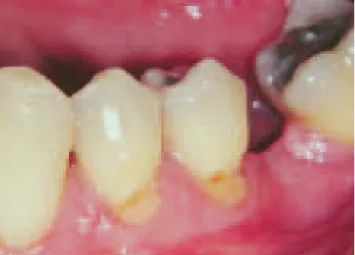
The patient, a 50-year old man, sought treatment due to the existence of non-carious cervical lesions in all the mandibular pre-molars that were causing him esthetic discomfort, as may be seen in Figure 1. The patient did not present a condition of hypersensitivity in any of these teeth,
did show a good condition of oral hygiene, conirmed by
the absence of carious lesions, existent restorations in good condition and good periodontal health.
The cervical lesions in teeth 34, 35, 44 and 45 showed
signiicant loss of enamel and dentin, indicating an initial
diagnosis of abfraction associated with wear caused by excessive tooth brushing. The degree of dentinal sclerosis
in all the pre-molars was classiied as type 3, which means
teeth with moderate amounts of dentinal sclerosis according to Swift Jr et al. (11).
The restorative proposal was to perform restorations of teeth
35 and 44 with the use of lowable composite resin as an
intermediate layer (Single Bond + Filtek Flow + Filtek Z250)
and of teeth 45 and 34 without the use of lowable composite
resin as a layer between the adhesive and microhybrid resin (Single Bond + Filtek Z250).
Restorative Procedure
The restorative procedures were as follows: anesthesia, cleaning with pumice stone and water with a rubber cup, followed by rinsing and drying; selection of the Filtek Z-250 microhybrid resin shade by means of the color scale provided by the manufacturer; all restorations were performed under absolute isolation. No additional retention or bevel was made. All materials used in the restorative procedures were applied in accordance with the manufacturer’s recommendations.
Restorations without Filtek-Flow: In teeth 44 and 36, the
Single Bond adhesive system was applied in the following way: a – acid etching (15 s); b – washing (15 s); c – drying with an air stream (30 s); d – re-wetting dentin with water (humid technique); e – a layer of adhesive was applied (10 s) by rubbing on the surface; f – air stream at a distance (20 s); g – application of another layer of adhesive by rubbing (10 s); h – air stream at a distance (20 s); i – light activation for 10 s with a VIP appliance at 600mW/cm2
(Bisco, Schaumburg, IL, USA). The lesions were illed with
Filtek Z-250 in increments (±3 increments). Each increment was light polymerized with the VIP halogen light appliance at 600 mW/cm2 (Bisco, Schaumburg, IL, USA) for 40 s.
Restorations with Filtek-Flow: Teeth 34 and 45 were
restored in a similar manner to teeth 44 and 35, with the exception that the FilteK-Flowable composite resin was used. After the adhesive was light activated, a thin layer of Filtek-Flowable composite resin (±1.5 mm) was inserted and light polymerized for 40 s.
The restorations were inished and polished with diamond
burs 1190F and 2135F (KG Sorensen, Barueri, SP, Brazil) with the aid of a spatula to protect the marginal gingiva (Fig. 2) and Sof-Lex Pop-On abrasive discs (3M ESPE, St. Paul, MN, USA).
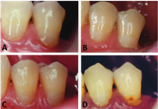
The patient was followed-up periodically, after 1 week, 18, 36 and 48 months (Fig. 3) and all the restorations were clinically evaluated in accordance with the USPHS criteria (United States Public Health Service – U.S. Public
Health Service) (12) modiied for the color criterion. The
following criteria were assessed: retention, anatomic shape, marginal discoloration, marginal desadaptation, secondary caries and post-operative sensitivity. The criterion for color combination used was that of Reusens et al. (13).
Fig. 2. Vestibular view of restoration of teeth 34 (restored with Single Bond + Filtek Z250, without Filtek-Flowable composite) and 35 (restored with Single Bond + Filtek Z250, with the use of Filtek-Flow as an intermediate layer). Note care taken when removing excesses in the cervical margin of the restoration.
In this criterion, the authors reclassiied Score A of the
USPHS system as A1 and A2. Traditionally, criterion A indicates that the restoration shows to be the same color as the tooth, and if there are differences in the color match and translucence between the restoration and the tooth, it is clinically acceptable. Reusens et al. (13) proposed the score of: A1 in which the restoration has an excellent color match to the point of not being perceptible, and A2 when the color match is good, but the difference with regard to the color between the tooth and the restoration is clinically perceptible.
Results
Up to 36 months of evaluation, there was no loss of retention, recurrent caries and loss of anatomic shape. After 18 months, the restorations began to present lack of marginal
adaptation classiied as “B”. The patient did not indicate
any spontaneous tooth sensitivity or sensitivity to an air stream.
All restorations showed a tendency to body discoloration, such as a type of yellowing, as from the period of 24 months (score A2). After 48 months, the restoration of tooth 35 was lost.
Discussion
Non-carious cervical lesions are considered the model for clinical evaluation of adhesive systems, in accordance with the recommendation of the ADA (6). To obtain partial approval from the ADA, the adhesive systems need to present less than 5% of marginal discoloration after six months, and
no loss of any restoration. Whereas to obtain inal approval,
the failure by loss of the restoration must not attain 10% after 18 months and less than 10% of the restorations may show marginal discolorations. The results of this clinical evaluation indicate that both groups tested would receive the seal of full approval.
One of the most important factors in the retention of non-carious cervical lesions is the bond to the cavity walls, since this type of cavity does not present any type of mechanical retention of the bond, it is provided exclusively by the adhesive system. Although a series of other factors may
directly inluence the retention of Class V restorations, such
as: occlusion; degree of dentinal sclerosis; and patient’s age (1,6), correct diagnosis is the most essential factor. It is well known that etiology of non-carious cervical lesions is of a multifactorial nature (1). Patients with a history of bruxism or clinical evidence of other forms of traumatic occlusion usually generate high occlusal stress on these
teeth. This creates an increase in lexure in the cervical
region due to the high occlusal stress that may result in the restoration falling out.
An alternative used to maximize retention rate of Class V restorations is using a material with a low modulus of elasticity. These materials can serve as type of cushion,
because they are lexible enough to resist the stresses
generated by polymerization shrinkage and facilitate the dissipation of these stresses produced by thermal variations, water sorption and occlusal load on the interface.
Some studies have shown an improvement in the performance of resin composite restorations when an additional layer of an intermediate material was placed between the resin composite and dentin substrate. Better dissipation of polymerization stresses (14), lower microleakage (14) and improved marginal adaptation (7) have been recorded.
As mentioned in the introduction, lowable composite
resins have a low modulus of elasticity (8) and thus they
may be used in cavities that undergo dental lexion (1). In addition, the low capacity of this material becomes a
desirable property due to good wetting, thus promoting better adaptation of the restorative material to the cavity walls.
However the nomenclature “lowable composite resin”
is used for materials of very different characteristics (8).
Flowable composite resins are more luid materials, and this
decrease in viscosity is always attributed to the reduction in the volume of inorganic material. In reality, the increase
in luidity may be achieved by modifying the monomer
composition of the material, not necessarily indicating a reduction in the modulus of elasticity of the material (8).
Nevertheless, recent indings have concluded that the use of a lowable composite resin as an intermediate layer did
not increase the retention rate of class V restorations after 12 and 24 months (15), indicating that other factors may be associated, irrespective of the presence of an intermediate layer absorbing stresses, to evaluate the retention rate of class V restorations.
Some studies have proposed the use of lowable composite
resin as the only material to restore non-carious cervical lesions (9). However, this technique has some disadvantages:
1) lowable composite resin present reduced mechanical
properties when compared with microparticle and microhybrid resins (9); 2) lower availability of shades to match the tooth structure (8); microparticle and microhybrid resins offer a variety of shades to restore dental elements
providing an excellent esthetic results; 3) it is more dificult to perform the sculpture of the restorations with lowable
composite resins (9); 4); the high organic content of the
lowable composite resins allow higher water sorption and
greater discoloration over time, as has been shown for the microparticle resins that have a higher organic content than the microhybrid resins (13,15).
Whereas in comparison with Adper Single Bond, the
48-month indings of the present case only conirm the good
results of this material in recent systematic review of literature (6)
It was concluded that: the use of Filtek-Flow as an intermediate layer did not improve clinical performance in comparison with restorations in which Filtek-Flowable composite resin was not used, after 48 months of clinical evaluation in non-carious cervical lesions. In the clinical case presented, the restoration that was lost was lined with
References
1. Levitch LC, Bader JD, Shugars DA, Heymann HO. Non-carious cervical lesions. J Dent 1994;22:195-207.
2. De Munck J, Van Landuyt K, Peumans M, Poitevin A, Lambrechts P, Braem M, et al. A critical review of the durability of adhesion to tooth tissue: methods and results J Dent Res 2005;84:118-32.
3. Browning WD, Brackett WW, Gilpatrick RO. Two-year clinical comparison of a microfilled and a hybrid resin based composite in non-carious class V lesions. Oper Dent 2000;25:46-50.
4. Baratieri LN, Canabarro S, Lopes GC, Ritter AV. Effect of resin viscosity and enamel beveling on the clinical performance of Class V composite restorations: Three-year results Oper Dent 2003;28:482-7. 5. Tay FR, Pashley DH. Resin bonding to cervical sclerotic dentin: A
review. J Dent 2004;32:173-96.
6. Peumans M, Kanumilli P, De Munck J, Van Landuyt K, Lambrechts P, Van Meerbeek B. Clinical effectiveness of contemporary adhesives: a systematic review of current clinical trials. Dent Mater 2005;21:864-81. 7. Heymann HO, Sturdevant JR, Bayne S, Wilder AD, Sluder TB, Brunson WD. Examining tooth flexure effects on cervical restorations: a two-year clinical study. J Am Dent Assoc 1991;122:41-7.
8. Reis A, Loguercio AD. Materiais Dentários Restauradores Diretos: dos fundamentos a aplicação clínica. Edt. Santos: São Paulo, 2007.
9. Bayne SC, Thompson JY, Swift Jr EJ, Ttamatiedes P, Wilkerson M. A characterization of first-generation flowable composites. J Am Dent Assoc 1998;129:567-77.
10. Unterbrink GL, Liebenberg WH. Flowable resin composites as “filled adhesives”: literature review and clinical recommendations. Quintessence Int 1999;30:249-57.
11. Swift Jr EJ, Perdigão J, Heymann HO, Wilder Jr AD, Bayne SC, May Jr KN, et al. Eighteen-month clinical evaluation of a filled and unfilled dentin adhesive. J Dent 2001;29:1-6.
12. Cvar JF, Ryge G. Reprint of criteria for the clinical evaluation of dental restorative materials. 1971. Clin Oral Investig 2005;9: 215-32.
13. Reusens B, D’hoore W, Vreven J. In vivo comparison of a microfilled and hybrid minifilled composite resin in class III restorations: 2-year follow-up. Clin Oral Invest 1999;3:62-9.
14. Choi KK, Condon JR, Ferracane JL. The effect of adhesive thickness on polymerization contraction stress of composite. J Dent Res 2000;79:812-7.