The aim of this study was to evaluate the efficacy of applying sonic energy on microtensile bond strength and microhardness after the restoration process. A total of 40 human third molars were extracted. Class II cavities were prepared and restored with composite SonicFill or Filtek Z350 XT with and without the application of sonic energy. After the teeth were stored in water for 24 h, the teeth were sectioned into sticks (1.0 mm2) and
subjected to tensile testing. For a depth Knoop hardness test, the samples were cut and indentations were made sequentially from the surface of the samples to the bottom of the samples in three intervals of 1 mm each. The samples were then subjected to a load of 50 g for 10 s. The results from the tensile (factors: placement system and composite) and hardness (factors: placement system, composite and depth) tests were subjected to the Kolmogorov-Smirnov normality test, followed by analysis of variance and Tukey’s test (5% significance). For the placement system factor, higher bond strength was observed for the cavities that were restored with sonic energy (p < 0.001). For depth Knoop hardness, the hardness at 1 mm depth was significantly greater than that at 3 mm depth just for the restorations with Filtek Z350 XT composite without the application of sonic energy. Therefore, the use of sonic energy during the restorative process improved bonding, yet it did not markedly affect the depth hardness for both composites.
Microtensile Bond Strength and
M i c r o h a r d n e s s o f C o m p o s i t e
R e s i n R e s t o r a t i o n s U s i n g a
Sonic-Resin Placement System
Victor Hugo Grandi1, Sandrine Bittencourt Berger1, Ana Paula Piovezan Fugolin2, Alcides Gonini-Júnior1, Murilo Baena Lopes1, Simonides Consani3, Ricardo Danil Guiraldo1
1Department of Restorative
Dentistry, School of Dentistry, UNOPAR – Universidade Norte do Paraná, Londrina, PR, Brazil
2Department of Restorative
Dentistry, Oregon Health & Science University, Portland, OR, USA
3Department of Restorative
Dentistry, Piracicaba Dental School, UNICAMP – Universidade Estadual de Campinas, Piracicaba, SP, Brazil
Correspondence: Ricardo Danil Guiraldo, Rua Marselha 183, 86041-140 Londrina, PR, Brazil. Tel: +55-43-3371-7820. e-mail: [email protected]
Key Words: composite resins, tensile strength, dentin; Knoop hardness.
Introduction
Resin composites have been extensively employed in restorative dentistry for several decades. In fact, more than five hundred million direct dental restorations are performed every year worldwide, thereby making it one of the most prevalent medical interventions for the human body (1). An incremental filling technique has been widely used for the placement of resin composite restorations (2). This technique consists of placing increments of resin-composite material in thickness of 2 mm or less followed by exposure to light curing from an occlusal direction and then repeating increments until the preparation is filled (3). Alternatively, bulk-fill techniques have the potential to substantially simplify restorative procedures and reduce chair time. For example, bulk-fill composites have been applied as a single increment up to 4 mm for class I and class II cavities, thus simplifying and reducing the clinical technique needed for bonded restorations (4).
The use of thicker increments for bulk-fill resin composites is possible due to advances in both photoinitiator dynamics and increased translucency of composite materials (5). The latter allows additional light to penetrate a composite, which allows a deeper cure to be achieved (6). Recently developed bulk-fill resin composites
have also exhibited reduced polymerization contraction stress and contraction rates compared with hybrid and flowable resin composites (7). However, a higher modulus of elasticity and increased plastic deformation of bulk-fill resin composites suggest that interfacial stress accumulation occurs, and this can lead to cuspal deflection and marginal gaps (6). Additionally, stress accumulation may be difficult to predict (7).
Restorations with Sonic placement system
ultrasonic energy to increase the flow of the compound and this provides better adaptation, less empty spaces, easier handling, and less clinical time (11). The objective of this study was to evaluate the application of ultrasonic energy to conventional and bulk-fill composites during restorations and to examine the resulting bond strength and hardness of the composites through microtensile and depth of hardness testing. The null hypotheses tested were that the use of ultrasonic energy does not interfere [1.] with the bond strength of the composites to tooth structure or [2.] with the hardness of the composites.
Material and Methods
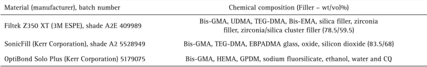
The restorative materials used for this study are described in Table 1.
Restorative Procedures
This study was approved by the Research Ethics Committee (protocol #1.345.279). A total of 40 non-restored, caries free human third molars were extracted. These molars also had no history of trauma, no bruxism, and no cracks. The molars were washed and stored in 0.1% thymol solution at 37 °C until they were used. The molars were used within three months of extraction. The root surface of each molar was embedded in Clássico acrylic resin (Clássico Dental Products, São Paulo, SP, Brazil) with the crown exposed (12).
Both mesial and distal surfaces of the class II cavities (6.0 mm wide x 2.0 mm deep x 4.0 mm tall) were prepared for each tooth with FG 1016HL spherical diamond burs (KG Sorensen, Cotia, SP, Brazil) in a high-speed hand piece (Dabi Atlante, Ribeirão Preto, SP, Brazil) that provided a copious spray of air and water. The preparations were finished with a FG 1092F finishing diamond bur (KG Sorensen). Each bur was replaced after five preparations. The inner angles of the cavities were rounded and the margins were not beveled. The molars were randomly divided into four groups (n = 10). Groups 1 and 2 included molars that were restored with SonicFill (Kerr Corporation) with and without the application of ultrasonic energy, respectively. Groups
3 and 4 included molars that underwent restorations with Filtek Z350 XT (3M ESPE, St. Paul, MN, USA) with and without the application of ultrasonic energy, respectively.
Each cavity was surrounded with a metal matrix band (Tofflemire Matrix Bands; Produits Dentaires SA, Vevey, Switzerland) before being incubated with dental conditioner gel (37% phosphoric acid; Dentsply, Petrópolis, RJ, Brazil) for 15 s. The cavities were then washed thoroughly with water and dried gently with air jets. OptiBond Solo Plus adhesive (Kerr Corporation) was applied for 15 s and then light cured for 20 s with a LED curing unit (Radii Cal; SDI, Bayswater, Victoria, VIC, Australia), according to the manufacturer’s directions. The output power (mW) of LED curing unit was measured with a power meter (Ophir Optronics Inc., Danvers, MA, USA) with a value of 538 mW. The light irradiance (mW/cm²) with a value of 1400 mW/cm2 was determined by dividing the output power by the tip area that was measured by calculating the area by formula πr2, where r was measured with a digital caliper (Mitutoyo, Tokyo, Japan) (13).
The composites were applied as follows: the Group 1 and Group 2 molars were restored with SonicFill composite that was applied in a single bulk increment with lightcuring for 20 s. The Group 2 molars using a Sonic-resin placement system. The Group 3 and Group 4 molars were restored with Filtek Z350 XT composite that was packed in empty dose tips and applied in two horizontal increments of 2 mm, with each increment lightcured for 20 s. The Group 4 molars using a Sonic-resin placement system. The same individual performed all of the restorations. After the restorations were completed, the molars were stored in distilled water for at 37 °C. After 24 h, the molars were subjected to microhardness and microtensile assays.
Microtensile Bond Strength Test
Specimens were sectioned perpendicular to the occlusal plane in the mesiodistal direction using the diamond saw (Isomet Diamond Wafering Blades; Buehler Ltd., Lake Bluff, IL, USA) of a cutting machine (Isomet 1000; Buehler Ltd.). The first section was removed and
Table 1. Restorative materials used in the study and composition according their manufacturers
Material (manufacturer), batch number Chemical composition (Filler – wt/vol%)
Filtek Z350 XT (3M ESPE), shade A2E 409989 Bis-GMA, UDMA, TEG-DMA, Bis-EMA, silica filler, zirconia
filler, zirconia/silica cluster filler (78.5/59.5)
SonicFill (Kerr Corporation), shade A2 5528949 Bis-GMA, TEG-DMA, EBPADMA glass, oxide, silicon dioxide (83.5/68)
OptiBond Solo Plus (Kerr Corporation) 5179075 Bis-GMA, HEMA, GPDM, sodium fluorsilicate, ethanol, water and CQ
V
. H. Grandi et al.
stored for microhardness testing at various depths. Briefly, cuts were made buccolingually with a stick to achieve a cross-sectional area of approximately 1.0 mm². The sticks were immersed in distilled water at 37 °C and were tested after 24 h.
Tensile testing was performed with a universal testing machine (EMIC DL2000; São José Dos Pinhais, PR, Brazil) under tension at 0.5 mm/min until failure occurred (14). The bonded surface area was calculated by using a digital caliper (Mitutoyo, Tokyo, Japan). Each stick was attached to the grips of a microtensile device with a cyanoacrylate resin (Super Glue; Henkel/Loctite, Westlake, OH, USA). The failure loads were recorded in newtons (N), and the
bond strength values were calculated in mega pascal (MPa) by dividing the failure load by the adhesive surface area (mm2). Fractured sticks were observed qualitatively under optical microscopy (Olympus Corp, Tokyo, Japan) at 40× magnification. Fractures were classified as cohesive (enamel or composite), adhesive (interface), or mixed (presence of composite and/or enamel in the same fragment). The percentages of the fracture modes and the percentage of specimens that were fractured before testing were recorded for each of the groups.
Hardness in Depth Test
Undisclosed sessions of the first cut were mounted in epoxy resin (20-8130-032; Buehler, Lake Bluff, IL, USA) and then were divided down the middle to expose the central region of each restoration. The specimens were flatted with SiC sandpapers (#600, #1200, #2000; Norton Abrasivos, Recife, PE, Brazil) to obtain polished and flattened surfaces. After the polishing step, all of the specimens were placed in an ultrasonic washer (Ultra Cleaner 1400; Unique, Indaiatuba, SP, Brazil) for 10 min to remove debris. Indentations were sequentially made using a hardness testing machine (HMV-G; Shimadzu, Kyoto, Japan). Three readings were taken from the top to bottom surfaces at 1 mm intervals under a load of 50 g for a dwell time of 10 s. The Knoop hardness for each depth was recorded as the average of three indentations made at the same depth.
Statistical Analysis
Statistical analyses were performed by using Minitab 16 for Windows 8 (Minitab, State College, PA, USA). Distributions of the measurements made were investigated with the Kolmogorov-Smirnov normality test, followed by parametric tests. Data for microtensile values (factors: placement system and composite) and hardness (factors: placement system, composite and depth) were evaluated statistically with analysis of variance (ANOVA), followed by Tukey’s test with a significance level of 5% (α= 0.05).
Results
Table 2 shows the percentages of premature failures that occurred for each experimental condition. Adhesive and mixed fractures were prevalent in all of the groups, indicating that the dentin/adhesive interfaces were tested at tensile.
Table 2. Premature failures that occurred in each group according to the fracture pattern for each experimental condition
Composite Application
of ultrasound
Premature failures (%)
Adhesive Mixed Cohesive
SonicFill Kerr
Yes 80 10 10
No 87.5 7.5 5
Filtek Z350 XT
Yes 80.5 13.0 6.5
No 79 12 9
Table 3. Mean microtensile bond strength values (MPa) for the composites with and without application of ultrasound
Composite
Technique
p value
With ultrasonic energy Without ultrasonic energy
SonicFill Kerr 35.01 (2.84) A 26.81 (4.70) B
<0.001
Filtek Z350 XT 33.17 (4.96) A 25.08 (5.80) B
p value 0.383
Statistically significant differences in the mean values at a 5% level according to Tukey’s test are indicated with different letters in each row. Standard deviation values are in parentheses.
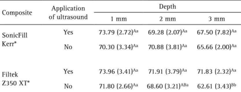
Table 4. Mean Knoop hardness values for the composites with and without sonic placement activation at different depths.
Composite Application
of ultrasound
Depth
1 mm 2 mm 3 mm
SonicFill Kerr*
Yes 73.79 (2.72)Aa 69.28 (2.07)Aa 67.50 (7.82)Aa
No 70.30 (3.34)Aa 70.88 (3.81)Aa 65.66 (2.00)Aa
Filtek Z350 XT*
Yes 73.96 (3.41)Aa 71.91 (3.79)Aa 71.83 (2.32)Aa
No 71.80 (2.66)Aa 68.60 (3.21)ABa 62.61 (3.43)Bb
Restorations with Sonic placement system
Microtensile bond strength data are presented in Table 3. There was no interaction between the type of factor activation and the composite used (p=0.977). Additionally, there were no statistically significant differences for the composite independent factor (p=0.383). However, the mean microtensile strength of the cavities that were restored with the application of ultrasonic energy was significantly higher than the hollows that were restored without ultrasonic energy (p<0.001).
Knoop hardness data at various depths are reported in Table 4. Interactions among the composites, techniques, and depth are indicated (p=0.021). For the Filtek Z350 XT composite, the hardness detected at a depth of 1 mm was more significant than the hardness detected at a depth of 3 mm. In contrast, at depths of 1 mm and 2 mm, there were no statistically significant differences between the composites and the different techniques.
Discussion
Composite resin has rapidly been replacing amalgam as the posterior restorative material of choice for many dental patients (15,16). However, the application of composite resin can involve a very complex and challenging procedure due to many material and clinical considerations (15,16). A goal of the present study was to assess the microtensile bond strength and hardness of a recently developed sonic-resin placement system. Despite being an in vitro study, careful attention was accorded to simulating a realistic clinical environment. Specifically, the aim of this study was to determine if use of the SonicFill composite resin system using an incremental-fill insertion technique had an effect on microtensile bond strength and depth of hardness in Class II cavities that was comparable to that of a universal, nanoparticle-filled composite, Filtek Z350 XT.
In many Class II cavity preparations, it is difficult to obtain proper contouring and adequate proximal contacts due to the lack of packing that is achieved with a composite resin (11). Thus, the flow of a composite material may play a major role in the ultimate success of a restoration (17). The need for composite resins to have certain flow characteristics has been addressed with the introduction of packable and more fluid composite resins. Packable composite resins were first introduced as an alternative to amalgam (11). They are characterized by a high filler load and a filler distribution that produces a consistency distinct from that of traditional composite resins. Regarding flow, composite resins that contain a lower concentration of filler are often characterized by a lower elastic modulus and viscosity (18). For the average clinician, the ideal composite resin material is sufficiently viscous to facilitate placement, yet has sufficient flow to achieve adequate marginal adaptation (19).
V
. H. Grandi et al.
adequate consistency between microtensile bond strength studies and the desire to clearly understand the correlation between a particular bond strength test and clinical performance have prompted recommendations to clarify the stick details (22,23). The current study show failures to bonding interfaces with fewer incidences of cohesive failures. Relevant fact for the credibility of the study.
It has been proposed that examining the microhardness of a composite at various depths is adequate for evaluating the depth of curing that occurs (7). A ratio > 0.80 for bottom-to-top microhardness has been reported to indicate an appropriate depth of cure (24). In the present study, all of the materials tested had a microhardness that exceeded 80% following polymerization. Using the ISO 4049 standard (25), the average depth of cure reported for SonicFill was 3.67 mm (11). Other studies reported similar depths of cure (3.46 mm and 3.43 mm, respectively) when the same ISO 4049 standard (25) was used (26,27). However, other studies have concluded that the ISO 4049 method (25) may overestimate the depth of curing that occurs compared to other techniques such as hardness or degree of conversion tests (27,28). To date, data regarding the depth of cure achieved with bulk-fill composite resin restorative materials with ultrasonic activation are limited. Therefore, hardness test to measure the depth of cure for bulk-fill versus conventional composites with ultrasonic activation was performed in the present study. The results showed that when ultrasonic waves were applied, an adequate depth of cure was achieved with both composites.
Depth of cure is dependent on the amount of light energy that is able to pass through resin-based composite specimens from scattered and absorbed light (29). Since dental resin-based composites consist of heterogeneous substances, including both resin and fillers, light is scattered at the resin-filler interface according to differences in the refractive indices of the individual compounds (29). Correspondingly, light transmittance in dental resin-based composites has been shown to decrease with increased filler content and for irregular filler shapes as a result of an increased specific surface area between the fillers and resin (30). Furthermore, for filler sizes ranging from 0.05–2 μm it has been demonstrated that reduced light transmission occurs due to the inability of the particles that are smaller than the wavelength of incident blue light to scatter the blue light (31). An additional aspect regarding the transmission of light through resin-based composites is the treatment of fillers (29). Silane-coated fillers have been found to enhance light transmission (29), while uncoated fillers decrease light transmission, due to the formation of a gap at the resin-filler interface during polymerization (29). Volumetric shrinkage that occurs during polymerization also reduces the optical path length, which according to
the Lambert-Beer law, increases light transmittance (32). However, in the present study, there was no statistically significant difference between the two composites that were examined.
Bulk-fill composites, such as Sonic Fill Kerr, have also been developed to reduce placement time and simplify the procedure. These materials are designed to be placed in 4 mm thick increments, without negatively affecting the mechanical and physical properties (33). The introduction of these new resin composites allows for an alteration in the restorative technique. Incremental layering has long been accepted as a standard technique for placement of resin-composite in cavity preparations (3) and it was used to Filtek Z 350 XT composite. The study limitation was that each composite was used as the technique recommended by its manufacturer, only adding the use of sonic activation to Filtek Z 350 XT composite. Thus, our results are inconsistent with first null hypothesis because a difference in the bond strength of the composites to the tooth structure was observed in the presence versus the absence of ultrasonic energy. In contrast, second null hypothesis was supported by the present results whereby hardness was not affected by the application of ultrasonic energy. Therefore, the following conclusions can be drawn: 1) The use of ultrasonic energy during the restorative process lead to greater bond strength to microtensile for both of the composites that were evaluated; 2) The use of ultrasonic energy during the restoring process generally resulted in similar hardness values for both of the composites.
Resumo
O objetivo deste estudo foi avaliar a eficácia da aplicação de energia sônica sobre a resistência de união à microtração e a microdureza após o processo de restauração. Um total de 40 terceiros molares humanos foram extraídos. Cavidades Classe II foram preparadas e restauradas com os compósitos SonicFill ou Filtek Z350 XT com e sem a aplicação de energia sônica. Após os dentes serem armazenados em água durante 24 h, foram seccionados em palitos (1,0 mm2) e submetidos a ensaio de
tração. Para um ensaio de dureza Knoop de profundidade, as amostras foram cortadas e as penetrações foram feitas sequencialmente a partir da superfície para o fundo das amostras em três intervalos de 1 mm cada. As amostras foram então submetidas a uma carga de 50 g durante 10 s. Os resultados dos testes de tração (fatores: sistema de inserção e compósito) e dureza (fatores: sistema de inserção x compósito x profundidade) foram submetidos ao teste de normalidade Kolmogorov-Smirnov, seguido da análise de variância e do teste de Tukey (significância de 5%). Para o fator sistema de inserção, observou-se maior resistência de união para as cavidades que foram restauradas com energia sônica (p < 0,001). Para a dureza Knoop de profundidade, a dureza a 1 mm de profundidade foi significativamente maior do que a profundidade de 3 mm apenas para as restaurações com o compósito Filtek Z350 XT sem a aplicação de energia sônica. Portanto, o uso de energia sônica durante o processo restaurador melhorou a união, mas não afetou acentuadamente a dureza de profundidade para ambos os compósitos.
References
Restorations with Sonic placement system
restorations - a meta-analysis. J Adhes Dent 2012;14:407-431. 2. Soares CJ, Bicalho AA, Tantbirojn D, Versluis A. Polymerization shrinkage
stresses in a premolar restored with different composite resins and different incremental techniques. J Adhes Dent 2013;15:341-350. 3. El-Safty S, Silikas N, Watts DC. Creep deformation of restorative
resin-composites intended for bulk-fill placement. Dent Mater 2012;28:928-935.
4. Orłowski M, Tarczydło B, Chałas R. Evaluation of marginal integrity of four bulk-fill dental composite materials: in vitro study. ScientificWorldJournal 2015;2015:701262.
5. Lassila LV, Nagas E, Vallittu PK, Garoushi S. Translucency of flowable bulk-filling composites of various thicknesses. Chin J Dent Res 2012;15:31-35.
6. Flury S, Hayoz S, Peutzfeldt A, Husler J, Lussi A. Depth of cure of resin composites: Is the ISO 4049 method suitable for bulk fill materials? Dent Mater 2012;28:521-528.
7. Ilie N, Hickel R. Investigations on a methacrylate- based flowable composite based on the SDR technology. Dent Mater 2011;27:348-355. 8. Ferracane JL. Correlation between hardness and degree conversion
during the setting reaction of unfilled dental restorative resins. Dent Mater 1985;1:11-14.
9. Asmussen E. Composite restorative resins: composition versus wallto-wall polymerization contraction. Acta Odontol Scand 1975;33:337-344.
10. Guiraldo RD, Consani S, Consani RL, Berger SB, Mendes WB, Sinhoreti MA, et al.. Comparison of silorane and methacrylate-based composite resins on the curing light transmission. Braz Dent J 2010;21:538-542. 11. Ibarra ET, Lien W, Casey J, Dixon SA, Vandewalle KS. Physical properties
of a new sonically placed composite resin restorative material. Gen Dent 2015;63:51-56.
12. Mushashe AM, Gonzaga CC, Cunha LF, Furuse AY, Moro A, Correr GM. Effect of Enamel and Dentin Surface Treatment on the Self-Adhesive Resin Cement Bond Strength. Braz Dent J 2016;27:537-542. 13. Segreto DR, Naufel FS, Brandt WC, Guiraldo RD, Correr-Sobrinho L,
Sinhoreti MA. Influence of photoinitiator and light-curing source on bond strength of experimental resin cements to dentin. Braz Dent J 2016;27:83-89.
14. Correa BC, Galo R, Scatena C, Borsatto MC, Spazzin AO, Corona SA, et al.. Effect of Metalloproteinase Inhibitors on the Microtensile Bond Strength of Composite Resin to Er:YAG Laser-Irradiated Dentin. Braz Dent J 2016;27:442-445.
15. Ferracane JL. Resin composite--State of the art. Dent Mater 2011;27:29-38.
16. Kalmowicz J, Phebus JG, Owens BM, Johnson WW, King GT. Microleakage of class I and II composite resin restorations using a sonic-resin placement system. Oper Dent 2015;40:653-661.
17. Ferracane JL, Moser JB, Greener EH. Rheology of composite restoratives. J Dent Res 1981;60:1678-1685.
18. Tanimoto Y, Nishiwaki T, Nemoto K. Dynamic viscoelastic behavior of
dental composites measured by Split Hopkinson pressure bar. Dent Mater J 2006;25:234-240.
19. Opdam NJ, Roeters JJ, Peters TC, Burgersdijk RC, Kuijs RH. Consistency of resin composites for posterior use. Dent Mater 1996;12:350-354. 20. Ilie N, Bucuta S, Draenert M. Bulk-fill resin-based composites: an
in vitro assessment of their mechanical performance. Oper Dent 2013;38:618-625.
21. Al-Harbi F, Kaisarly D, Michna A, ArRejaie A, Bader D, El Gezawi M. Cervical interfacial bonding effectiveness of class ii bulk versus incremental fill resin composite restorations. Oper Dent 2015;40:622-635.
22. Eckert GJ1, Platt JA. A statistical evaluation of microtensile bond strength methodology for dental adhesives. Dent Mater 2007;23:385-391.
23. Armstrong S, Geraldeli S, Maia R, Raposo LH, Soares CJ, Yamagawa J. Adhesion to tooth structure: a critical review of “micro” bond strength test methods. Dent Mater 2010;26:e50-62.
24. Bouschlicher MR, Rueggeberg FA, Wilson BM. Correlation of bottom-to-top surface microhardness and conversion ratios for a variety of resin composite compositions. Oper Dent 2004;29:698-704.
25. ISO 4049 “Dentistry: polymer-based restorative materials” Geneva Switzerland, 2009.
26. Garcia D, Yaman P, Dennison J, Neiva GF. Polymerization shrinkage and depth of cure of bulk-fill flowable composite resins. Oper Dent 2014;39:441-448.
27. Benetti AR, Havndrup-Pedersen C, Honore D, Pedersen MK, Pallesen U. Bulk-fill Resin composites: polymerization contraction, depth of cure, and gap formation. Oper Dent 2014;40:190-200.
28. Moore BK, Platt JA, Borgess G, Chu TG, Katsilieri I. Depth of cure of dental resin composites: ISO 4049 depth and microhardness of types of materials and shades. Oper Dent 2008;33:408-412.
29. Bucuta S, Ilie N. Light transmittance and micro-mechanical properties of bulk fill vs. conventional resin based composites. Clin Oral Invest 2014;18;1991-2000.
30. Arikawa H, Kanie T, Fujii K, Takahashi H, Ban S. Effect of filler properties in composite resins on light transmittance characteristics and color. Dent Mater J 2007;26:38-44.
31. Fujita K, Ikemi T, Nishiyama N. Effects of particle size of silica filler on polymerization conversion in a light-curing resin composite. Dent Mater 2011;27:1079-1085.
32. Feng L, Suh BI, Shortall AC. Formation of gaps at the filler-resin interface induced by polymerization contraction stress: gaps at the interface. Dent Mater 2010;26:719-729.
33. Czasch P, Ilie N. In vitro comparison of mechanical properties and degree of cure of bulk fill composites. Clin Oral Investig 2013;17:227-235.