Severe lower urinary tract symptoms due to anteriorly
located midline prostatic cyst arising from the bladder
neck in a young male: case report
Sintomas graves do trato urinário inferior em decorrência de cisto anteriormente
localizado na linha mediana da próstata proveniente do colo vesical em um jovem
do sexo masculino: relato de caso
Ali Gürağaç
I, Zafer Demirer
II, Bilal Fırat Alp
III, Emin Aydur
IIIDepartment of Urology, School of Medicine, Gulhane Military Medical Academy, Ankara, Turkey
ABSTRACT
CONTEXT: Prostatic cysts are uncommon. These cysts are usually asymptomatic and are diagnosed incidentally during ultrasonographic examination. On rare occasions, they may cause drastic symptoms.
CASE REPORT: We report on a case of severely symptomatic anteriorly located prostatic cyst arising from the bladder neck in a 30-year-old man presenting with lower urinary tract symptoms, without clinical evi-dence of benign prostatic hyperplasia. Transrectal ultrasonography (TRUS), computed tomography (CT) and cystourethroscopy demonstrated a projecting prostatic cyst that occupied the bladder neck at the precise twelve o’clock position. It was acting as a ball-valve, such that it obstructed the bladder outlet. Transurethral unrooing of the cyst was performed and the patient’s obstructive symptoms were success-fully resolved. Histopathological examination indicated a retention cyst.
CONCLUSIONS: It should be borne in mind that midline prostate cysts can be a reason for bladder outlet obstruction in a young male. Such patients may have tremendous improvement in symptoms through transurethral unrooing of the cyst wall.
RESUMO
CONTEXTO: Cistos prostáticos são incomuns. Esses cistos são geralmente assintomáticos e são diagnostica-dos incidentalmente durante o exame ultrassonográico. Raramente podem causar sintomas importantes.
RELATO DE CASO: Relatamos um caso sintomático de grave cisto prostático de localização anterior, origi-nário do colo da bexiga de um homem de 30 anos de idade, que apresentou sintomas do trato uriorigi-nário in-ferior, sem evidência clínica de hiperplasia prostática benigna. Ultrassonograia transretal (TRUS), tomograia computadorizada (CT) e cistouretroscopia demonstraram um cisto prostático saliente que ocupou o colo da bexiga na posição exata de 12 horas. O cisto estava agindo como uma válvula de esfera, obstruindo a saída da bexiga. Retirada da cobertura do cisto foi realizada por via transuretral e os sintomas obstrutivos do paciente foram resolvidos com sucesso. O exame histopatológico indicou um cisto de retenção.
CONCLUSÕES: Deve ser lembrado que a linha média do cisto de próstata pode ser motivo de obstrução da saída da bexiga em um jovem do sexo masculino. Esses pacientes podem ter notável melhoria nos sintomas com retirada da cobertura por via transuretral da parede do cisto.
IMD. Specialist, Department of Urology, Tatvan Military Hospital, Bitlis, Turkey.
IIMD. Specialist, Department of Urology, Eskişehir Military Hospital, Eskişehir, Turkey.
IIIMD. Associate Professor, Department of Urology, School of Medicine, Gulhane Military Medical Academy, Ankara, Turkey.
KEY WORDS: Cysts. Prostate. Prostatic diseases. Urinary bladder.
Urinary bladder neck obstruction.
PALAVRAS-CHAVE: Cistos.
Próstata.
Doenças prostáticas. Bexiga urinária.
INTRODUCTION
Over recent years, the widespread availability of transrectal ultrasound (TRUS), computed tomography (CT) and magnetic resonance imaging (MRI) has led to increased frequency of diagnoses of incidental prostatic cysts. he majority of prostatic cysts are asymptomatic and originate in the posterior area of the prostate, such as in the Müllerian ducts and the utricle, as an embryological remnant; these cysts are observed in 0.5% to 7.9% of patients.1,2 However, these improved imaging techniques
have increased the incidental determination of midline prostatic cysts (MPCs) in adult males, and the frequency of these indings is currently estimated to be 5-14%.3
Although the majority of the patients are symptom-free, enlarged prostatic cysts can compress adjacent structures, such as the posterior urethra, bladder neck or seminal ves-icles, and then the patients may suffer obstructive or irrita-tive voiding symptoms, recurrent urinary tract infections, epi-didymitis, chronic pelvic pain syndrome, hematospermia, low semen volume, or even infertility.2-4 Prostatic retention cysts
rarely become symptomatic, but they may cause symptoms when the cyst enlarges to more than 3 cm. However, symp-toms may occur even with smaller cysts if the location is just beside the bladder neck, and such cases are often misdiag-nosed or confused with benign prostatic hyperplasia (BPH) or neuropathic bladder.2-5
We report on the case of a severely symptomatic, anteri-orly located prostatic cyst arising from the bladder neck in a 30-year-old man who presented with lower urinary tract symptoms (LUTS), without any clinical evidence of benign prostatic hyperplasia or any endoscopic management. To our knowledge, symptomatic MPCs are generally located poste-riorly and are rare. In fact, there are seven published reports of anteriorly positioned symptomatic MPCs arising from and obstructing the bladder neck with a ball-valve action during voiding.6-12
CASE REPORT
A 30-year-old healthy man came to our outpatient clinic with a two-year history of severe LUTS, including frequent void-ing, hesitancy, weak urinary stream and the sensation of resid-ual urine. Despite alpha blocker drug treatment, his symp-toms had worsened. His medical history was not significant in terms of previous urethral catheterization, urinary tract infec-tion, pelvic/perineal trauma or neurological deficit. He had two children and did not have any ejaculatory complaints or infertility.
His International Prostate Symptom Score (IPSS) was 20, and his quality-of-life (QoL) score was 5. Digital rectal examination revealed a normal firm and nontender prostate
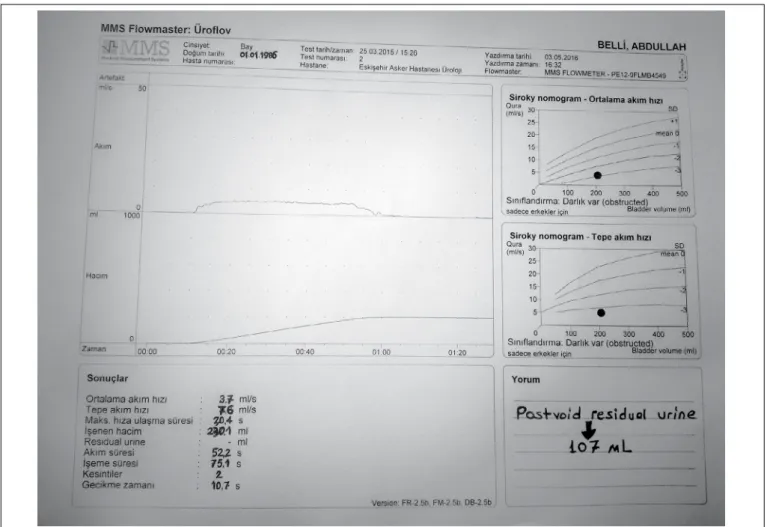
without palpable nodules. A routine urine examination was normal and the culture was sterile. The urine cytology was not suggestive of malignancy, the serum prostatic specific antigen (PSA) level was 0.82 ng/ml (the reference value for PSA for the age range of 30-40 years is 0-2 ng/ml), and routine biochemical laboratory examinations were within normal limits. Uroflowmetry (Figure 1) demonstrated a peak flow rate (Qmax) of 7.6 ml/sec with a voided volume of 230 ml, and the postvoid residual urine volume was 107 ml (the reference values are voided volume > 150 ml and Qmax >15 ml/s). TRUS showed an anteriorly positioned prostatic cyst arising from the bladder neck and obstructing the bladder outlet. The cyst diameter was approximately 13 x 10 mm, inside a prostate with a volume of 22 ml. CT urography was performed to identify the origin of the cystic lesion and to rule out ectopic ureter or ureterocele (Figure 2).
he patient then underwent cystourethroscopy examina-tion under general anesthesia. his showed that the posterior wall of the prostatic urethra was normal and that there was cys-tic hemispherical bulging based on the anterior portion of the prostate. he bulge was located on the bladder neck at precisely twelve o’clock and was entirely compressing the bladder outlet. It was acting like a ball-valve, without lateral lobe hyperplasia
(Figure 3). Moderate trabeculation due to gross back pressure
change was also noted in the bladder. Transurethral marsupial-ization of the prostatic cyst to release the anatomical obstruction was performed, and milky luid was expelled during the unroof-ing procedure.
A urethral catheter was let in place for two days, and the patient was then discharged. Histopathological examination revealed that the cyst wall was lined with benign lattened pros-tatic glandular epithelium without any preneoplastic change, which was consistent with the diagnosis of a prostatic reten-tion cyst. At a return visit in the third postoperative month, a subjective dramatic improvement in symptoms was noted. IPSS was 5 and QoL was 2 at three months ater the operation. Urolowmetry showed an increased Qmax (18 ml/sec with a voided volume of 300 ml) and no residual urine. Furthermore, the patient had no symptoms suggestive of erectile dysfunction or ejaculation disorders.
DISCUSSION
Figure 1. Preoperative urolowmetry showing obstructed voiding.
Urolowmetry demonstrating average low rate: 3.7 ml/sec; peak low rate (Qmax): 7.6 ml/sec; time to reach maximum low: 30.4 sec; voided volume: 230.1 ml; postvoid residual urine: 107 ml; low time: 52.2 sec; voiding time: 75.1 sec; deductions: 2; delay time: 10.7 sec.
Figure 2. Sagittal (A) and axial (B) computed tomography urography images showing low-density small nodular lesion in the bladder neck that represents the anteriorly positioned midline prostatic cyst (arrow).
Figure 3. (A) and (B): anteriorly positioned midline prostatic cyst obstructing the bladder neck with action like a ball-valve; (C) and (D): transurethral unrooing of the cyst.
A
C
B
D
Galosi et al. reported that MPCs are seen by TRUS in 9.8% of cases.1 It has been reported that MPCs were observed in
7.6% of healthy men and 5% of symptomatic outpatients.2 Over
recent years, new imaging techniques such as TRUS, CT and MRI have increased the incidental determination of MPCs in adult males, and their frequency is currently estimated to be 5-14%.3,13,14
However, the majority of prostatic cysts are asymptom-atic. They can be categorized as symptomatic when the cyst’s presence is accompanied by infection or if its size and ana-tomical relationships affect the adjacent structures, which are most often located laterally.14 An analysis on 34 patients with
symptomatic prostatic cysts by Tambo et al.7 revealed that
40% of the patients suffered from obstructive urinary symp-toms, 33% from urinary retention, 9% from urodynia and 6% from infertility.
In 2009, Galosi et al.1 classiied prostatic cysts into six distinct
types based on TRUS and pathological correlation: isolated medial cysts, cysts of the ejaculatory duct, simple or multiple parenchymal
cysts, complicated cysts (infectious or hemorrhagic), cystic tumors and secondary cysts relating to parasitic disease. MPCs are less common and are generally located posteriorly. hey have traditionally been classiied as Müllerian duct cysts and as enlarged prostatic utricles (mega-utricles), ejaculatory ducts, seminal vesicles and prostatic retention cysts.15,16 Furuya et al.17 classiied
MPCs, with or without the presence of sperm in the luid content, using concomitant TRUS-guided opaciication and dye injection. If there is communication with the seminal tract, then sperm can be found in the luid content. hey further classiied MPCs into four categories: Type 1 MPC with no communication into the urethra (traditional prostatic utricle cyst); Type 2a MPC with no communication into the urethra [cystic dilatation of prostatic utricle (CDU)]; Type 2b CDU in communication with the seminal tract; and Type 3 cystic dilatation of the ejaculatory duct. hey also found that the location, shape and volume of the MPC and the PSA level of the MPC luid did not inluence the classiication.18 his
Table 1. Search of the literature in medical databases for case reports on “Severe lower urinary tract symptoms due to anteriorly located midline prostatic cyst arising from the bladder neck in a young male”. The search was conducted on April 12, 2016
Database Search strategies Papers found Related papers
MEDLINE (via PubMed)
((Prostatic cyst[Title]) AND lower urinary tract symptoms[Title]) 2 2 ((Prostatic cyst[Title]) AND midline[Title]) AND “case reports”[Publication Type] 3 1
((Prostatic cyst[Title]) AND midline[Title]) 7 2
Embase (via Elsevier) ((“Prostate”) or “Prostatic”) AND “Cysts’” [Title Words] AND “case reports” [Publication Type] 0 0 LILACS (via Bireme) ((“Prostate”) or “Prostatic”) AND “Cysts’” [Title Words] AND “case reports” [Publication Type] 2 0
because of the similarities of symptoms and primary treatment among prostate cysts.
According to the results from the pathological examina-tion, our patient had a retention cyst of the prostate. his type of cyst is totally diferent from Müllerian duct cysts and pros-tatic utricle cysts, which are always lined with cuboidal or columnar epithelial cells. Retention cysts of the prostate gland are true acquired cysts and result from obstruction of pros-tatic glandular ductules, thus causing dilatation of the glan-dular acini. hey can be located within any glanglan-dular zone of the prostate.16,17 Moreover, there are no sperm cells in the luid
obtained from prostatic retention cysts, and they usually do not cause symptoms. However, on rare occasions, they may cause obstructive symptoms if located close to the bladder neck, as in the case of our patient.16,17
Although approximately 35 patients with symptomatic pros-tatic cysts have been reported, there are only seven published reports on anteriorly located MPCs.11 Furthermore, to the best
of our knowledge, only seven such cases of MPC of the bladder neck have been reported in the literature, as in our case. Also, this is the irst case of an anteriorly located MPC of the bladder neck found in Turkey.7,11
Multiple therapeutic options have been described for man-agement of symptomatic prostatic cysts, including transrec-tal aspiration with or without sclerotherapy, marsupialization with a transurethral technique and open surgery.7,18,19 Although
recurrences of cysts that were incompletely excised during open surgery have been reported, recurrence-free results have been reported for medial prostatic cysts treated with the transure-thral technique.2,6 Chang et al.10 previously reported successful
results from transurethral resection of an MPC presenting with LUTS. Zhang et al.20 also recommended transurethral
unroof-ing of small prostatic cysts (< 2 cm x 2 cm) that are close to the bladder cavity or urethra, because of the simplicity of the tech-nique, the low risk of complications and the shorter convales-cence period.
We searched for similar cases in diferent databases (PubMed, Embase and LILACS databases) using the terms: “prostatic cyst” AND “lower urinary tract symptoms” AND “midline” (Table 1). We found that few cases have been published.
Our patient was sufering from obstructive voiding problems rather than irritative symptoms, and transurethral unrooing of the prostatic cyst, which was located anteriorly in the midline position, provided satisfactory results for his complaints. In this case, standard transurethral resection of the prostate was avoided in order to prevent antegrade ejaculation and erectile dysfunc-tion, in the absence of the lateral lobe of prostatic hyperplasia.
CONCLUSION
Although symptomatic anteriorly located MPCs of the bladder neck, as described in the case presented here, are uncommon, they should be borne in mind in the diferential diagnosis of obstructive voiding symptoms, especially in young patients and patients who do not respond to medical therapy such as use of alpha-blockers. Transurethral unrooing of the cyst may pro-vide safe treatment with successful and satisfactory results in selected cases.
REFERENCES
1. Galosi AB, Montironi R, Fabiani A, et al. Cystic lesions of the prostate
gland: an ultrasound classiication with pathological correlation. J
Urol. 2009;181(2):647-57.
2. Dik P, Lock TM, Schrier BP, Zeijlemaker BY, Boon TA. Transurethral
marsupialization of a medial prostatic cyst in patients with
prostatitis-like symptoms. J Urol. 1996;155(4):1301-4.
3. Furuya S, Hisasue S, Kato H, Shimamura S. Novel insight for midline
cyst formation in prostate: The involvement of decreased prenatal
testosterone suggested by second-to-fourth digit ratio study. Int J
Urol. 2015;22(11):1063-7.
4. Mayersak JS. Urogenital sinus-ejaculatory duct cyst: a case report
with a proposed clinical classiication and review of the literature. J
Urol. 1989;142(5):1330-2.
5. Pillai RG, Al Naieb ZA. Successful Endoscopic Laser De-rooing
of Simple Prostatic Cyst Causing Bladder Outlet Obstruction -
A Case Study. Med Surg Urol. 2013;2(1). Available from: http://
www.omicsonline.org/successful-endoscopic-laser-de-rooing-of-
simple-prostatic-cyst-causing-bladder-outlet-obstruction-a-case-study-2168-9857.1000107.pdf. Accessed in 2016 (Jul 5).
6. Nayyar R, Dogra PN. Anteriorly placed midline intraprostatic cyst. J
7. Tambo M, Okegawa T, Nutahara K, Higashihara E. Prostatic cyst arising
around the bladder neck-cause of bladder outlet obstruction: two
case reports. Hinyokika Kiyo. 2007;53(6):401-4.
8. Issa MM, Kalish J, Petros JA. Clinical features and management of
anterior intraurethral prostatic cyst. Urology. 1999;54(5):923.
9. Yildirim I, Kibar Y, Sümer F, et al. Intraurethral prostatic cyst:
a rare cause of infravesical obstruction. Int Urol Nephrol.
2003;35(3):355-6.
10. Chang SG, Hwang IC, Lee JH, Park YK, Lim JW. Infravesical obstruction
due to benign intraurethral prostatic cyst. J Korean Med Sci.
2003;18(1):125-6.
11. Lee JY, Kang DH, Park HY, et al. An anteriorly positioned midline
prostatic cyst resulting in lower urinary tract symptoms. Int Neurourol
J. 2010;14(2):125-9.
12. Diaz RR, Lee JY, Choi YD, Cho KS. Unroofed midline prostate cyst
misled into a stricture with obliterative bladder neck contracture
following a laser prostatectomy. Int Neurourol J. 2013;17(1):34-7.
13. Ishikawa M, Okabe H, Oya T, et al. Midline prostatic cysts in healthy
men: incidence and transabdominal sonographic indings. AJR Am J
Roentgenol. 2003;181(6):1669-72.
14. Nayyar R, Wadhwa P, Dogra PN. Midline intraprostatic cyst: An
unusual cause of lower urinary tract symptoms. Indian J Urol.
2008;24(1):109-11.
15. Juárez Soto A, Ribè Subirà N, Manasia P, et al. Classiication of cystic
structures located at the midline of the prostate: our experience.
Arch Ital Urol Androl. 2004;76(2):75-9.
16. Nghiem HT, Kellman GM, Sandberg SA, Craig BM. Cystic lesions of the
prostate. Radiographics. 1990;10(4):635-50.
17. Furuya R, Furuya S, Kato H, et al. New classification of midline
cysts of the prostate in adults via a transrectal
ultrasonography-guided opacification and dye-injection study. BJU Int.
2008;102(4):475-8.
18. Stricker HJ, Kunin JR, Faerber GJ. Congenital prostatic cyst causing
ejaculatory duct obstruction: management by transrectal cyst
aspiration. J Urol. 1993;149(5):1141-3.
19. Saito S. Transrectal ultrasound-guided puncture, drainage, and
minocycline hydrochloride sclerotherapy for the symptomatic
prostatic cyst. J Endourol. 2002;16(9):693-5.
20. Zhang H, Qi F, Wang J, Chen M, Li Z, Zu X. Midline Prostatic Cysts
Presenting with Chronic Prostatitis or Secondary Infertility and
Minimally Invasive Treatment: Endoscopic or Laparoscopic Approach?
Surgical Science. 2011;2:285-9. Available from: http://ile.scirp.org/pdf/
SS20110500013_35824482.pdf. Accessed in 2016 (Jun 8).
Sources of funding: None
Conlict of Interest: None
Date of irst submission: March 15, 2016
Last received: May 17, 2016
Accepted: May 28, 2016
Address for correspondence:
Zafer Demirer
Eskişehir Military Hospital, Department of Urology, Eskişehir, Turkey
Tel. +90 222 2204530-4250/+90 505 4628289
Fax. +90 2222303433
E-mail: [email protected]