ABSTRACT
c
ase repor
t
Bijan Khademi
Seied Mohammad Owji
Khadije Jamshidi Khosh
Mohammad Mohammadianpanah
Behrooz Gandomi
Description of a neural sheath
tumor of the trigeminal nerve:
immunohistochemical and
electron microscopy study
Shiraz University of Medical Sciences, Shiraz, Iran
CONTEXT: Malignant neural sheath tumors of the trigeminal nerve affecting the nasal cavity and the paranasal sinuses are extremely rare. With conventional optical microscopy, their identifi cation is diffi cult, and it is necessary to confi rm them by means of electron microscopy and immunohistochemical techniques. CASE REPORT: The patient was a 41-year-old woman with a ten-month progressive history of pain followed by painful edema in the left facial region, and with symptoms of bleeding, secre-tion and nasal obstrucsecre-tion. Studies with imaging methods suggested the presence of an expansive process in the left nasal and paranasal cavities. In the biopsy, the histopathological fi ndings from optical microscopy were suggestive of a tumor of neural origin in the trigeminal nerve. Immunohis-tochemical and electron microscopy studies con-fi rmed that it was a malignant tumor of the neural sheath of the trigeminal nerve. We describe the clinical, radiological, and histological features of this tumor and review the literature. KEY WORDS: Nerve sheath neoplasms. Para-nasal sinuses. Trigeminal nerve. Radiotherapy. Chemotherapy.
INTRODUCTION Sarcomas of the head and neck region are a rare and diverse group of neoplasms, accounting for less than 1% of all neoplasms that occur in this area.1 Primary neurogenic tumor of the nose
and paranasal sinuses account for only 4% of all neural tumors of the head and neck region.2
Ma-lignant peripheral nerve sheath tumor (MPNST) of the trigeminal nerve affecting the sinonasal region is rare, and a search in PubMed revealed only 20 cases reported so far. The extremi-ties, trunk, chest and retroperitoneum are the most common primary sites for this aggressive neoplasm.3,4
Here, we describe a case with malignant peripheral nerve sheath tumor (MPNST) of the maxillary sinus, studied by means of electron microscopy.
CASE REPORT A 41-year-old woman presented with a 10-month history of progressive toothache that was followed by painful swelling of the left cheek, epistaxis, nasal obstruction and discharge. On examination, a tender swelling of the left cheek was found. Intraoral exami-nation showed an ulcerative, hard mass in the left upper alveolar ridge. There was some left orbital edema.
Computed tomography (CT) scans showed a space-occupying lesion arising from the left maxillary sinus, causing ex-pansion of the left maxillary sinus. This mass extended medially to the nasal cavity, superiorly to the fl oor of the left orbit, and inferiorly to the left upper alveolar ridge (Figure 1). A biopsy from the maxillary sinus revealed spindle cell sarcoma, suggestive of peripheral nerve sheath tumor (Figure 2). Electron microscopy revealed elongated ovoid tumor cells with thin cytoplasmic processes within a collagen matrix (Figure 3). The presence of basal lamina around the
cells, tonofi laments in the cytoplasm and branching of the nucleus were diagnostic of MPNST (Figure 4). Immunohistoche-mistry studies demonstrated moderate focal positivity for S100 (Figure 5) and vimentin (Figure 6), thus confi rming the diagnosis of MPNST of the trigeminal nerve.
The patient underwent left maxillectomy with curative intent. However, the disease was too extensive: gross residual tumor was left be-hind and complete tumor clearance could not be obtained surgically. Because of the persistent gross residual disease, the patient received a combination of chemotherapy and radiation therapy. The chemotherapy course consisted of 4 cycles of cyclophosphamide (1000 mg/m2),
vincristine sulfate (1.4 mg/m2) and doxorubicin
(50 mg/m2) once every three weeks. By the end
of the fourth cycle of treatment, there had still not been any signifi cant clinical response to the chemotherapy. Therefore, the patient was treated with a radical radiation therapy using an anterior and lateral paired wedged fi eld technique, and a dose of 70 Gy was delivered in 35 daily fractions. Complete response was achieved by the end of the radiation therapy. During 26 months of follow-up after terminat-ing the radiotherapy, there was no evidence of locoregional and distant failure.
DISCUSSION Sarcomas account for 24% of all nonsqua-mous cell malignant tumors of the sinonasal region.5 MPNST, also known as malignant
schwannoma, malignant neurilemoma, neu-rogenic sarcoma, and neurofi brosarcoma, is a rare neoplasm that originates in Schwann’s cells of the nerve sheath.3 Most nerve sheath
tumors of the nose and paranasal sinuses arise from the ophthalmic and maxillary branches of the trigeminal nerve and its terminal branches, although it is often difficult to identify exactly which nerve is involved. The
334
most common sites for these tumors are the ethmoid and maxillary sinuses. Other sites are the nasal cavity and the sphenoid sinus. The frontal sinus is the least common site.6,7
MPNSTs can arise de novo or as a result of the malignant transformation of a neurofi broma (Von Recklinghausen’s disease). These tumors occur in all age groups and in both sexes, but the peak incidence is between the second and fourth decades of life. The tumor has no predilection for sex and race.8
The clinical presentation differs according to the site involved, but the most common symptoms are unilateral nasal obstruction, epistaxis, pain in the facial region and swell-ing of the facial and orbital region. The other
complaints presented include mucopurulent rhinorrhea, hyposmia and headache.9 It must be
remembered that nasal obstruction has a wide variety of clinical differential diagnoses and, in cases of spontaneous extrusion of the teeth in the maxillary alveolar arch, the pathological lesion in the maxillary sinus must be investigated.
Histopathological
Histopathological characteristics characteristics
Histologically, MPNST has no classical appearance and it is often confounded with other tumors such as fi brosarcoma, rhabdo-myosarcoma, esthesioneuroblastoma, poorly differentiated lymphoma and amelanotic melanoma.10,11
It is diffi cult to use optical microscopy alone to differentiate MPNST from other spindle cell sarcomas, especially if its origin from a nerve trunk cannot be identifi ed. The interpretation is based on optical microscopy sampling as well as the clinical and gross anatomical contexts of each case, while immunohistochemistry fi ndings of S100 and vimentin are diagnostic. Electron microscopic investigation may yield important clues for fi nal diagnosis of MPNST. Intracyto-plasmic tonofi laments and collections of dense-core granules12,13 have been found in most cases
of MPNST. The latter was not seen in the present case. Long-spaced collagen has been seen in other benign or malignant neurogenic tumors. The overall ultrastructural features that differentiate
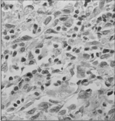
Figure 2. Interlacing fascicles of tightly packed ovoid to spindle cells, bearing hyperchromatic nuclei and indistinct cy-toplasm with high mitotic rate in a biopsy from the maxillary sinus (hematoxylin-eosin, 400 X).
Figure 3. Electron microscopy of tumor cells in a peripheral nerve sheath tumor, demonstrating elongated ovoid cells with thin cytoplasmic processes within a col-lagen matrix (uranium acetate and lead citrate, 10,000 X).
Figure 1. Coronal computed tomogra-phy scan from the nose and paranasal sinuses, showing a large mass in the left maxillary sinus with extension to the nasal cavity, in a 41-year-old woman.
Figure 4. Presence of basal lamina around the cells, showing tonofi laments in the cytoplasm and branching of the nucleus of a peripheral nerve sheath tumor (uranium acetate and lead citrate, 10,000 X).
Figure 5. Tumor cells with positive reacti-vity for S100 (1,000 X) in immunochemis-try of a peripheral nerve sheath tumor.
Figure 6. Tumor cells with positive reacti vity for vimentin (220 X) in im-munochemistry of a peripheral nerve sheath tumor.
335
Schwann cell origin from other spindle cells are characterized by long, slender cytoplasmic processes enveloping each other and other cells. These processes are outlined by a discrete basal lamina and also tonofi laments and dense core granules in the cytoplasm of tumor cells.12,13
On the other hand, the electron microscopic fi ndings from other tumors that are diagnosed differentially from MPNST are histologically distinct and well defi ned.
Clinical course and
Clinical course and treatment
treatment
Local recurrence is the major cause of treatment failure and poor overall survival
in these patients.5 Regional metastasis is
infrequent. Sarcomas involving the sinonasal region may have an indolent course for long-lasting periods, and may present with only nasal obstruction or drainage in the absence of orbital involvement. Combined therapy is the cornerstone of treatment for most soft tissue sarcomas of the head and neck region.14
Wide surgical resection is the primary treat-ment for neurogenic sarcomas. Because of the tendency for neurogenic sarcomas to recur locally, postoperative radiation therapy is recommended by many physicians.15
Radia-tion therapy signifi cantly improves survival for patients with T4 lesions.14
Doxorubicin-based adjuvant chemothera-py appears to improve disease-free survival and probably improves overall survival in adults with localized resectable head and neck soft tissue sarcoma. However, the role of adju-vant chemotherapy in neurogenic sarcomas remains poorly defi ned.15,16
CONCLUSION The paranasal sinuses are not a common location for MPNST, but if the clinical and radiological investigations and the gross and histological fi ndings are consistent with MPNST, electron microscopy is suggested for confi rmation of the fi nal diagnosis.
1. Colville RJ, Charlton F, Kelly CG, Nicoll JJ, McLean NR. Multidisciplinary management of head and neck sarcomas. Head Neck. 2005;27(9):814-24.
2. Dublin AB, Dedo HH, Bridger WH. Intranasal schwannoma: magnetic resonance and computed tomography appearance. Am J Otolaryngol. 1995;16(4):251-4.
3. Younis RT, Gross CW, Lazar RH. Schwannomas of the para-nasal sinuses. Case report and clinicopathologic analysis. Arch Otolaryngol Head Neck Surg. 1991;117(6):677-80. 4. Mannan AA, Singh MK, Bahadur S, Hatimota P, Sharma
MC. Solitary malignant schwannoma of the nasal cavity and paranasal sinuses: report of two rare cases. Ear Nose Throat J. 2003;82(8):634-6, 638, 640.
5. Spiro JD, Soo KC, Spiro RH. Nonsquamous cell malignant neoplasms of the nasal cavities and paranasal sinuses. Head Neck. 1995;17(2):114-8.
6. DiNardo LJ, Rumsey RL. Management of malignant schwanno-mas of the paranasal sinuses and anterior skull base. Ear Nose Throat J. 1996;75(6):377-80.
7. Mosharrafa TM, Kuppersmith RB, Porter JP, Donovan DT. Pathologic quiz case 1. Malignant peripheral nerve sheath tumor of the ethmoidal sinus. Arch Otolaryngol Head Neck Surg. 1997;123(6):654, 656-7.
8. Ross C, Wright E, Moseley J, Rees R. Massive schwannoma of the nose and paranasal sinuses. South Med J. 1988;81(12):1588-91. 9. Nagayama I, Nishimura T, Furukawa M. Malignant schwannoma
arising in a paranasal sinus. J Laryngol Otol. 1993;107(2):146-8. 10. Ghosh BC, Ghosh L, Huvos AG, Fortner JG. Malig-nant schwannoma. A clinicopathologic study. Cancer. 1973;31(1):184-90.
11. White HR Jr. Survival in malignant schwannoma. An 18-year study. Cancer. 1971;27(3):720-9.
12. Taxy JB, Battifora H, Trujillo Y, Dorfman HD. Electron mi-croscopy in the diagnosis of malignant schwannoma. Cancer. 1981;48(6):1381-91.
13. Chen KT, Latorraca R, Fabich D, Padgug A, Hafez GR, Gilbert EF. Malignant schwannoma: a light microscopic and ultrastruc-tural study. Cancer. 1980;45(7):1585-93.
14. Bhattacharyya N. Factors affecting survival in maxillary sinus cancer. J Oral Maxillofac Surg. 2003;61(9):1016-21. 15. Sturgis EM, Potter BO. Sarcomas of the head and neck region.
Curr Opin Oncol. 2003;15(3):239-52.
16. Sarcoma Meta-analysis Collaboration (SMAC). Adjuvant che-motherapy for localised resectable soft tissue sarcoma in adults. Cochrane Database Syst Rev. 2000;(4):CD001419.
Sources of funding: Not declared
Confl ict of interest: Not declared
Date of fi rst submission: November 25, 2005
Last received: May 5, 2006
Accepted: October 17, 2006
REFERENCES
AUTHOR INFORMATION
Bijan Khademi, MD. Associate professor of Otolaryngology,
Department of Otolaryngology - Head and Neck Surgery, Khalili Hospital, Shiraz University of Medical Sciences, Shiraz, Iran.
Seied Mohammad Owji, MD. Assistant professor of Pathology,
Department of Pathology, Faghihi Hospital, Shiraz University of Medical Sciences, Shiraz, Iran.
Khadije Jamshidi Khosh, MD. Resident of Otolaryngology,
Department of Otolaryngology - Head and Neck Surgery, Khalili Hospital, Shiraz University of Medical Sciences, Shiraz, Iran.
Mohammad Mohammadianpanah, MD. Assistant professor of
Radiation Oncology, Department of Radiation Oncology, Nemazee Hospital, Shiraz University of Medical Sciences, Shiraz, Iran.
Behrooz Gandomi, MD. Assistant professor of Otolaryngology,
Department of Otolaryngology - Head and Neck Surgery, Khalili Hospital, Shiraz University of Medical Sciences, Shiraz, Iran.
Address for correspondence:
Mohammad Mohammadianpanah
Department of Radiation Oncology, Nemazee Hospital, Shiraz University of Medical Sciences, Shiraz, Iran.
Post code: 71935 Fax: ++98- 711-6260135 E-mail: [email protected]
Copyright © 2006, Associação Paulista de Medicina
RESUMO Descrição de um tumor maligno da bainha do nervo trigêmeo: estudo por meio de microscopia eletrônica e imunoistoquímica
CONTEXTO: O tumor maligno da bainha neural do trigêmeo que acomete a cavidade nasal e os seios paranasais é extremamente raro. À microscopia óptica habitual sua identifi cação é difícil, sendo necessária a confi rmação através de microscopia eletrônica e de técnicas imunoistoquímicas.
RELATO DE CASO: A paciente era uma mulher de 41 anos com história de 10 meses de progressiva dor seguida de edema álgico em região facial à esquerda, e de sintomas de sangramento, secreção e obstrução nasal. Estudos com métodos de imagem sugeriam a presença de processo expansivo em cavidades nasais e paranasais à esquerda. À biopsia, os achados histopatológicos à microscopia óptica foram sugestivos de tumor de origem neural no trigêmeo. Estudos imunoistoquímico e por microscopia eletrônica confi rmaram se tratar de neoplasia maligna da bainha neural do trigêmeo. Descrevemos ascaracterísticas clínicas, radiológicas e histológicas deste tumor, e revisamos a literatura.
PALAVRAS-CHAVE: Neoplasias da bainha neural. Seios paranasais. Nervo trigêmeo. Radioterapia. Quimioterapia.