Retrospective analysis of oral peripheral
nerve sheath tumors in Brazilians
Abstract: Traumatic neuroma, neuroi broma, neurilemmoma, palisad-ed encapsulatpalisad-ed neuroma and malignant peripheral nerve sheath tumor (MPNST) are peripheral nerve sheath tumors and present neural origin. The goal of this study was to describe the epidemiological data of oral peripheral nerve sheath tumors in a sample of the Brazilian population. Biopsies requested from the Oral Pathology Service, School of Den-tistry, Federal University of Minas Gerais (MG, Brazil), between 1966 and 2006 were evaluated. Lesions diagnosed as peripheral nerve sheath tumors were submitted to morphologic and to immunohistochemical analyses. All cases were immunopositive to the S-100 protein. Thirty-i ve oral peripheral nerve sheath tumors were found, representing 0.16% of all lesions archived in the Oral Pathology Service. Traumatic neuroma (15 cases) most frequently affected the mental foramen. Solitary neuroi broma (10 cases) was more frequently observed neuroin the palate. Neuroneuroi -broma associated with neuroi -bromatosis type I (2 cases) was observed in the gingival and alveolar mucosa. Neurilemmoma (4 cases) was more commonly observed in the buccal mucosa. Malignant peripheral nerve sheath tumors (3 cases) occurred in the mandible, palate, and tongue. Palisaded encapsulated neuroma (1 case) occurred in the buccal mucosa. The data coni rmed that oral peripheral nerve sheath tumors are uncom-mon in the oral region, with some lesions presenting a predilection for a specii c gender or site. This study may be useful in clinical dentistry and oral pathology practice and may be used as baseline data regarding oral peripheral nerve sheath tumors in other populations.
Descriptors: Neuroma; Neuroi broma; Nerve sheath neoplasms; Mouth; Epidemiology.
Juliana Tito Salla(a)
Aline Cristina Batista Rodrigues Johann(b)
Bruna Gonçalves Garcia(a)
Maria Cássia Ferreira Aguiar(c)
Ricardo Alves Mesquita(c)
(a) DDS; (b)MSc; (c)PhD, Professor – Department
of Oral Surgery, Medicine and Pathology, School of Dentistry, Federal University of Minas Gerais, MG, Brazil.
Corresponding author:
Ricardo Alves Mesquita
Universidade Federal de Minas Gerais Faculdade de Odontologia
Av. Antônio Carlos, 6667, sala 3204 – Pampulha
Belo Horizonte - MG - Brazil CEP: 31270-901
E-mail: [email protected]
Introduction
Peripheral nerve sheath tumors may be classii ed in benign and malignant. The i rst category includes traumatic neuroma, neuroi broma, neurilemmoma, and palisaded encapsulated neuroma (PEN) whereas the second category consists of malignant peripheral nerve sheath tumor (MPNST). These tumors share a common neural origin but present microscopic and clinical heterogeneity.1,2
The traumatic neuroma is a non-neoplastic le-sion characterized by the proliferation of schwann cells and nerve i bers that arise following injury to a nerve.3-5 Jones, Franklin6 (2006), in a series of
44,007 oral and maxillofacial pathologies, observed that 0.34% of the cases were of traumatic neuroma. In studies of oral peripheral nerve sheath tumor cas-es, the following frequencies of traumatic neuroma were observed: 16%7 and 29.9%.1
Neuroi broma is a circumscribed but non-encap-sulated tumor consisting of a mixture of schwann cells, perineurial cells, and endoneurial i bro-blasts.1,2,4 Jones, Franklin6 (2006) found the
fre-quency of this lesion to be 0.43%. In oral peripheral nerve sheath tumors, the frequencies identii ed for neuroi broma were 20.8%1 and 32%.7
Neurilemmoma is an encapsulated neoplasm composed of schwann cells arranged in two pat-terns: Antoni type A and Antoni type B.1,5 Although
they can occur in any region, 25 to 48% of the le-sions occur in the head and neck region.8 One study
regarding oral and maxillofacial pathologies report-ed a 0.1% frequency for this lesion.6 The
frequen-cies of 16.9%1 and 22%7 for neurilemmoma were
re-corded in studies of the oral peripheral nerve sheath tumors.
PEN is characterized by a bundle of nerves inter-posed to schwann cells, which characteristically ag-gregate in palisades.1,2,9 This is a well-circumscribed
tumor that represents 0.05% of the oral biopsy specimens.10 The frequency of PEN found in a study
of oral peripheral nerve sheath tumors was 20.8%.1
MPNSTs are spindle-cell sarcomas that arise from nerve, neuroi broma, neurilemmoma, or tis-sue containing nerves.2,11 Although 8 to 16% of
MPNSTs develop in the head and neck region, this lesion is extremely rare in the oral cavity.12 A
simi-lar frequency (8%) of this lesion was described in a study on oral peripheral nerve sheath tumors.7
Epidemiologic studies on oral lesions have been conducted, being important sources of knowledge and contributing to the practice of clinical dentistry and oral pathology.13,14 However, few authors have
assessed the epidemiological features of oral pe-ripheral nerve sheath tumors to date.1,5-7 The aim of
this paper was to describe the epidemiological data on the peripheral nerve sheath tumors recorded in the archives of the Oral Pathology Service, School of Dentistry, Federal University of Minas Gerais (UFMG), MG, Brazil.
Material and Methods
The protocol of this study was approved by the Committee for Bioethics in Research, UFMG (016/03).
All i les regarding oral peripheral nerve sheath tumors submitted to the Oral Pathology Service, School of Dentistry, UFMG (FO-UFMG) from 1966 to 2006 were reviewed. These i les were classii ed in accordance with histological and immunohisto-chemical criteria. Histological sections stained with haematoxilin and eosin were analyzed using an optic microscope (Axiostar 1122-110, Carl Zeiss, Oberkochen, Baden-Wüttemberg, Germany). Patient clinical data and lesion location were recorded. The histological criteria used to classify the peripheral nerve sheath tumors were in accordance with Enz-inger, Weiss2 (1995).
Immunohistochemistry staining to detect S-100 proteins was performed for all lesions using the standard streptavidin-biotin-peroxidase method on deparafi nized tissue sections. The sections were incubated with an S-100 primary antibody (Z0311, Dako Corporation® Carpinteria, CA, USA) at a
1:700 dilution for 18 hours at 4°C. Mayers hema-toxilin was used as a counterstain. All lesions were positive for S-100. In MPNST lesions, immunohis-tochemistry staining for AE1/AE3 (M3515, Dako Corporation® Carpinteria, CA, USA; at a 1:100
dilution), HHF35 (M0635, Dako Corporation®
Carpinteria, CA, USA; at a 1:100 dilution), and α- smooth muscle actin (M0851, Dako Corporation®
performed. An antigen retrieval was also performed using a 10 mM citrate buffer (pH = 6.0, 30 min at 98°C) in stains testing AE1/AE3 and α- smooth muscle actin. The primary antibody was also incu-bated for 18 hours at 4°C. MPNST showed a nega-tive stain for AE1/AE3, HHF35, and α- smooth muscle actin.
Results
Oral peripheral nerve sheath tumors represented 35 (0.2%) of 21,476 specimens biopsied from 1966 to 2006 in the Oral Pathology Service, FO-UFMG. The cases diagnosed included 15 (42.9%) traumatic neuromas, 10 (28.6%) solitary neuroi bromas, 2 (5.6%) neuroi bromas associated with neuroi bro-matosis type I, 4 (11.4%) neurilemmomas, 1 (2.9%) PEN, and 3 (8.6%) MPNSTs. The clinical data are presented in Table 1.
The neurilemmomas presented 50% of the le-sions composed of Antoni type A pattern and 50% composed of a mixture of both patterns, Antoni type A and type B. All of these tumors were encap-sulated.
Discussion
The oral peripheral nerve sheath tumors were considered rare as they occurred in only 35 cases among 21,476 biopsied lesions from FO-UFMG, representing 0.2% of all lesions. Studies regarding oral peripheral nerve sheath tumors reported the frequencies of these lesions, without immunohisto-chemical evaluation.6,7 However, Chrysomali et al.1
(1997) evaluated 77 oral peripheral nerve sheath tumors using immunohistochemistry to detect the S-100 protein, CD57, epithelial membrane antigen (EMA), factor XIIIa, CD34, CD68, and collagen IV. Likewise, Magnusson10 (1996) studied 12 cases
of oral PEN evaluating the expression of S-100, neu-roi lament and EMA.
Used as an auxiliary method in the diagnosis of the peripheral nerve sheath tumors, the most com-mon immunohistochemical markers described in the literature include: S-100, CD34, and EMA. In the present study, S-100 was used because it repre-sented an easy marker, has been widely used in the identii cation of schwann cells, and aids greatly in the diagnostic process.1,15-17 The neural origin of all
lesions of this study was coni rmed by the
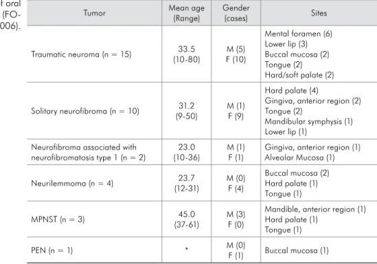
immuno-Tumor Mean age (Range)
Gender
(cases) Sites
Traumatic neuroma (n = 15) 33.5 (10-80)
M (5) F (10)
Mental foramen (6) Lower lip (3) Buccal mucosa (2) Tongue (2) Hard/soft palate (2)
Solitary neurofibroma (n = 10) 31.2 (9-50)
M (1) F (9)
Hard palate (4)
Gingiva, anterior region (2) Tongue (2)
Mandibular symphysis (1) Lower lip (1)
Neurofibroma associated with neurofibromatosis type 1 (n = 2)
23.0 (10-36)
M (1) F (1)
Gingiva, anterior region (1) Alveolar Mucosa (1)
Neurilemmoma (n = 4) 23.7 (12-31)
M (0) F (4)
Buccal mucosa (2) Hard palate (1) Tongue (1)
MPNST (n = 3) 45.0 (37-61)
M (3) F (0)
Mandible, anterior region (1) Hard palate (1)
Tongue (1)
PEN (n = 1) * M (0)
F (1) Buccal mucosa (1)
F: Female; M: Male; *: no data; MPNST: malignant peripheral nerve sheath tumor, PEN: palisaded encapsu-lated neuroma.
Table 1 - Clinical data of oral peripheral nerve sheath tumors
histochemical tests for the S-100 protein, since all samples were positive to this antigen. In 100% of the cases of traumatic neuroma, schwann cells with-in the nerve fascicles were immunoreactive toward S-100. This is also reported in the i ndings from Chrysomali et al.1 (1997) and Weiss et al.17 (1983).
The positive stain for S-100 was also observed in 100% of the neuroi broma cases, which is a rate similar to that found in the literature.1,15,17 However,
as indicated by Enzinger, Weiss2 (1995), the S-100
immunostain in neuroi broma is not as notable as in neurilemmoma. In the present study, all cases of neurilemmoma also showed positive staining for S-100, which is also reported by Hirose et al.15 (2003).
However, Weiss et al.17 (1983) found positive
immu-nostains for S-100 in 91% of the cases of neurilem-moma, whereas Chrysomali et al.1 (1997) observed
this in only 31% of the cases. The case of PEN, on the other hand, stained positively for S-100. Arg-enyi3 (1990) found 100% of the cases to be positive
for S-100, in contrast with Chrysomali et al.1 (1997)
who found 25% of the cases to be immunopositive. S-100 immunostaining was observed in 100% of the MPNST cases presented in this study, however, the literature described this as varying from 50 to 90% of the cases.2,17
Traumatic neuroma is most frequently located in the tongue, lips,1,18 and mental nerve area.5 We also
found the same neuroma in areas including the buc-cal mucosa and palate. These tumors affected adults and children from 10 to 80 years of age, with a mean age of 33.5, which is in accordance with Chrysoma-li et al.1 (1997). The female preponderance noticed
in this study is also reported in the i ndings from Chrysomali et al.1 (1997).
In the solitary neuroi broma, the most common sites found by Shklar, Meyer7 (1963) and Wright,
Jackson5 (1980) were the tongue, the palate, the
buc-cal mucosa and the l oor of the mouth. In contrast, the main site observed by Ellis et al.19 (1977) was the
posterior region of the mandible, while Chrysomali et al.1 (1997) reported it in the alveolar mucosa and the
palate. In the present study, the palate and the gin-giva were the most frequently intraoral and paraoral tissues involved. Also, one case located in the man-dibular symphysis was observed in this study.
Like-wise, neuroi bromas of the jaw have been described in the literature.20,21 As to neuroi bromas, it was
ob-served that these lesions affected adults and children from 9 to 50 years of age, with a mean age of 31.2, which is in accordance with the literature.2,5,11,19 As
observed by other authors,1,5,19 our study also
point-ed out a female preponderance. However, Enzinger, Weiss2 (1995) and Pilavaki et al.11 (2004) related no
gender dominance to this lesion.
Neuroi bromatosis type I is a disorder character-ized by the presence of two or more of the following i ndings: six or more “café au lait” macules (> 5 mm in diameter in puberty and > 15 mm in postpuberty patients), two or more neuroi bromas of any type or one plexiform, freckling in the axillary or inguinal regions, optic glioma, two or more “Lish” nodules, and a distinctive osseous lesion.22 Two cases of
neu-roi broma were associated with neuneu-roi bromatosis type I. Association of the oral neuroi broma and neuroi bromatosis type I is uncommon.5 The lesions
in the oral soft tissues have been identii ed with a prevalence of less than 10%,23 26%20 and up to 72%
of patients.21 In addition, neuroi bromas associated
with neuroi bromatosis type I are most frequently found in the tongue, but have also been identii ed in the gingiva, the alveolar mucosa, the palate, the buc-cal mucosa, the lip, the l oor of the mouth, and the buccal mucosa.20,22 In this study, these lesions were
identii ed in the gingiva and in the alveolar mucosa and presented no gender predilection.
The cases of neurilemmoma presented a predilec-tion for the buccal mucosa although other authors observed it in the lips,2 the tongue, the palate19 and
the mandible.5 These most often occurred in
chil-dren and young adults with a mean age of 23.7 years. Most reports suggested that the majority of tumors arise between 10 and 4024,25 and between 20 and 50
years of age.11 Some studies reported that males and
females are equally affected. Shklar, Meyer7 (1963)
reported a male preponderance while other studies showed a female dominance.24,25 In this study, only
women were affected.
5th, 6th, and 7th decades of life, and that both genders
are equally affected.5,10,26 PEN rarely appears in the
oral cavity, and when it was diagnosed, it occurred more commonly in the palate and maxillary mu-cosa.10,26 This runs in direct contrast with our case,
which was found in the buccal mucosa.
Although only three cases of MPNST were ob-served, it is interesting to note that all occurred in male patients. Enzinger, Weiss2 (1995) also
report-ed that more males than females were affectreport-ed by MPNST. The mean age of 45 years was observed in our study. Other authors reported that this malig-nant lesion affects, most frequently, patients from 20 to 50 years of age.2 The present lesions occurred
in the tongue, the palate and the mandible. The cas-es of this entity have been reported mostly in soft tissues and less commonly in bones.27 It can occur
in the palate, lips, buccal mucosa, mental foramen,
and submandibular triangle.28
Conclusions
This paper provides epidemiological data that can be an important auxiliary in clinical dentistry and oral pathology practice, mainly in the diagnosis of the oral peripheral nerve sheath tumors, which rarely occur in the oral cavity. This study may also be used as a data reference of oral peripheral nerve sheath tumors for other populations.
Acknowledgements
This study was supported by grants from the National Council for Scientii c and Technological Development (CNPq - 484974/2006-8), FAPEMIG (CDS 895/05), and CAPES, Brazil. RA Mesquita and MCF Aguiar are research fellows of CNPq.
References
1. Chrysomali E, Papanicolaou SI, Dekker NP, Regezi JA. Benign neural tumors of the oral cavity: a comparative immunohisto-chemical study. Oral Surg Oral Med Oral Pathol Oral Radiol Endod. 1997;84(4):381-90.
2. Enzinger FM, Weiss SW. Soft tissue tumors. 3rd ed. St. Louis: Mosby-Year Book; 1995.
3. Argenyi ZB. Immunohistochemical characterization of pali-saded, encapsulated neuroma. J Cutan Pathol. 1990;17(6):329-35.
4. Requena L, Sangueza O. Benign neoplasms with neural differ-entiation: a review. Am J Dermatopathol. 1995;17(1):75-96. 5. Wright BA, Jackson D. Neural tumors of the oral cav-ity. Oral Surg Oral Med Oral Pathol Oral Radiol Endod. 1980;49(6):509-22.
6. Jones AV, Franklin CD. An analysis of oral and maxillofacial pathology found in adults over a 30-year period. J Oral Pathol Med. 2006;35(7):392-401.
7. Shklar G, Meyer I. Neurogenic tumors of the mouth and jaws. Oral Surg Oral Med Oral Pathol. 1963;9:1075-93.
8. Williams HK, Cannell H, Silvester K, Williams DM. Neuri-lemmoma of the head and neck. Br J Oral Maxillofac Surg. 1993;31(1):32-5.
9. Tomich CE, Moll MC. Palisaded, encapsulated neuroma of the lip. J Oral Surg. 1976;34(3):265-8.
10. Magnusson B. Palisaded encapsulated neuroma (solitary cir-cumscribed neuroma) of the oral mucosa. Oral Surg Oral Med Oral Pathol Oral Radiol Endod. 1996;82(3):302-4.
11. Pilavaki M, Chourmouzi D, Kiziridou A, Skordalaki A, Zarampouzas T, Dervelengas A. Imaging of peripheral nerve
sheath tumors with pathologic correlation: pictorial review. Eur J Radiol. 2004;52(3):229-39.
12. Eversole LR, Schwartz DW, Sabes WR. Central and peripheral fibrogenic and neurogenic sarcoma of the oral regions. Oral Surg Oral Med Oral Pathol. 1973;36(1):49-62.
13. Corrêa PH, Nunes LC, Johann AC, Aguiar MC, Gomez RS, Mesquita RA. Prevalence of oral hemangioma, vascular mal-formation and varix in a Brazilian population. Braz Oral Res. 2007;21(1):40-5.
14. Santos JN, Pinto LP, Figueiredo CR, Souza LB. Odonto-genic tumors: analysis of 127 cases. Pesqui Odontol Bras. 2001;15(4):308-13.
15. Hirose T, Tani T, Shimada T, Ishizawa K, Shimada S, Sano T. Immunohistochemical demonstration of EMA/Glut1-positive perineurial cells and CD34-positive fibroblastic cells in periph-eral nerve sheath tumors. Mod Pathol. 2003;16(4):293-8. 16. Johnson MD, Glick AD, Davis BW. Immunohistochemical
evaluation of Leu-7, myelin basic-protein, S100-protein, glial-fibrillary acidic-protein, and LN3 immunoreactivity in nerve sheath tumors and sarcomas. Arch Pathol Lab Med. 1988;112(2):155-60.
17. Weiss SW, Langloss JM, Enzinger FM. Value of S-100 protein in the diagnosis of soft tissue tumors with particular reference to benign and malignant Schwann cell tumors. Lab Invest. 1983;49(3):299-308.
18. Sist TC, Greene GW. Traumatic neuroma of the oral cavity. Oral Surg Oral Med Oral Pathol. 1981;51(4):394-402. 19. Ellis GL, Abrams AM, Melrose RJ. Intraosseous benign neural
review of the literature. Oral Surg Oral Med Oral Pathol. 1977;44(5):731-43.
20. D’Ambrosio JA, Langlais RP, Young RS. Jaw and skull changes in neurofibromatosis. Oral Surg Oral Med Oral Pathol. 1988;66(3):391-6.
21. Shapiro SD, Abramovitch K, Van Dis ML, Skoczylas LJ, Lan-glais RP, Jorgenson RJ et al. Neurofibromatosis: oral and radiographic manifestations. Oral Surg Oral Med Oral Pathol. 1984;58(4):493-8.
22. Bongiorno MR, Pistone G, Aricó M. Manifestations of the tongue in neurofibromatosis type I. Oral Dis. 2006;12(2):125-9.
23. Baden E, Jones JR, Khedekar R, Burns WA. Neurofibroma-tosis of the tongue: a light and electron microscopic study with review of the literature from 1849 to 1981. J Oral Med. 1984;39(3):157-64.
24. Cherrick HM, Eversole LR. Benign neural sheath neoplasms of the oral cavity. Report of thirty-seven cases. Oral Surg Oral Med Oral Pathol. 1971;32(6):900-9.
25. Hatziotia JC, Asprides H. Neurilemoma (Schwannoma) of the oral cavity. Oral Surg Oral Med Oral Pathol. 1967;24(4):510-26.
26. Chauvin PJ, Wysocki GP, Daley TD, Pringle GA. Palisaded encapsulated neuroma of oral mucosa. Oral Surg Oral Med Oral Pathol. 1992;73(1):71-4.
27. Bullock MJ, Bedard YC, Bell RS, Kandel R. Intraosse-ous malignant peripheral nerve sheath tumor. Report of a case and review of the literature. Arch Pathol Lab Med. 1995;119(4):367-70.