198
Case Report
REVISTA PAULISTA DE MEDICIN AEwing’s sarcoma of the he ad and ne ck
Discipline of O torhinolaryngology, Department of O torhinolaryngology and O phthalmology,
Universidade Estadual de Campinas, Campinas, Brazil
a b s t r a c t
CO N TEX T: Ewing ’s sarco ma is a rare neo plasm, which usually arises in lo ng bo nes o f the limbs and in flat bo nes o f the pelvis, with the invo lvement o f head and neck bo nes being very unusual.
CASE REPO RT: a case o f Ewing ’s sarco ma o ccurring in the man-dible o f a 3 5 -year-o ld female. Pain and swelling o f the tumo r were the main co mplaints. The early hypo thesis was an undifferentiated malig nant neo plasm, po ssibly a sarco ma. The CT scan depicted an expansive lesio n, encapsulated, with septa and characteristics o f so ft tissue, invo lving the left side o f the mandible and extending to the surro unding tissues. The patient underwent surg ical excisio n o f the lesio n, the definitive diag no sis o f Ewing ’s sarco ma was estab-lished, and the patient co mmenced o n radio therapy.
KEY W O RDS: Sarco ma. Ewing ’s. Pharynx. Mandible. Head and N eck Surg ery. O to laryng o lo g y.
• Adriano Santana Fo nseca • Raquel Mez z alira • Ag rício N ubiato Crespo • Antô nio Emílio Bo rto leto Junio r • Jo rg e Riz z ato Pascho al
INTRODUCTION
Ewing’s sarco m a is an unco m m o n m alignant ne o plasm, lo cally aggre ssive , which o ccurs mo re o fte n in m ale s than fe m ale s, and in the first thre e de -cades o f life.1
Lo ng b o nes are the mo st co mmo n site. The tum o r is ve ry rare in o to laryngo lo gy practice , b ut it m ay b e fo und invo lving the m andib le , ce rvi-cal ve rte b rae and the te m po ral b o ne s.
CASE REPORT
A thirtyfive ye aro ld Caucasian fe m ale pre -se nte d swe lling o n the le ft side o f the m andib le , in August 1994, whic h had b e e n p re se nt fo r se ve n mo nths. She also co mplained o f painful no dulatio ns in the surro unding gum s, which we re co m pro m is-ing he r m asticatio n and spe e ch, and causis-ing o tal-gia and o do ntaltal-gia. Fifteen years earlier she had had he r te e th e xtracte d and she was using o rtho do ntic pro sthe se s o n the lo we r and uppe r arcade s.
On examinatio n a firm painful lesio n invo lving the angle and b o dy o f the m andib le , as we ll as the le ft sub m andib ular re gio n and the flo o r o f the o ral cavity, was no te d. Pare sthe sia o f the le ft lo we r ar-cade was also ide ntifie d.
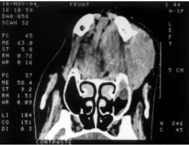
The b io psy sho wed a pattern suggestive o f un-differentiated malignant neo plasm. The immuno his-to che m ical stains po inte d the diagno sis his-to wards a sarco m a. The CT scan de picte d an e xpansive le sio n with o pacity, co m patib le with so ft tissue s, capsu-late d and with se pta, invo lving the le ft po rtio n o f the m andib le and e xte nding to the surro unding
199
sue s ( Figures 1 and 2).
The patie nt unde rwe nt le ft he m im andib ule cto m y and trache o scto m y in the sam e surgical pro ce -dure. The histo lo gical findings sho wed a small ro und ce ll m alignant ne o plasm o f the b o ne , co m patib le with Ewing’s sarco m a with ne o plastic invasio n o f the adjace nt so ft tissue s. The surgical m argins we re no t co m pro m ise d.
Afte rwards, the patie nt was tre ate d with ra-dio therapy. The cervical regio n and then the left side o f the face we re tre ate d to a to tal do se o f 50 Grays, in twe nty-five e xpo sure s o f two Grays. The patie nt has b e e n in fo llo w-up fo r fo ur ye ars and is fre e o f dise ase and fe e ling we ll.
DISCUSSION
Ewing’s sarco m a is an unusual dise ase co m -prising ab o ut 4 to 6% o f all prim ary b o ne tum o rs. It o riginate s in the m arro w cavity and is fo und in the e piphyse s o f lo ng and flat b o ne s. Invo lve m e nt o f the he ad and ne c k in Ewing ’s Sarc o m a is ve ry unusual, acco unting fo r appro xim ate ly 1 to 4% o f case s. The skull and mandib le are the mo st fre q ue nt site s.1,2 It is rare afte r the third de cade and o ccurs
mo st o ften in the seco nd decade. It is mo re co mmo n in m ale s than fe m ale s.3
Generally it presents with pain and lo cal swell-ing, dilated veins, hyperthermia, anemia, increased erythro cyte sedimentatio n rate and leuko cyto sis.2,3,4
A histo ry o f previo us trauma is present in many re-po rted cases. The initial evaluatio n includes radio -graphic study o f the suspected area, which sho ws ar-eas o f b o ne rarefactio n, freq uently asso ciated with increased density, perio steal reactio n, and bo ne neo
-fo rmatio n resulting in an “o nio n layers” appearance.3
The d iagno sis is e stab lishe d b y b io p sy, in which the tum o r is se e n as laye rs o f sm all ro und cells, similar to lympho cytes, b ut larger. Mito tic cells are rare , inte rce llular stro m a is scarce and a large po rtio n o f the tum o r m ay b e ne cro tic. Tum o r ce lls place d aro und a cle ar ce ntral are a fo rm ing ro se tte s m ay b e se e n, re se m b ling Ho m e r-Wrigt ro se tte s, typ i c a l o f n e u ro b l a s to m a s . In tra c yto p l a s m i c glyco ge n is a de finitive aspe ct, b ut no t patho gno -m o nic b e cause it is also pre se nt in o the r pri-m itive tum o r ce lls such as o ste o sarco m as, rhab do m yo sa-rco m as and ne uro b lasto m as.3,4 He nce , the diffe re
n-tial diagno sis o f Ewing’s sarco m a e nco m passe s a wide num b e r o f dise ase s including o ste o sarco m as, rh a b d o m yo s a rc o m a s , n e u ro b l a s to m a s , m e s e n c h ym al c h o n d ro s arc o m a an d m alig n an t lym pho m a.
The ap p ro p riate tre atm e nt fo r Ewing’s sar-co m a has b e e n the surgical e xcisio n o f the tum o r asso ciate d with radio the rapy and che m o the rapy.
Due to the high lo cal re o ccurre nce rate (20%) fo llo wing radiatio n the rapy alo ne , radical surgical re m o val m ust b e atte m pte d to incre ase lo cal co n-tro l whenever feasib le. The same applies to the mandib le , since to day’s re co nstructio n te chniq ue s alle -viate e sthe tical and functio nal im pairm e nt to the patie nt. Radio the rapy must b e use d as ne o adjuvant the rapy o r in no n-re se ctab le prim ary radio se nsitive tum o rs. Che m o the rapy m ust b e re se rve d to pre ve n-tio n and tre atm e nt o f m e tastasis.
The asso ciatio n o f surge ry, radio the rapy and che mo the rapy has significantly impro ve d the 5-ye ar survival ratio , no w re aching 40 to 75%.3
The single m o st im po rtant indicato r is the prim ary site , and
Figure 1. CT scan showing an expansive lesion on the left portion of the mandible, causing deviation of the tongue away from the midline.
Figure 2. CT scan of a capsulated mass involving the mandible with septa and deviation of the surrounding tissues.
200
r e s u m o
CO N TEX TO : O Sarco ma de Ewing é uma do ença que g eralmente aco mete o s o sso s lo ng o s e a pelve, sendo rara a o co rrência em cabeça e pesco ço .
RELATO DE CASO : Caso de Sarco ma de Ewing co meçando na mandíbula em uma paciente de 3 5 ano s, sendo a tumo ração e a do r lo cal o s principais sinto mas referido s. O diag nó stico inicial fo i de neo plasia malig na indiferenciada, sug estivo de sarco ma e a to mo g ra fia c o mp uta d o riz a d a mo stro u le sã o e xp a nsiva c o m características de tumo r de partes mo les, septada e encapsulada, aco metendo a mandíbula esquerda e apresentando extensão para as partes mo les adjacentes. O tratamento realiz ado fo i a remo ção cirúrg ica da lesão , co mplementado co m radio terapia pó s-o perató ria. O diag nó stico definitivo fo i de Sarco ma de Ewing .
PALAVRAS-CHAVE: Sarco ma de Ewing . Mandíbula. Cirurg ia de Cabeça e Pesco ço . O to rrino laring o lo g ia.
Adriano Santana Fonse ca, MD. Seco nd-year Resident, Department o f Oto laryngo lo gy and Ophthalmo lo gy, Universidade Estadual de Campinas, Campinas, Brazil.
Raque l Me zzalira, MD.Do cto r, Department o f Oto laryngo lo gy and Ophthalmo lo gy, Universidade Estadual de Campinas, Campinas, Brazil.
Agrício Nubiato Cre spo, MD, PhD. Chairman, Discipline o f Oto rhino laryngo l-o gy Surgery, Department l-o f Otl-o laryngl-o ll-o gy and Ophthalml-o ll-o gy, Universidade Estadual de Campinas, Campinas, Brazil.
Antônio Emílio Bortole to Junior.UndergraduateMedical Student, Medical Sciences, Universidade Estadual de Campinas, Campinas, Brazil.
Jorge Rizzato Paschoal, MD, PhD. Assistant Pro fesso r, Discipline o f Oto rhino laryngo lo gy Surgery, Department o f Oto laryngo lo gy and Ophthalmo l-o gy, Universidade Estadual de Campinas, Campinas, Brazil.
Source s of funding: No t declared
Conflict of inte re st: No t declared
Last re ce ive d: 14 Octo ber 1999
Acce pte d: 14 December 1999
Addre ss for corre sponde nce :
Adriano Santana Fo nseca Rua Irmã Serafina, 657 - Apto . 95 Campinas/SP - Brasil - CEP 13015-201 E-mail: adrisf@ co rreio net.co m.br
p u b lis hin g in fo r m a t io n 1. Prindull G, Willert HG, No tter G. Lo cal therapy o f rhabdo myo sarco ma,
o steo sarco ma and Ewing’s Sarco ma o f children and ado lescents. Eur J Pediatr 1985;144(2):120-4.
2. Siegal P, Oliver WR, Reinus WR, et al.. Primary Ewing’s sarco ma invo lving the bo nes o f the head and neck. Cancer 1987;60(11):2829-40.
3. Sneige N, Batsaki JG. Ewing’s sarco ma o f bo ne and so ft tissues. Ann Oto l Rhino l Laryngo l 1989;98(5 Pt 1):400-2.
4. Yalcin S, Turo glu HT, Ozdamar S, Sadiko glu Y, Gurbuzer B, Yenici O. Ewing’s sarco ma o f the mandible. Oral Surg Oral Med Oral Patho l 1993;76(3):362-7.
REFERENCES
thus, prim ary tum o rs o f the he ad and ne ck, e spe -cially the m andib le , have a significantly highe r sur-vival ratio .2 An e xtre m e ly high e rythro cyte se dim e
n-Sao Paulo Med J/Rev Paul Med 2000; 118(6):198-200.