○ ○ ○ ○ ○ ○ ○ ○ ○ ○ ○ ○ ABST RAC T○ ○ ○ ○ ○ ○ ○ ○ ○ ○ ○ ○ ○ ○ ○ ○ ○IN T RO D U C T IO N○ ○ ○ ○ ○ ○ ○ ○ ○ ○
Head and neck paragangliomas are rare and are true neuroendocrine neoplasias. T hey originate from various paraganglia,1 members of a complex and intriguing family of endo-crine cells that present centripetal distribution with a tendency towards symmetry, extend-ing from the middle ear and skull base region to the pelvic floor.2
All these cells are derived from the neural crest and form the diffuse dispersed neuroen-docrine system. T hese kinds of cells are found in the central and peripheral nervous system and in many classic endocrine organs, for ex-ample the adrenal gland. T hey are distributed in the majority of body tissues, organized as isolated cells or in groups of 3 or 4 cells, ex-hibiting a heterogeneity of phenotypes that makes for structural diversity. T here are basi-cally two kinds of cells: the principal cells and the sustentacular cells. T he former are rounded, with a dark central nucleus, eccen-tric, with eosinophilic granules in their cyto-plasm, identified as neurosecretory granules by means of histochemical or immunohisto-chemical reactions, or using electron microscopy, with a halo around the membrane rim also being noted. T hey are grouped in nests and can be classified as light, dark and pyknocytic. T he sustentacular cells, located on the periphery of these nests of principal cells (Zellballen model) are pale, with elongated nuclei and indistinct cytoplasm. Near to the principal cells, axonal processes known as the mesaxonia can be observed. Endothelial and pericytic cells, and occasionally mast cells, are also found in paraganglia.3
Head and neck paraganglia have an inti-mate relationship with vascular and neural structures.4,5 T hey have a strategic localization that allows the development of a chemorecep-tor function in response to alterations in gas concentrations in arterial blood.6 Another important aspect is the chemoreceptor-sensi-tive function of the carotid bodies, which are sensitive to p02, pH and pCO2 alterations in the blood, in which the chronic hypoxemia stimulates the carotid body hyperplasia.7
T hese paragangliomas present approxi-mately the same architecture as a normal para-ganglion, with some variation in size and shape, but the main difference in this neopla-sia is the proliferation of principal cells in nests surrounded by sustentacular cells that repre-sent 1 to 5 % of the cells of a paraganglioma and form a prominent vascular network.8 T hree architectural patterns have been de-scribed:9 1) the normal or Zellballen model; 2) angiomatous, with large spindle or crescent-shaped principal cells and the appearance of capillaries; and 3) adenomatous, with a marked similarity between the principal cells and the epithelial cells, i.e. polyhedral cells with abundant cytoplasm and columnar ar-rangement.
Carotid body paragangliomas are differ-ent from hyperplasia in that they presdiffer-ent pro-liferation of the principal cells whereas in hy-perplasia there is proliferation of the princi-pal and sustentacular cells.10 T he majority of head and neck paragangliomas contain neu-rosecretory granules with vasoactive substances like epinephrine, norepinephrine, dopamine and serotonin.11 T he fact that these catecholamines are present does not signify
O
ri
gi
n
a
l
A
rt
ic
• Abrão Rapoport• Venâncio Avancini Ferreira Alves
• O dilon Victor Porto Denardin
• Josias de Andrade Sobrinho
• M arcos Brasilino de Carvalho
neck: clinical, morphological and
immunohistochemical aspects
Head and Neck Service of Heliópolis Hospital (Hosphel), and Pathology
Service of Oswaldo Cruz Hospital, São Paulo, Brazil
CO N TEX T: Pro tein marker po sitivity can assist in the definitio n o f the therapeutic appro ach to wards head and neck parag ang lio mas. The establish-ment o f the therapeutic appro ach sho uld inco r-po rate the results o f such an investig atio n.
O BJECTIVE: To establish criteria fo r benig nancy and malig nancy o f vag al and jug ular-tympanic para-g anpara-g lio mas, via the study o f the relatio nships o f sex, ag e, tumo r size, duratio n o f co mplaints, site, family histo ry, presence o f metastases, treatment, histo lo g ical architecture and cell type with the immuno histo chemical reactio ns to S1 0 0 pro tein, chro mo g ranin and Ag Ki6 7 .
DESIGN : A retro spective study o f histo lo g ical and clini-cal reco rds.
SETTIN G: The Helió po lis and O swaldo Cruz tertiary g eneral ho spitals, São Paulo .
SAM PLE: 8 cases o f head and neck parag ang lio mas.
M AIN M EASUREM EN TS: Determinatio n o f deg ree o f po sitivity to parag ang lio mas via immuno histo -chemical reactio ns.
RESULTS: 1 ). The pro tein markers fo r the principal cells (Ag Ki6 7 and chro mo g ranin) were sensitive in 1 0 0 % o f the tumo rs when used to g ether. 2 ). S1 0 0 pro tein was well identified in the cyto plasm and nucleus o f sustentacular cells and underwent reductio n in the neo plasias.
CO N CLUSIO N S: Chro mo g ranin was pro ven to be a g eneric marker fo r neuro endo crine tumo rs; S1 0 0 pro tein was po sitive in all 8 cases and the Ag Ki6 7 had lo w po sitivity in all cases.
that there is a biological effect.12 Nevertheless, the measurement of vanil-mandelic acid is rec-ommended as a screening method when these tumors are suspected.13, 14
The clinical presentation is usually asymp-tomatic. Pain, hoarseness, dysphagia, Horner’s syndrome, tinnitus and hearing loss may oc-casionally be presented. T here is evidence that these paragangliomas present as family histo-ries,15, 16 which has led to studies of oncogene identification.17,18 Most paragangliomas are benign, but 6 to 9% of head and neck para-gangliomas present histologically and biologi-cally malignant behavior,19 with the appear-ance of mitotic cells, cell pleomorphism and central necrosis in Zellballen areas). Malig-nancy, widely discussed by many authors, is only fully characterized by distant metastasis,20 with there being concern regarding the lack of identified histopathological factors (i.e. the cell atypia, nuclear polymorphism and local invasion factors that are typical of malig-nancy).21 Histological comparison between benign and malignant paragangliomas does not demonstrate any differences,22 and the most important factor among these histologi-cal factors seems to be the site of the tumor itself. T he incidence of metastasis in para-aor-tic paragangliomas ranges from 28 to 42%. T he knowledge available at present still does not allow the reasons for this difference in re-lation to head and neck paragangliomas to be explained.21
Using electron microscopy, a pattern of dimi-nution or absence of sustentacular cells was found in the tumor architecture of malignant paragang-liomas.10 This was the first histological observa-tion step towards predicting the clinical behavior of such tumors. It was followed by the introduc-tion of protein markers using immunohistochemi-cal methods, in order to identify prognostic fac-tors in head and neck paragangliomas.
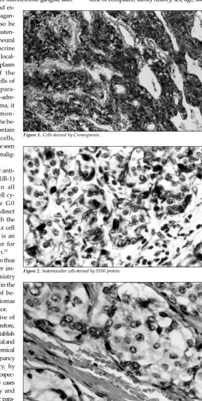
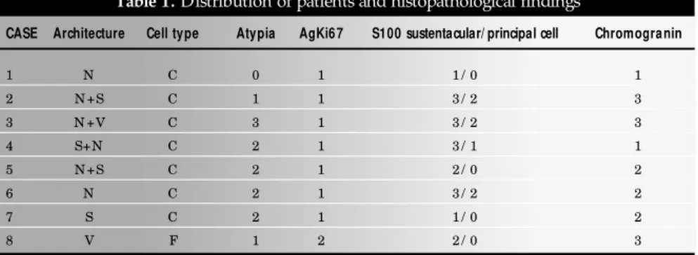
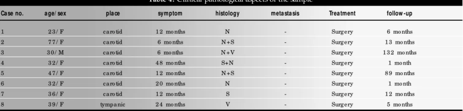
Various tumor markers have been tested. Some have confirmed the neuroendocrine origin of the neoplasia and others have shown up strong suspicions of malignant behavior.4 In these latter cases, even in the absence of distant metastases, there was greater tumor aggressiveness, either with the invasion of ad-jacent structures or with disease recurrence. In the present study, three immunohisto-chemical markers were used: chromogranin (Figure 1), S100 protein (Figure 2) and AgKi67 (Figure 3).
Chromogranin, the main marker for neu-roendocrine cells, is a structural protein found in neurosecretory granules of principal cells. Its function is to stabilize the intra-cellular matrix of neurosecretory granules, thereby
showing itself to be an excellent indicator of neuroendocrine differentiation. Well-differen-tiated tumors contain more neurosecretory granules and the undifferentiated ones have less of them.4
S100 protein, a dimeric 21-Kd protein bonding with calcium, was isolated from the nervous system. It has been identified in sus-tentacular cells of autonomous ganglia, adre-nal medullas and
ex-tra-adrenal paragan-glia. It may also be identified in susten-tacular cells of neural and neuroendocrine neoplasias. It is local-ized in the cytoplasm and nuclei of the sustentacular cells of extra-adrenal para-ganglia. In extra-adre-nal paraganglioma, it has been demon-strated that all the be-nign tumors contain sustentacular cells, whereas these are seen to be absent in malig-nant tumors.4
T he nuclear anti-gen AgKi67 (MiB-1) is a protein in all phases of the cell cy-cle except the G 0 phase, and has a direct relationship with the growth rate of a cell population. It is an excellent marker for cell proliferation.23
The question thus arises of whether im-munohistochemistry could be helpful in the identification of be-nign paragangliomas and their behavior.
T he objective of this work was therefore, in summary, to establish clinical, histological and immunohistochemical criteria for benignancy and malignancy, by means of the retrospec-tive analysis of 8 cases of carotid body and jugular-tympanic para-gangliomas.
○ ○ ○ ○ ○ ○ ○ ○ ○ ○ ○ ○ ○ ○M ET H O D S○ ○ ○ ○ ○ ○
From an analysis of the records of the Head and Neck Service at Heliópolis Hospital, 8 cases of head and neck paragangliomas were found to have been submitted to surgical procedure at the Head and Neck Service between 1977 and 1995. The following data were analyzed: time of complaint, family history, sex, age, site
Figure 1. Cells stained by Cromogranin.
Figure 2. Sustentocullar cells stained by S100 protein.
of the tumor, its size (considering sizes as greater or less than 5 cm) and metastases.
For the present study, new histological sections of thickness 3 micrometers were ob-tained from the old paraffin blocks. T he slides were stained with hematoxylin and eosin and other slides were also prepared for the immu-nohistochemical reactions. All slides were re-viewed by the same pathologist. T he archi-tecture of the paraganglioma was evaluated and classified as follows:
a) N = nest pattern (classic or Zellballen pat-tern), with sustentacular cells (elongated cells with indistinct cytoplasm) surround-ing groups of principal cells (rounded or polyhedral cells that are eccentric, with granular cytoplasm) and vascular stro-mata;
b) A = Adenomatous: principal cells with marked similarity to epithelial cells, i.e. polyhedral cells with abundant cytoplasm organized in columns, surrounded by sus-tentacular cells and vascular stromata; c) V = angiomatous: spindle-shaped
princi-pal cells, similar to endothelial cells, sur-rounded by sustentacular cells and vascu-lar stromata;
d) S = solid: basically consisting of various polymorphic principal cells, with few vas-cular stromata.
T he predominant cell type in paragang-liomas is the principal cell, accounting for 50 to 90% of the total number of cells. T hese cells are rounded, large, with prominent nu-clei, eosinophilic cytoplasm rich in granules
and ranging from 2 to 10 micrometers in di-ameter. T hese cells may vary in size and shape and were classified as follows: a) C = principal cells: 2 to 10 micrometers in diameter; b) D = small cells: less than 2 micrometers in diam-eter; c) E = giant cells: more than 10 microm-eters in diameter; d) F = fusiform cells: spin-dle-shaped cells, similar to vascular endothe-lium.
Cell atypia were evaluated and defined in terms of variation in shape and cell volume, the increase in the nucleus-cytoplasm relation-ship, irregularities in nuclear rims, nuclear hy-perchromatism and the presence of nucleoli. T hey were semi-quantified as 0 (absent), 1 (mild), 2 (moderate) and 3 (accentuated).
In the immunohistochemical methods, three protein markers were evaluated: AgKi67, chromogranin and S100 protein with ampli-fication using the streptavidin-biotin-peroxi-dase complex. T he immunohistochemical re-activity for AgKi67 (AcMiB-1) was evaluated by quantitative analysis: a) 1 = rare cells (less than one per field at 400x magnification); b) 2 = moderate number of positive cells (up to 25% of cells); c) 3 = high proportion of posi-tive cells (greater than 25%).
As it was already known that there would not be any neoplasias unaccompanied by cell proliferation, then if there were any case with a negative reaction to chromogranin, this would not be considered valid and would therefore act as a further control for the reac-tion. T he immunohistochemical reactivity to chromogranin (for principal cells) and S100
protein (for principal and sustentacular cells) was evaluated by the following semi-quantifi-cation: a) 0 = negative; b) 1 = rare stained cells (less than 1 per field at 400x magnification); c) 2 = moderate positivity (up to 25% of the cells); d) 3 = numerous positive cells (more than 25% of the cells).
○ ○ ○ ○ ○ ○ ○ ○ ○ ○ ○ ○ ○ ○ ○R ESU LT S○ ○ ○ ○ ○
T he distribution according to sex was 7 (87.5%) of women and 1 (12.5%) man, with a mean age of 39.5 years (range 23 – 77 years). At the time of the complaint, six patients (75%) reported that the time of onset of the lesion had been more than 1 year earlier and 2 patients (25%) reported it as being less than 1 year earlier. T he main complaint in 7 cases (87.5%) was a painless cervical tumor and in 1 case (12.5%) it was hearing loss and ear pain. The distribution of patients in relation to the size of the tumor showed that 4 of them had a lesion of more than 5 cm and 4 less than 5 cm. The follow-up ranged from 1 to 132 months and no case of distant metastasis was noted.
T he main histopathological and immu-nohistochemical findings are shown in Table 1 and Figures 1, 2, and 3.
There was a predominance of the nest pat-tern (5 cases: nos. 1, 2, 3, 5 and 6) and in 3 of them this was associated with other patterns (cases 2 and 5 with solid patterns, and case 3 with an angiomatous pattern). The predominant cell type was principal cells (7 cases: nos. 1, 2, 3, 4, 5, 6 and 7) and the remaining case had spin-dle-shaped cells as the main type (case 8).
Table 2 shows the relationships between tumor size and cell atypia, chromogranin re-activity, AgKi67 reactivity and positivity to S100 protein reaction for sustentacular cells. T he relationships between cell prolifera-tion among principal cells and chromogranin, and between sustentacular cells and S100 pro-tein, are shown in Table 3.
T he clinical-pathological characteristics are presented in Table 4, in which it can be noted that 5 cases (62.5%) had a moderate to numerous quantity of cells reactive to S100 and chromogranin (cases 2, 3, 5, 6 and 8). Table 1. Distribution of patients and histopathological findings
CASE Architecture Cell type Atypia AgKi6 7 S100 sustentacular/ principal cell Chromogranin
1 N C 0 1 1 / 0 1
2 N +S C 1 1 3 / 2 3
3 N +V C 3 1 3 / 2 3
4 S+N C 2 1 3 / 1 1
5 N +S C 2 1 2 / 0 2
6 N C 2 1 3 / 2 2
7 S C 2 1 1 / 0 2
8 V F 1 2 2 / 0 3
N = nest pattern; S = so lid pattern; V = ang io mato us, C = principal cells; F = spindle cells.
Table 2.Relation between tumor size and cell atypia, chromogranin and AgKi67 reactivity, and sustentacular cell positivity to S100 protein reaction.
Tumor (size) Atypia Chromogranin AgKi67 S1 00 protein
-/ + 2 (2 5 %) 1 (1 2 .5 %) 3 (3 7 .5 %) 0
< 5
++/ +++ 2 (2 5 %) 3 (3 7 .5 %) 1 (1 2 .5 %) 4 (5 0 %)
-/ + 1 (1 2 .5 %) 1 (1 2 .5 %) 4 (5 0 %) 2 (2 5 %)
> 5
++/ +++ 3 (3 7 .5 %) 3 (3 7 .5 %) 0 2 (2 5 %)
- = neg ative; + = rare staining cells; ++ = mo derate po sitivity; +++ = hig h po sitivity.
Table 3. Principal cell positivity to chromogranin and sustentacular cell
reactivity to S100 protein
Chromogranin S1 0 0 (sustentacular cells)
(principal cells) -/ + ++/ +++
-/ + 1 (1 2 .5 %) 1 (1 2 .5 %)
++/ +++ 1 (1 2 .5 %) 5 (6 2 .5 %)
○ ○ ○ ○ ○ ○ ○ ○ ○ ○ ○ ○ D ISC U SSIO N○ ○ ○ ○ ○ ○ ○ ○
T he uncertain nature of head and neck paragangliomas makes their therapeutic man-agement controversial. The clinical and epide-miological understanding still allows for some liberty of choice that discourages standardiza-tion of the approach to this disease. It is not clear why certain paragangliomas with same histopathological pattern follow different courses. These continuing uncertainties have justified new research ranging from epidemio-logical to cytogenetic studies, with the aim of identifying the biological behavior of the tumor. The symptoms of these tumors are volume-dependent, such that their slow growth leads to the characteristics of an insidious disease. In a study among inhabitants of higher altitudes,24 it was found that the tumors evolved more rap-idly. In that study, the female to male ratio was 8.3:1 with a mean age of 49 years. Tumors of the carotid body represented 79% of the cases of tumors of the para-pharyngeal space and the incidence of malignancy was 3.3%. The size of the tumor ranged from 2 to 12 cm (mean of 5.4 cm) and 1% of the cases had a family his-tory.24 Such results have been confirmed by other authors.19
In the present study, the mean evolution time for the disease was 17.5 months, with women predominating (7:1) and a mean age of 39.5 years old. Most of the tumors (87.5%) were from the carotid body and 12.5% from the tympanic cavity. T he mean size of the tumor was 4.81 cm and no reports of a family history were found. T he female predominance differs from reports in the literature, except those from high altitudes. T he mean age, size of tumor and site distribution were similar to those reported in the literature. T here were no cases of multicentricity, family history, re-lapse and malignancy. T he low number of cases could have led to a bias.
Cell atypia were found in cases 2, 3, 4, 5, 6, 7 and 8, of which five (cases 3, 4, 5, 6 and 7) had moderate to accentuated atypia. In
Table 4. Clinical-pathological aspects of the sample
Case no. age/ sex place symptom histology metastasis Treatment follow -up
1 2 3 / F caro tid 1 2 mo nths N - Surg ery 6 mo nths
2 7 7 / F caro tid 6 mo nths N +S - Surg ery 1 3 mo nths
3 3 0 / M caro tid 6 mo nths N +V - Surg ery 1 3 2 mo nths
4 3 2 / F caro tid 4 8 mo nths S+N - Surg ery 1 mo nth
5 4 7 / F caro tid 1 2 mo nths N +S - Surg ery 8 9 mo nths
6 3 2 / F caro tid 2 0 mo nths N - Surg ery 1 mo nth
7 3 6 / F caro tid 1 2 mo nths S - Surg ery 1 2 mo nths
8 3 9 / F tympanic 2 4 mo nths V - Surg ery 5 mo nths
Classic Pattern = Zellballen; V = ang io mato us; S = so lid.
addition, cellular necrosis was found in cases 3 and 5. T he cell atypia data were correlated to the size of the tumor and this size was re-lated to the reaction to AgKi67, chromogranin and S100 protein. In tumors bigger than 5 cm, 37.5% presented moderate to accentu-ated atypia. T he relationship between size of tumor and tumor markers was not significant. In studies using optical and electron microscopy,10 an absence of sustentacular cells has been noted in malignant paragangliomas24 and their metastases. T his has triggered a new era in the study of paragangliomas. Susten-tacular cells, previously less easily identifiable under the optical microscope because of con-fusion with vascular pericytes, have become better understood using electron microscopy. Nevertheless, the task of identification has remained difficult and slow. We have simplified the task via immunohistochemical techniques, enhancing diagnoses and providing correlations with other aspects that assist in understanding the biological behavior of these tumors.
In a study using protein markers,4 two excel-lent markers for principal cells (of which there are greater quantities than of sustentacular cells) were detected that, when used in combination, gave 100% sensitivity for neuroendocrine neoplasms. These markers are divided into two groups: (1) enzymatic markers, represented by AgKi67, and (2) specific protein granules, represented by chromogranin. Their reactions can differentiate the true cases of paragangliomas from carcino-matous tumors. Chromogranin and AgKi67 cor-relate with the cell differentiation patterns of these tumors. Tumors that are more differentiated are more similar to normal tissue and, in consequence, they contain more cytoplasmic granules and re-act more intensely to these markers. On the other hand, undifferentiated tumors have less reactivity to chromogranin and AgKi67. In that same study, 4 an absence of S100 protein was noted in malig-nant paragangliomas. Some studies have found different degrees of reactivity to these markers for principal cells, with the loss of reactivity being re-lated to malignancy.25 Controversy persists
regard-ing the real decrease in sustentacular cells in ma-lignant tumors. One important point is that there is no case of malignant paraganglioma with sus-tentacular cells.24
S100 protein is well identified in the cy-toplasm and nuclei of sustentacular cells and undergoes diminution in neoplasias. T he pres-ence of numerous sustentacular cells is highly associated with benign paragangliomas. T he inverse is also true: a lack of sustentacular cells is associated with more undifferentiated and therefore malignant tumors. T his consump-tion or disappearance of sustentacular cells still does not have any explanation.
Comparing the results of the present study, the immunohistochemical reactions were unspecific and variable. On the other hand, in the relationship between chromogranin in principal cells and S100 in sustentacular cells, 62.5% of the reactions were moderate to accentuated. T hese data may rep-resent the course of the cases of benign dis-ease, i.e. an absence of recurrence and metas-tasis. T his affects the treatment because, de-pending on the site of tumor, its size and the patient’s age, a more aggressive adjuvant treat-ment may be avoidable. If this information were available before treatment, we would cer-tainly be more conservative and might even contra-indicate elective surgical treatment.
analysis, imaging studies and anatomo-pathological studies of the surgical specimen, we can make use of immunohistochemical methods in assisting us to differentiate between benign and malignant tumor cases.
○ ○ ○ ○ ○ ○ ○ ○ ○ ○ ○ C O N C LU SIO N○ ○ ○ ○ ○ ○ ○ ○ ○
We can confirm that chromogranin proved to be a generic marker for neuroendocrine tumors, the S100 protein was positive in all cases and the
cell proliferation marker AgKi67 showed low positivity in all cases but one. Finally, 62.5% of the cases showed moderate to strong reaction to chromogranin and S100 protein, which was con-sistent with the benign evolution of these tumors.
1. van der MA, Cornelisse C, Hermans J, et al. DNA flow cytometry
of hereditary and sporadic paragangliomas (glomus tumours). Br J Cancer1991;63:298-302.
2. Bosq F, Micheau C, Nivet P, Luboinski B. Paragangliomas of the head and neck: immunohistochemical analysis of 16 cases
in comparison with neuroendocrine carcinomas. Path Res Pract 1991;187:814-23.
3. Grimley P, Glenner G. Histology and ultrastructure of carotid body paragangliomas: comparison with the normal gland.
Can-cer 1967;20:1473-88.
4. Kliewer K, Wen D, Cancilla P, Cochran A. Paragangliomas:
assessment of prognosis by histological, immunohistochemi-cal, and ultrastructural techniques. Human Pathology
1989;20:29-39.
5. Fruhwirth J, Koch G, Hauser H, et al. Paragangliomas of the
carotid bifurcation: oncological aspects of vascular surgery. Eur J Surg Oncol1996;22:88-92.
6. Heymans C, Bouckaert C. Les chémo-récepteurs du sinus
carotidien. Ergebn Physiol 1939;41:28-55.
7. Arias-Stella J, Valcarcei J. Chief cell hyperplasia in the human carotid body at high altitudes. Hum Physiol1976;7:361-73.
8. Capella C, Riva C, Cornaggia M, Chiaravalli M, Frigerio B. His-topathology, cytology and cytochemistry of pheochromocytomas
and paragangliomas including chemodectomas. Path Res Pract 1988;183:176-87.
○ ○ ○ ○ ○ ○ ○ ○ ○ ○ ○ ○ ○ ○ ○ ○ ○ ○ ○ ○ ○ ○ ○ ○ ○ ○ ○ ○ ○ ○ ○ ○ ○ ○ ○ ○ ○ ○ ○ ○ ○ ○ ○ ○ ○ ○ ○ ○ ○ ○ ○ ○ ○ ○ ○ ○R EFER EN C ES○ ○ ○ ○ ○ ○ ○ ○
9. Lecompte P. Tumors of the carotid body. Am J Pathol
1948;24:305-21.
10. Robertson D, Cooney T. Malignant carotid body paraganglioma:
light and electron microscopic study of the tumor and its metas-tasis. Cancer 1980;46:2623-33.
11. Ryse-Davies J, Dawson I, Westbury G. Some morphological, histo-chemical, and chemical observations on chemodectomas and the
normal carotid body, including a study of the chromaffin reaction and possible ganglion cell elements. Cancer1964;17:185-202.
12. Lloyd R, Sisson J, Shapiro B, Verhofstad A. Immunohistochemi-cal loImmunohistochemi-calization of epinephrine, norepinephrine,
catecholamine-synthesizing enzymes and chromogranin in neuroendocrine cells and tumors. Am J Pathol 1986;125:45-54.
13. Levit S, Sheps S, Espinosa R, Remine W, Harrison E. Catecho-lamine-secreting paraganglioma of glomus-jugular region
resem-bling pheochromocytoma. New Engl J Med1969;281:805-11. 14. Glasscock M, Jackson C, Nissen A, Smith P. Diagnosis and
man-agement of catecholamine secreting glomus tumors. Laryngo-scope 1984;94:1008-15.
15. McCaffrey T, Meyer F, Michels V, Peipgras D, Marion, M. Fa-milial paragangliomas of the head and neck. Arch Otolaryngol
Head Neck Surg1994;120:1211-6.
16. Zaslav A, Myssiorek D, Mucia C, Fox J. Cytogenetic analysis of
tissues from patients with familial paragangliomas of the head and neck. Head & Neck 1995;17:102-7.
17. Wang D, Barros A, Johnston C, Buchanan K. Oncogene
expres-sion in carotid body tumors. Cancer 1996;77:2581-7. 18. Wang D, Johnston C, Barros A, Buchanan K. Expression of
apoptosis-suppressing gene bcl-2 in human carotid body tumors. J Pathol1997;183:218-21.
19. Lack E, Cibilla A, Woodruff J, Farr H. Paragangliomas of the head and neck region: a clinical study of 69 patients. Cancer
1977;39:397-409.
20. Shamblin W, Remine W, Sheps S, Harrison E. Carotid body
tumor (chemodectoma): clinicopathological analysis of ninety cases. Am J Surg 1971;122:732-9.
21. Moberg A. Malignant carotid body tumor with metastases in the lungs.Acta Otolaryngol1961;53:590-4.
22. Romanski R. Chemodectoma (non-chromaffin paraganglioma) of the carotid body with distant metastases. Am J Pathol1954;30:1-9.
23. Schwarting R. Little missed markers and Ki67. Lab Invest 1993;68:597-9.
24. Rodríguez-Cuevas S, López-Garza J, Labastida-Almendaro S. Carotid body tumors in inhabitants of altitudes higher than 2000
meters above sea level. Head & Neck1998;20:374-8. 25. Kliewer K, Cochran A, Wen D, Cheng L, Cancilla P. An
immu-nohistochemical study of 37 paragangliomas. Med Sci Res 1987;15:87-8.
26. Silverstone S. Radiation therapy of glomus jugular tumors. Arch Otolaryngol1973;97:43-8.
CONTEXTO:. A marcação positiva pode auxiliar na escolha da terapêutica dos paragangliomas de cabeça e pescoço. A definição do tratamento deve ser realizada incorporando o resultado desses testes.
OBJETIVO: Por meio do estudo da relação do sexo, idade, tamanho de lesão, tempo de queixa, localização, história familiar, metástase, tratamento, arquitetura, tipo celular com as reações imunohistoquímicas para proteína S1 0 0 , cromogranina e AgKi67, estabelecimento de critérios de benignidade e malignidade dos paragang-liomas vagais e júgulo-timpânicos.
TIPO DE ESTUDO: Estudo retrospectivo de arquivos histológicos e clínicos.
LOCAL: Hospital Heliópolis e Oswaldo Cruz, São Paulo.
○ ○ ○ ○ ○ ○ ○ ○ ○ ○ ○ ○ ○ ○ ○ ○ ○ ○ ○ ○ ○ ○ ○ ○ ○ ○ ○ ○ ○ ○ ○ ○ ○ ○ ○ ○R ESU M O○ ○ ○ ○ ○ ○
PARTICIPANTES: Oito casos de paraganglio-mas de cabeça e pescoço.
VARIÁVEIS EST U DADAS: Grau de positividade para paragangliomas com reação imunohistoquímica.
RESULTADOS: Os marcadores protéicos para as células principais (AgKi67 e cromogranina) foram sensíveis em 100% das neoplasias quando usados conjuntamente; A proteína S100 foi bem identificada no citoplasma e núcleo das células sustentaculares e sofre diminuição nas neoplasias.
CONCLUSÕES: a cromogranina comprovou ser um marcador genérico para os tumores neuro-endócrinas, a proteína S100 foi positiva nos oito casos e o Ki67 foi baixo em todos os casos.
PALAVRAS-CHAVE: Paragangliomas. Marcadores. AgKi67. Cromogranina e proteína S100.
Pedro de Alcâ nta ra de Andra de Filho, M D. Head and N eck Surg ery, Ho spital Helió po lis, São Paulo , Braz il.
Abrã o Ra poport, M D, PhD. Head and N eck Surg ery, Ho spital Helió po lis (Ho sphel), São Paulo , Braz il.
Venâ ncio Ava ncini Ferreira Alves, M D, PhD. Patho l-o gy, Faculdade de Medicina da Universidade de Sãl-o Paull-o , São Paulo , Braz il.
Odilon Victor Porto Denardin, PhD. Head and Neck Sur-gery, Ho spital Helió po lis, São Paulo, Brazil.
Josia s de Andra de Sobrinho, PhD. Head and Neck Sur-gery, Ho spital Helió po lis, São Paulo , Brazil.
M a rcos Bra silino de Ca r va lho, PhD. Head and N eck Surg ery, Ho spital Helió po lis, São Paulo , Braz il.
Sources of funding: MEC/ CAPES DS 0 6 6 / 9 6
Conflict of interest: N o t declared
La st received: 2 8 April 2 0 0 0
Accepted: 0 3 May 2 0 0 0
Address for correspondence
Abrão Rapo po rt
Praça Amadeu Amaral, 4 7 – cj. 8 2 São Paulo / SP – Brazil – CEP 0 1 3 2 7 -0 1 0 E-mail: cpg cp.ho sphel@ attg lo bal.net
CO PYRIG HT© 2 0 0 1 , Asso ciação Paulista de Medicina