Copyright
© ABE&M todos os dir
eitos r
eser
vados.
Analysis of glucose-dependent
insulinotropic peptide receptor
(
GIPR
) and luteinizing hormone
receptor (
LHCGR
) expression in
human adrenocortical hyperplasia
Análise da expressão dos receptores do peptídeo insulinotrópico
dependente de glicose (GIPR) e do hormônio luteinizante
(LHCGR) nas hiperplasias adrenocorticais humanas
Marcia Helena Soares Costa1, Sorahia Domenice1, Ana Claudia Latronico1,
Regina Matsunaga Martin1, Mirian Yumie Nishi1, Antonio Marmo Lucon2,
Berenice Bilharinho Mendonca1, Maria Candida Barisson Villares Fragoso1
ABSTRACT
Objective: To analyze the aberrant expression of the GIPR and LHCGR in different forms of
adrenocortical hyperplasia: ACTH-independent macronodular adrenal hyperplasia (AIMAH), primary pigmented nodular adrenocortical disease (PPNAD) and diffuse adrenal hyperplasia secondary to Cushing’s disease (DAHCD). Methods: We quantified GIPR and LHCGR
expres-sions using real time PCR in 20 patients with adrenocortical hyperplasia (seven with AIMAH, five with PPNAD, and eight with DAHCD). Normal adrenals tissues were used as control and the relative expression was compared with β-actin. Results: GIPR and LHCGR expressions
were demonstrated in all tissues studied. Median GIPR and LHCGR mRNA levels were 1.6;
0.4; 0.5 and 1.3; 0.9; 1.0 in adrenocortical tissues from AIMAH, PPNAD and DAHCD respecti-vely. There were no differences between GIPR and LHCGR expressions in all tissues studied.
Conclusions: GIPR and LHCGR overexpression were not identified in the studied cases, thus
suggesting that this molecular mechanism is not involved in adrenocortical hyperplasia in our patients. Arq Bras Endocrinol Metab. 2009;53(3):326-31.
Keywords
Adrenal hyperplasia; gene expression; G-protein coupled receptors (GPCRs)
RESUMO
Objetivo: Analisar a expressão aberrante do GIPR e do LHCGR em diferentes formas de
hiperplasias adrenocorticais: hiperplasia adrenal macronodular independente de ACTH (AIMAH), doença adrenocortical nodular pigmentada primária (PPNAD) e hiperplasia adre-nal difusa secundária à doença de Cushing (DAHCD). Métodos: Quantificou-se por PCR em tempo real a expressão desses receptores em 20 pacientes: sete com AIMAH, cinco com PPNAD e oito com DAHCD. Adrenais normais foram utilizadas como controle e a expressão relativa desses receptores foi comparada à expressão da β-actina. Resultados: A expressão desses receptores foi demonstrada em todos os tecidos estudados. A mediana da expres-são do GIPR e do LHCGR foi de 1,6; 0,4; 0,5 e de 1,3; 0,9; 1,0 nos tecidos dos pacientes com
AIMAH, PPNAD e DAHCD, respectivamente. Não houve diferença significativa na expressão desses receptores nos tecidos estudados. Conclusões: Hiperexpressão do GIPR e do LHCGR
não foi observada, sugerindo que esse mecanismo não está envolvido na patogênese mole-cular da hiperplasia adrenal nesses pacientes. Arq Bras Endocrinol Metab. 2009;53(3):326-31.
Descritores
Hiperplasia adrenal; expressão gênica; receptores de membrana 1 Unidade de Endocrinologia do
Desenvolvimento, Laboratório de Hormônios e Genética Molecular LIM/42, Divisão de Endocrinologia e Metabologia 2 Divisão de Urologia, Hospital das Clínicas, Faculdade de Medicina, Universidade de São Paulo (HC-FMUSP), São Paulo, SP, Brazil
Correspondence to:
Marcia Helena Soares Costa e Maria Candida Barisson Villares Fragoso
Disciplina de Endocrinologia e Metabologia, HC-FMUSP Av. Dr. Enéas de Carvalho Aguiar, 155 – 2° andar, bloco 6
05403-900 – São Paulo, SP, Brasil, [email protected]
Copyright
© ABE&M todos os dir
eitos r
eser
vados.
INTRODUCTION
A
CTH-independent Cushing’s syndrome may oc-cur due to adrenocortical tumors and different kinds of hyper plasia: ACTH-independent macronodu-lar adrenal hyperplasia (AIMAH), primary pigmented nodular adrenocortical disease (PPNAD) and its vari-ant subtype, non-pigmented micronodular hyperpla-sia (1-3).The pathways involved in ACTH-independent hor-mone secretion and cell proliferation in these disor-ders have not been completely elucidated (4-6). The cortisol production in AIMAH has been shown to be regulated by eutopic and ectopic aberrant expression of G-protein coupled receptors (1,2,7-9).
In PPNAD, presented frequently as part of Carney com-plex syndrome (10), germline mutations of PRKAR1A
and, recently, PDE11A mutations were related to the
etiology of the disease (11,12).
The aberrant expression of G-protein coupled receptor has been related to some cases of adrenal hyperplasia. The glucose-dependent insulinotropic peptide receptor (GIPR), a member of the secretin-vasoactive intestinal peptide receptor family, is widely distributed in peripheral organs as well as in the brain (13). During the last decade, GIPR overexpression has been identified in the adrenals of patients with Cushing’s syndrome due to AIMAH and adrenal adenoma (2,14-18). Its expression has also been de-scribed in adrenal hyperplasia secondary to Cushing’s disease, however, the form how GIPR overexpression stimulates the steroidogenesis pathway remains un-certain (19,20).
The luteinizing hormone receptor (LHCGR), a G protein-coupled receptor mainly involved in the regulation of gonadal functions (21), is normally ex-pressed in the human zona reticularis of the adrenal gland (22). The aberrant LHCGR adrenal expression
was first identified in a French-Canadian woman with transient Cushing’s syndrome during pregnancies that reappeared after post-menopausal LH increase (23). Overexpression of this receptor was then identified in several in vitro studies of steroid-secreting AIMAH and
adrenocortical tumors (24-26).
The aim of this study is to investigate, by using real time PCR, if the aberrant expression of these receptors,
GIPR and LHCGR, would be involved in adrenal
en-largement in PPNAD and Cushing’s disease, as well as in our cases of AIMAH.
METHODS
The study was approved by the Ethical Committee of Hospital das Clínicas, São Paulo, Brazil, and written informed consent was obtained from all patients. We studied 20 Brazilian patients with adrenocortical dis-orders (18 females and 2 males; age ranged from 18 to 69 years old). Seven patients had AIMAH, five had PP-NAD and eight patients had Cushing’s disease. Com-plete clinical and molecular features of these patients are shown in table 1. The pre-surgical hormonal evalu-ation of the patients included peripheral blood determi-nation of electrolytes, LH, FSH, testosterone, estradi-ol, ACTH, dehydroepiandrosterone sulfate (DHEAS), dehydroepiandrosterone (DHEA), androstenedione, 11-deoxycortisol, aldosterone, plasmatic renin activity, cortisol levels in basal condition and after overnight ad-ministration of 1 and 8 mg of dexamethasone. Urinary cortisol of 24 hours was also determined.
Six of seven cases of AIMAH (patients 2-7, Table 1) were previously submitted to an in vivo screening
pro-tocol for the aberrant receptor presence (27) and two siblings (cases 5 and 7) have presented a cortisol incre-ment > 50% after cisapride test, suggesting an abnormal response due to 5-HT4 receptor.
Quantitative expression of GIPR and LHCGR
All patients underwent bilateral or unilateral adrenalec-tomy, except for two patients (cases 5 and 6 of Table 1), in whom adrenal biopsies were performed. Adrenal tis-sue was obtained after surgical proceedings. Tumor sam-ples were obtained from the core of the excised tumors to minimize possible contamination by the surrounding normal tissue. Necrotic and hemorrhagic areas were also avoided. Nodule tissue fragments were immediately stored in liquid nitrogen until RNA extraction.
RNA extraction, DNA synthesis and RT-PCR
Total RNA was isolated from frozen tissue using Trizol Reagent (Invitrogen, Grand Island, NY, USA). Reverse transcription (RT) was performed in 5 µg of total RNA of each sample using Multiscribe from a High-capacity cDNA Archive Kit (Applied Biosystems, Foster City, CA, USA) in a 50 µL total reaction.
Copyright
© ABE&M todos os dir
eitos r
eser
vados.
Table 1. Clinical and molecular data of 20 patients with adrenocortical hyperplasia
Patients Age
(years) Sex Phenotype Genetic alterations Diagnosis
GIPR *Expression
LHCGR *Expression
1 34 F Cushing’s syndrome - AIMAH 138.5 0.9
2 53 F Cushing’s syndrome - AIMAH 13 1.3
3 26 F Cushing’s syndrome - AIMAH 1.6 3.0
4 69 F Cushing’s syndrome - AIMAH 0.8 1.3
5 51 F Cushing’s syndrome - AIMAH 0.6 0.8
6 45 F Cushing’s syndrome - AIMAH 3.0 2.1
7 44 M Cushing’s syndrome - AIMAH 0.2 0.05
8 18 M Cushing’s syndrome Y21X(PRKAR1A) PPNAD 0.2 0.9
9 55 F Cushing’s syndrome - PPNAD 0.05 0.4
10 29 F Cushing’s syndrome - PPNAD 0.4 0.9
11 35 F Cushing’s syndrome - PPNAD 0.5 1.8
12 23 F Cushing’s syndrome - PPNAD 0.4 2.1
13 31 F Cushing’s syndrome DAHCD 0.3 0.5
14 34 F Cushing’s syndrome DAHCD 0.9 0.7
15 35 F Cushing’s syndrome DAHCD 0.7 0.7
16 32 F Cushing’s syndrome DAHCD 0.5 0.8
17 31 F Cushing’s syndrome DAHCD 0.2 1.1
18 27 F Cushing’s syndrome DAHCD 1.5 1.3
19 33 F Cushing’s syndrome DAHCD 0.3 1.4
20 32 F Cushing’s syndrome DAHCD 0.4 1.7
Normal adrenal 0.9 1.3
Normal adrenal 0.9 1.5
Normal adrenal 0.9 1.8
Normal adrenal 0.5 1.9
Normal adrenal 1.4 3.0
Normal adrenal 0.4 3.4
Normal adrenal 0.1 3.7
Normal adrenal 0.5 9.6
Control tissue
(pancreas) 74.4
Control tissue (testes) 45.3
F: female; M: male; DAHCD: diffuse adrenal hyperplasia secondary to Cushing disease. * Relative expression levels compared to β-actin.
the LHCGR. The GIPR amplification was performed
using available commercial primers and a probe (Assay ID Hs006092_m1, Applied Biosystems Foster City, CA, USA). The LHCGR amplification was performed with
the following pair of primers, 5’ GCACAATGGAGC-CTTCCGT 3’; 5’ GGCCTGCAATTTGGTGGAA 3’ and the probe 5’ CCGAAAACCTTGGATATTT 3’.
β-actin (assay ID-4326315E, Applied Biosystems Foster City, CA, USA) was chosen as the internal control. Mul-tiplex reactions consisted of 12.5 µL 2x TaqMan Uni-versal PCR master mix, 1.25 µL of each 20x assay on
demand, 1.5 µL of cDNA and water to complete 25 µL final volume. PCR parameters were 50˚C for two min-utes, 95˚C for ten minutes followed by 50 cycles at 95˚C for 15 seconds and 60˚C for 1 minute.
Validation experiments were performed to verify that the amplification efficiency of the controls was similar to that of the target genes.
Copyright
© ABE&M todos os dir
eitos r
eser
vados.
where the ΔΔCT is the difference between the selected
ΔCT value of a particular sample and the ΔCT of a pool using 61 normal adrenals from autopsies (Clontech, Palo Alto, CA, USA) (28). The mean expression of the target genes in the normal adrenals pool was as-signed an expression value of 1.0 and the fold increase or decrease in the expression levels in each hyperplasia sample was determined by comparison.Pancreas and testis were obtained during the surgical resections of kidney tumors, pancreatic cysts and gonads, being used as positive expression controls for GIPR and LHCGR,
respectively.
Statistical analysis
GIPR and LHCGR expressions from all tissues
ana-lyzed were compared by Kruskal Wallis test. The value of p < 0.05 was considered statistically significant. In each group of patients, data are presented as median and range. The Spearman test was used to establish correlation between the receptor expression, clinical aspects and hormonal levels of patients.
RESULTS
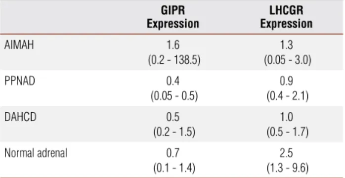
The expression of GIPR and LHCGR was
demon-strated in all tissues studied. GIPR expression was very
low in all normal adrenal tissues studied (median level: 0.7, ranging from 0.1 to 1.4) while LHCGR
expres-sion levels in normal adrenal tissues were more vari-able (median level: 2.5, ranging from 1.3 to 9.6). Median GIPR mRNA levels were 1.6, 0.4 and 0.5 in
adrenocortical tissues from patients with AIMAH, PPNAD and Cushing’s disease respectively (Figure 1). Median LHCGR mRNA levels were 1.3, 0.9
and 1.0 in adrenocortical tissue from patients with AIMAH, PPNAD and Cushing’s disease respectively (Figure 2). The median and ranges of both receptor expressions are shown in table 2.
No significant difference in GIPR expression was
observed among these forms of adrenocortical hy-perplasia and normal adrenals, while it was observed that the LHCGR expression was lower in AIMAH
(p = 0.02) and Cushing’s disease (p = 0.02), but not statistically significant in PPNAD when compared to normal adrenals (p = 0.06). We did not observe any statistical difference between GIPR and LHCGR
expressions in the different forms of hyperplasia stu-died (Table 2).
Figure 1. Expression levels of GIPR mRNA of 20 adrenal hyperplasia cases: five PPNAD, eight hyperplasia cases secondary to CD (Cushing’s disease) and seven AIMAH compared to eight normal adrenals.
Figure 2. Expression levels of LHCGR mRNA of 20 adrenal hyperplasia cases: five PPNAD, eight hyperplasia cases secondary to CD (Cushing’s disease) and seven AIMAH, compared to eight normal adrenals.
Table 2. GIPR and LHCGR relative expression levels (median and range) in
adrenocortical hyperplasia
GIPR Expression
LHCGR Expression
AIMAH 1.6
(0.2 - 138.5)
1.3 (0.05 - 3.0)
PPNAD 0.4
(0.05 - 0.5)
0.9 (0.4 - 2.1)
DAHCD 0.5
(0.2 - 1.5)
1.0 (0.5 - 1.7)
Normal adrenal 0.7 (0.1 - 1.4)
2.5 (1.3 - 9.6)
DAHCD: diffuse adrenal hyperplasia secondary to Cushing’s disease.
We did not find a correlation between GIPR and LHCGR expression levels in adrenocortical tissues and pre-surgical hormonal levels (p > 0.05).
DISCUSSION
Cushing’s syndrome secondary to aberrant hormone re-ceptors has been described by several authors in the last decade (7,29). This condition has been largely identified in AIMAH and adenomas, and some sporadic cases of hyperplasia secondary to Cushing’s disease and PPNAD were also related to this mechanism (2,8,19). Recently,
1000
100
N PPNAD CD AIMAH
10
1
0,1
0,01
GIPR Expression (Log transformation)
(Fold of normal adrenal pool)
10
N PPNAD CD AIMAH
1
0,1
0,01
LHCGR Expression (Log transformation)
Copyright
© ABE&M todos os dir
eitos r
eser
vados.
new patterns of LHCGR and GIPR expressions have been described, implicating both receptors in the pathophysi-ology of aldosterone-secreting tumors and androgen se-cretion resulting in hirsutism, suggesting a larger role of G-protein receptors in adrenocortical disease (26,30).
To investigate the potential role of GIPR and LH-CGR expression in adrenocortical hyperplasia we stud-ied a group of patients (20 cases) with adrenal enlarge-ment due to several etiologies.
Despite the molecular mechanism responsible for the aberrant expression of these receptors, they still need to be clarified (2,31-34); experimental studies have demonstrated that abnormal expression of GIPR and LHCGR in adrenocortical cells provoke phenotyp-ic changes in these cells, leading to the deregulation in their proliferation fate and eliciting adrenocortical tum-origenesis. This hyperproliferative adrenocortical tissue lead to GIP or LH-dependent secretion of cortisol and low ACTH levels (35,36).
The abnormal adrenal expression of the functional LHCGR has been identified in some cases of AIMAH and adrenocortical tumors (23-25,37); this receptor ex-pression has been documented in normal adrenal gland (38), although it was not well investigated in other forms of adrenocortical hyperplasia; We therefore ex-amine the LHCGR expression in eight cases of adreno-cortical hyperplasia secondary to Cushing’s disease and five cases of PPNAD that have presented lower LHCGR expression when compared to normal adrenal tissues. Our results do not support the role of this receptor in the adrenal enlargement due to such disorders.
The hypothesis that chronic stimulation or activa-tion of the ACTH signaling pathway may be associated to GIPR expression suggested by the results of Swords and cols.(19) in all five patients with diffuse adrenocor-tical hyperplasia secondary to Cushing’s disease and one case with PPNAD has not been confirmed (20,39). In our study, no difference in GIPR expression was ob-served in the different forms of adrenocortical hyper-plasia studied: adrenal hyperhyper-plasia secondary to Cush-ing’s disease; AIMAH and PPNAD, as well as between adrenocortical hyperplasia and normal adrenals, which did not confirm the previously described find-ings. However, the patient 1 showed a high expression level (138.5) bringing into evidence the great variability of GIPR expression in the adrenal hyperplasia (40,41).
In conclusion, we ruled out GIPR and LHCGR
overexpression as being related to adrenocortical hy-perplasia due to AIMAH and PPNAD in our cases. Our
results suggest that GIPR is not involved in the mo-lecular mechanisms implicated in the development of diffuse adrenocortical hyperplasia of Cushing’s disease. These data support the idea that the role of ACTH stimulation in the regulation of GIPR ectopic expres-sion might be really reduced.
Acknowledgements: the authors thank to the staff of Laboratório de Hormônios e Genética Molecular LIM/42, and particularly, Emilia Modolo Pinto and Maria Aparecida Medeiros for providing excellent technical support. We also thank Doctor Alexander Au-gusto Jorge and Doctor Silvia Correa Souza Leão for the statistical analysis. This research was supported in part by Fundação de Ampa-ro a Pesquisa do Estado de São Paulo (Fapesp Grants 03/07449-1 to M.H.S.C and 04/15046-7 to B.B.M.) and by Conselho Nacio-nal de Desenvolvimento Científico e Tecnológico (CNPq Grants 300828/2005-5 to B.B.M. and 300469/2005-5 to A.C.L.).
Disclosure: there is no conflict of interest between the authors and the funding agencies that would prejudice the impartiality of this work.
REFERENCES
Lacroix A, Ndiaye N, Tremblay J, Hamet P. Ectopic and abnormal 1.
hormone receptors in adrenal Cushing’s syndrome. Endocr Rev. 2001;22(1):75-110.
Lacroix A, Baldacchino V, Bourdeau I, Hamet P, Tremblay J. 2.
Cushing’s syndrome variants secondary to aberrant hormone re-ceptors. Trends Endocrinol Metab. 2004;15(8):375-82.
Stratakis CA, Boikos SA. Genetics of adrenal tumors associa-3.
ted with Cushing’s syndrome: a new classification for bilateral adrenocortical hyperplasias. Nat Clin Pract Endocrinol Metab. 2007;3(11):748-57.
Bourdeau I, D’Amour P, Hamet P, Boutin JM, Lacroix A. Aberrant 4.
membrane hormone receptors in incidentally discovered bilate-ral macronodular adrenal hyperplasia with subclinical Cushing’s syndrome. J Clin Endocrinol Metab. 2001;86(11):5534-40. Bourdeau I, Antonini SR, Lacroix A, Kirschner LS, Matyakhina 5.
L, Lorang D, et al. Gene array analysis of macronodular adre-nal hyperplasia confirms clinical heterogeneity and identifies several candidate genes as molecular mediators. Oncogene. 2004;23(8):1575-85.
Bourdeau I, Matyakhina L, Stergiopoulos SG, Sandrini F, Boikos 6.
S, Stratakis CA. 17q22-24 chromosomal losses and alterations of protein kinase a subunit expression and activity in adrenocorti-cotropin-independent macronodular adrenal hyperplasia. J Clin Endocrinol Metab. 2006;91(9):3626-32.
Bertherat J, Contesse V, Louiset E, Barrande G, Duparc C, Grous-7.
sin L, et al. In vivo and in vitro screening for illegitimate recep-tors in adrenocorticotropin-independent macronodular adrenal hyperplasia causing Cushing’s syndrome: identification of two cases of gonadotropin/gastric inhibitory polypeptide-dependent hypercortisolism. J Clin Endocrinol Metab. 2005;90(3):1302-10. Costa MH, Lacroix A. Cushing’s syndrome secondary to ACTH-8.
independent macronodular adrenal hyperplasia. Arq Bras Endo-crinol Metabol. 2007;51(8):1226-37.
Bourdeau I, Lampron A, Costa MH, Tadjine M, Lacroix A. Adre-9.
nocorticotropic hormone-independent Cushing’s syndrome. Curr Opin Endocrinol Diabetes Obes. 2007;14(3):219-25.
Stratakis CA, Kirschner LS, Carney JA. Clinical and molecular 10.
recom-Copyright
© ABE&M todos os dir
eitos r
eser
vados.
mendations for patient evaluation. J Clin Endocrinol Metab. 2001;86(9):4041-6.
Kirschner LS, Carney JA, Pack SD, Taymans SE, Giatzakis C, Cho 11.
YS, et al. Mutations of the gene encoding the protein kinase A type I-alpha regulatory subunit in patients with the Carney com-plex. Nat Genet. 2000;26(1):89-92.
Horvath A, Boikos S, Giatzakis C, Robinson-White A, Groussin L, 12.
Griffin KJ, et al. A genome-wide scan identifies mutations in the gene encoding phosphodiesterase 11A4 (PDE11A) in individuals with adrenocortical hyperplasia. Nat Genet. 2006;38(7):794-800. Usdin TB, Mezey E, Button DC, Brownstein MJ, Bonner TI. Gastric 13.
inhibitory polypeptide receptor, a member of the secretin-vasoac-tive intestinal peptide receptor family, is widely distributed in peri-pheral organs and the brain. Endocrinology. 1993;133(6):2861-70. Lacroix A, Bolté E, Tremblay J, Dupré J, Poitras P, Fournier H, 14.
et al. Gastric inhibitory polypeptide-dependent cortisol hyper-secretion--a new cause of Cushing’s syndrome. N Engl J Med. 1992;327(14):974-80.
Herder WW, et al. Food-dependent Cushing’s syndrome resul-15.
ting from abundant expression of gastric inhibitory polypeptide receptors in adrenal adenoma cells. J Clin Endocrinol Metab. 1996;81:3168-72.
Chabre O, Liakos P, Vivier J, Chaffanjon P, Labat-Moleur F, Martinie M, 16.
et al. Cushing’s syndrome due to a gastric inhibitory polypeptide-de-pendent adrenal adenoma: insights into hormonal control of adreno-cortical tumorigenesis. J Clin Endocrinol Metab. 1998;83(9):3134-43. Lebrethon MC, Avallet O, Reznik Y, Archambeaud F, Combes J, 17.
Usdin TB, et al. Food-dependent Cushing’s syndrome: characte-rization and functional role of gastric inhibitory polypeptide re-ceptor in the adrenals of three patients. J Clin Endocrinol Metab. 1998;83(12):4514-9.
N’Diaye N, Hamet P, Tremblay J, Boutin JM, Gaboury L, Lacroix A. 18.
Asynchronous development of bilateral nodular adrenal hyper-plasia in gastric inhibitory polypeptide-dependent cushing’s syn-drome. J Clin Endocrinol Metab. 1999;84(8):2616-22.
Swords FM, Aylwin S, Perry L, Arola J, Grossman AB, Monson JP, 19.
et al. The aberrant expression of the gastric inhibitory polypepti-de (GIP) receptor in adrenal hyperplasia: does chronic adrenocor-ticotropin exposure stimulate up-regulation of GIP receptors in Cushing’s disease? J Clin Endocrinol Metab. 2005;90(5):3009-16. Antonini SR, Baldacchino V, Tremblay J, Hamet P, Lacroix A. Ex-20.
pression of ACTH receptor pathway genes in glucose-dependent insulinotrophic peptide (GIP)-dependent Cushing’s syndrome. Clin Endocrinol (Oxf). 2006;64:29-36.
Ascoli M, Fanelli F, Segaloff DL. The lutropin/choriogonadotropin 21.
receptor, a 2002 perspective. Endocr Rev. 2002;23(2):141-74. Abdallah MA, Lei ZM, Li X, Greenwold N, Nakajima ST, Jauniaux 22.
E, et al. Human fetal nongonadal tissues contain human chorionic gonadotropin/luteinizing hormone receptors. J Clin Endocrinol Metab. 2004;89(2):952-6.
Lacroix A, Hamet P, Boutin JM. Leuprolide acetate therapy in 23.
luteinizing hormone--dependent Cushing’s syndrome. N Engl J Med. 1999;341(21):1577-81.
Feelders RA, Lamberts SW
24. , Hofland LJ, van Koetsveld PM, Verho-ef-Post M, Themmen AP, et al. Luteinizing hormone (LH)-res-ponsive Cushing’s syndrome: the demonstration of LH receptor messenger ribonucleic acid in hyperplastic adrenal cells, which respond to chorionic gonadotropin and serotonin agonists in vi-tro. J Clin Endocrinol Metab. 2003;88(1):230-7.
Goodarzi MO, Dawson DW, Li X, Lei Z, Shintaku P, Rao CV, et 25.
al. Virilization in bilateral macronodular adrenal hyperplasia controlled by luteinizing hormone. J Clin Endocrinol Metab. 2003;88(1):73-7.
Saner-Amigh K, Mayhew BA, Mantero F, Schiavi F, White PC, Rao 26.
CV, et al. Elevated expression of luteinizing hormone receptor
in aldosterone-producing adenomas. J Clin Endocrinol Metab. 2006;91(3):1136-42.
Lacroix AM, H. Hammet, P. Clinical evaluation of the presence of 27.
abnormal hormone receptors in adrenal Cushing’s syndrome. The Endocrinologist. 1999;(9):9-15.
Livak KJ, Schmittgen TD. Analysis of relative gene expression 28.
data using real-time quantitative PCR and the 2(-Delta Delta C(T)) Method. Methods. 2001;25(4):402-8.
Mircescu H, Jilwan J, N’Diaye N, Bourdeau I, Tremblay J, Hamet 29.
P, et al. Are ectopic or abnormal membrane hormone receptors frequently present in adrenal Cushing’s syndrome? J Clin Endo-crinol Metab. 2000;85(10):3531-6.
Tsagarakis S, Tsigos C, Vassiliou V, Tsiotra P, Pratsinis H, Kletsas D, 30.
et al. Food-dependent androgen and cortisol secretion by a gastric inhibitory polypeptide-receptor expressive adrenocortical adenoma leading to hirsutism and subclinical Cushing’s syndrome: in vivo and in vitro studies. J Clin Endocrinol Metab. 2001;86(2):583-9. Antonini SR, N’Diaye N, Hamet P, Tremblay J, Lacroix A. Analy-31.
sis of the putative promoter region of the GIP receptor gene (GIPR) in GIP-dependent Cushing’s syndrome (CS). Endocr Res. 2002;28(4):755-6.
Antonini SR, N’Diaye N, Baldacchino V, Hamet P, Tremblay J, La-32.
croix A. Analysis of the putative regulatory region of the gastric inhibitory polypeptide receptor gene in food-dependent Cushing’s syndrome. J Steroid Biochem Mol Biol. 2004;91(3):171-7. Baldacchino V, Oble S, Décarie PO, Bourdeau I, Hamet P, Tremblay 33.
J, et al. The Sp transcription factors are involved in the cellular ex-pression of the human glucose-dependent insulinotropic polypep-tide receptor gene and overexpressed in adrenals of patients with Cushing’s syndrome. J Mol Endocrinol. 2005;35(1):61-71. Lampron A, Bourdeau I, Hamet P, Tremblay J, Lacroix A. Whole 34.
genome expression profiling of glucose-dependent insulinotro-pic peptide (GIP)- and adrenocorticotropin-dependent adrenal hyperplasias reveals novel targets for the study of GIP-dependent Cushing’s syndrome. J Clin Endocrinol Metab. 2006;91(9):3611-8. Mazzuco TL, Chabre O, Feige JJ, Thomas M. Aberrant expression 35.
of human luteinizing hormone receptor by adrenocortical cells is sufficient to provoke both hyperplasia and Cushing’s syndrome features. J Clin Endocrinol Metab. 2006;91(1):196-203.
Mazzuco TL, Chabre O, Sturm N, Feige JJ, Thomas M. Ectopic ex-36.
pression of the gastric inhibitory polypeptide receptor gene is a sufficient genetic event to induce benign adrenocortical tumor in a xenotransplantation model. Endocrinology. 2006;147(2):782-90. Wy LA, Carlson HE, Kane P, Li X, Lei ZM, Rao CV. Pregnancy-asso-37.
ciated Cushing’s syndrome secondary to a luteinizing hormone/ human chorionic gonadotropin receptor-positive adrenal carcino-ma. Gynecol Endocrinol. 2002;16(5):413-7.
Pabon JE, Li X, Lei ZM, Sanfilippo JS, Yussman MA, Rao CV. 38.
Novel presence of luteinizing hormone/chorionic gonadotropin receptors in human adrenal glands. J Clin Endocrinol Metab. 1996;81(6):2397-400.
N’Diaye N, Tremblay J, Hamet P, De Herder WW, Lacroix A. Adre-39.
nocortical overexpression of gastric inhibitory polypeptide recep-tor underlies food-dependent Cushing’s syndrome. J Clin Endo-crinol Metab. 1998;83(8):2781-5.
Croughs RJ, Zelissen PM, van Vroonhoven TJ, Hofland LJ, N’Diaye 40.
N, Lacroix A, et al. GIP-dependent adrenal Cushing’s syndrome with incomplete suppression of ACTH. Clin Endocrinol (Oxf). 2000;52(2):235-40.
Groussin L, Perlemoine K, Contesse V, Lefebvre H, Tabarin A, 41.