ABSTRACT
BACKGROUND AND OBJECTIVES: Temporomandibular dysfunction is deined as a set of dysfunctions that afect the masticatory muscles, the temporomandibular joint and asso-ciated structures. he objective of this study was to systematize scientiic evidence on the techniques of physiotherapeutic treat-ment for temporomandibular disorders.
CONTENTS: he search was performed on the Medline, LI-LACS and Scielo databases, as well as the Pubmed search tool for articles published in the last 10 years, from August 2006 to August 2016. he survey was carried out with the following descriptors: “temporomandibular joint” and “physiotherapy”, “temporomandibular joint disorders” and “physiotherapy”, “temporomandibular joint” and “physiotherapy techniques”, “temporomandibular joint disorders” and “physiotherapy tech-niques”. We included randomized trials and case reports, com-posed only of patients with temporomandibular disorders who underwent physical therapy. he search totaled 32 studies and 11 of them were selected. he pain was assessed by unanimity. he articles did the same amount of sessions.
CONCLUSION: Several resources such as ultrasound, laser, ca-thodic current; or manual therapies, as muscle stretching, and joint mobilization bring remarkable beneits to temporomandi-bular dysfunction. However, studies with higher methodological quality with follow-up are necessary.
Keywords: Physiotherapy, Temporomandibular dysfunction, Temporomandibular joint, Temporomandibular joint disorders.
Physiotherapeutic treatment in temporomandibular disorders
Tratamento fisioterapêutico nas desordens temporomandibulares
Marcelo Pelicioli1, Rafaela Simon Myra1, Vivian Carla Florianovicz1, Juliana Secchi Batista2
1. Universidade do Estado Santa Catarina, Faculdade de Fisioterapia, Florianópolis, SC, Brasil. 2. Universidade de Passo Fundo, Disciplina de Fisioterapia, Carazinho, RS, Brasil.
Submitted in May 03, 2017.
Accepted for publication in November 08, 2017. Conlict of interests: none – Sponsoring sources: none
Correspondence to:
BR Km 292, Av. Brasil Leste, 285 - São José 99052-900 Passo Fundo, RS, Brasil. E-mail: [email protected]
© Sociedade Brasileira para o Estudo da Dor
RESUMO
JUSTIFICATIVA E OBJETIVOS: A disfunção temporomandi-bular é deinida como um conjunto de disfunções que acometem os músculos mastigatórios, a articulação temporomandibular e estruturas associadas. O objetivo deste estudo foi sistematizar evidências cientíicas sobre técnicas de tratamento isioterapêu-tico para as desordens temporomandibulares.
CONTEÚDO: A busca foi realizada a partir da consulta às ba-ses de dados Medline, LILACS e Scielo, além da ferramenta de busca Pubmed dos artigos publicados nos últimos 10 anos, de agosto 2006 à agosto de 2016. O levantamento foi realizado com os seguintes descritores: “articulação temporomandibular” e “isioterapia”, “transtornos da articulação temporomandibular” e “isioterapia”, “temporomandibular joint” and “physiotherapy techniques”, “temporomandibular joint disorders” and “physio-therapy techniques”. Foram incluídos ensaios randomizados e relatos de casos, compostos apenas por pacientes com desordens temporomandibulares que realizaram tratamento isioterapêuti-co. A busca totalizou 32 estudos e destes, foram selecionados 11 artigos. A dor foi avaliada por unanimidade. Os artigos realiza-ram a mesma quantidade de sessões.
CONCLUSÃO: Diversos recursos como o ultrassom, laser, cor-rente catódica, ou ainda, terapias manuais como alongamento muscular e mobilização articular trazem benefícios notáveis na dor da disfunção temporomandibular. Porém, estudos com maior qualidade metodológica com follow-up são necessários Descritores: Articulação temporomandibular, Disfunção tem-poromandibular, Fisioterapia, Transtornos da articulação tempo-romandibular.
INTRODUCTION
Temporomandibular joint (TMJ) is considered the most com-plex structure of the human body. TMJ performs rotational and translational movements due to the double articulation of the temporal bone condyle. he fact that TMJ presents two joints (condyles) connected to the mandible requires that they work synchronously between dental occlusion, neuromuscular bal-ance and the joint itself. his joint is vulnerable to functional or pathological alterations, leading to disorders such as temporo-mandibular disorder (TMD)1.
muscle and TMJ sensitivity to palpation, limitation and/or dis-turbances of mandibular movement and joint noises. It is esti-mated that 40 to 75% of the population has at least one TMD signal, such as noise, and at least one symptom, such as facial pain or TMJ (33%)2.
TMD afects a large part of the world’s population. Due to this fact, it is essential to develop therapeutic techniques for its treat-ment. Physiotherapy contributes to lessening the TMD symp-toms, as it stimulates proprioception, production of synovial luid in the joint, improves the elasticity of adhered muscle ibers and pain3.
hus, to minimize the efects caused by TMD, physiotherapy becomes a fundamental and integral part of these patients’ treatment.
Given the above, this study aimed to organize the scientiic evi-dence on the physiotherapeutic treatments used in patients with temporomandibular disorders.
CONTENTS
he systematic review was carried out from a retrospective con-sultation with Scielo, Pubmed and LILACS databases. Articles collection was carried out in September 2016, and the search strategy was formulated through the descriptors crossing (DeCS and MeSH). Only the researches with patients diagnosed with temporomandibular dysfunction or disorder and treated with physiotherapy techniques were included. Also, the studies should be in Portuguese or English, published from August 2006 to August 2016. In Scielo and LILACS (DeCS) bases, the fol-lowing crossings were used: “temporomandibular joint” AND “physiotherapy” OR “temporomandibular joint disorders” AND
through crossings between “temporomandibular joint” AND “physiotherapy techniques” OR “temporomandibular joint dis-orders” AND “physiotherapy techniques”. In the initial phase, titles and abstracts were independently identiied and evaluated by two reviewers to select those meeting the eligibility criteria. Articles that did not it the criteria described were excluded by the title review, followed by exclusion by the abstract, and inally, the potentially relevant studies were retained for further analysis of the full text. he relevant information was presented in the form of descriptive tables, considering the following variables: year, country, sample, evaluated outcomes, methodological de-sign, intervention, and efects found.
In the initial search in the databases were found 32 articles. After the irst selection by title, 13 articles were excluded, remaining 16 for abstracts analysis. From these, 11 articles were selected that it the established inclusion criteria. Figure 1 shows the se-lecting process of the included articles. Table 1 presents the list of selected studies that used physiotherapeutic techniques for temporomandibular disorders treatment.
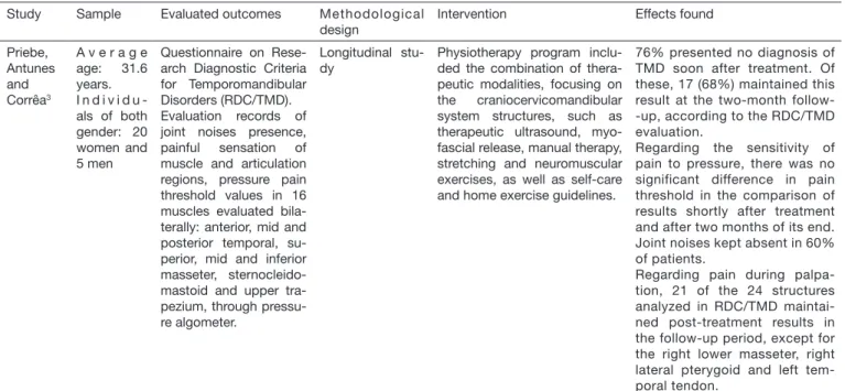
Table 1. Description of selected studies that used physiotherapeutic techniques for temporomandibular disorders treatment
Study Sample Evaluated outcomes Methodological design
Intervention Effects found
Priebe, Antunes and Corrêa3
A v e r a g e age: 31.6 years. I n d i v i d u -als of both gender: 20 women and 5 men
Questionnaire on Rese-arch Diagnostic Criteria for Temporomandibular Disorders (RDC/TMD). Evaluation records of joint noises presence, painful sensation of muscle and articulation regions, pressure pain threshold values in 16 muscles evaluated bila-terally: anterior, mid and posterior temporal, su-perior, mid and inferior masseter, sternocleido-mastoid and upper tra-pezium, through pressu-re algometer.
Longitudinal stu-dy
Physiotherapy program inclu-ded the combination of thera-peutic modalities, focusing on the craniocervicomandibular system structures, such as therapeutic ultrasound, myo-fascial release, manual therapy, stretching and neuromuscular exercises, as well as self-care and home exercise guidelines.
76% presented no diagnosis of TMD soon after treatment. Of these, 17 (68%) maintained this result at the two-month follow--up, according to the RDC/TMD evaluation.
Regarding the sensitivity of pain to pressure, there was no significant difference in pain threshold in the comparison of results shortly after treatment and after two months of its end. Joint noises kept absent in 60% of patients.
Regarding pain during palpa-tion, 21 of the 24 structures analyzed in RDC/TMD maintai-ned post-treatment results in the follow-up period, except for the right lower masseter, right lateral pterygoid and left tem-poral tendon.
Figure 1. Data search
Databases: Scielo, Pubmed and LILACS (n=32)
Deleted articles (n=13) Reading titles
Deleted articles (n=5) Abstracts analysis (n=16)
Selected articles (n=11)
Study Sample Evaluated outcomes Methodological design
Intervention Effects found
Franco et al.16
A 35-year--old female patient 10 ses-sions, 1 time per week
Physiotherapeutic eva-luation sheet, composed of ROM evaluation, ins-pection, palpation, phy-sical exams.
Case report, eva-luated before and after intervention and reassessed 15, 30 and 60 days after inter-vention.
Performed passive stretching of ECOM and trapezium, low--intensity laser application of gallium arsenide (AS-GA) 4J parameters for the area of the joint in a punctual form and 8J in the muscular area in the punctual form and scanning with 1mm, with pulsatile mode 1 min per point. Facial relaxa-tion with slip techniques, guide-lines for complementary home exercises, active stretching of the cervical muscles, extensors and lexors of the head and neck. MTP deactivation techni-que. Night maintenance of the myorelaxing occlusal plaque
There was a gradual reduction of painful sensations through VAS, the relief average of pain symp-toms was 20% per session, rea-ching zero in the last sessions.
Freire et al.6
A v e r a g e age: 34.5 years
24 individu-als (21 fe-males and 3 males)
Questionnaire on Rese-arch Diagnostic Criteria for Temporomandibular Disorders (RDC/TMD) Temporomandibular in-dex (TMI) and its sub--indexes
Longitudinal stu-dy, 10 sessions. Evaluated before treatment (AV1), immediately after treatment (AV2) and two months after the end of treatment (AV3)
Continuous 3 MHz ultrasound, with the intensity of 1.3 W/cm2, for 3 minutes for chronic pain; in pulsed mode with an intensi-ty of 0.5 W/cm 2, for 3 minutes for acute pain.
Supericial thermotherapy with infrared radiation for 20 minutes. Myofascial release and stre-tching bilaterally.
Techniques of distraction and therapeutic massage in the cer-vical spine and the TMJ. Exercise with silicone rubber tube
Reduction in diagnoses number in all subgroups and diagnosis absence in 41.7% of the 24 par-ticipants after treatment.
Signiicant reduction of TMI in the comparison between AV1 and AV2 (p = 0.000). There was no di-fference between AV2 and AV3 (p = 0.204) in 13 participants evalu-ated two months after the end of treatment.
Amaral et al.7
A v e r a g e age: 25.6 years.
50 individu-als of both gender
Questionnaire on Rese-arch Diagnostic Criteria for Temporomandibular Disorders (RDC/TMD) Stabilometric evaluation on a force platform, with eyes open and closed.
Longitudinal stu-dy DTM Group (presenting TMD, mandibular de-viation or delec-tion) and control group (not pre-senting TMD)
Non-speciic mandibular mo-bilization (MMI). The patient is positioned in dorsal decubitus and disposable gloves were used by the therapist; the ifth chierodactyl positioned on top of the second or third molar (if present) to perform the MMI in a small degree intermittently for one minute, with ive repli-cates being performed. Betwe-en each mobilization, a buccal opening with tongue was per-formed ten times on the inci-sive papilla, to promote local relaxation.
Statistically signiicant difference was only for the TMD group at the center of pressure oscillation (p <0.03) in the mediolateral displa-cement (p <0.006), in the medio-lateral amplitude (p <0.01) and in the velocity variable in the antero-posterior directions, (p <0.03) and mediolateral (p <0.03).
Gomes et al.5
A v e r a g e age: 22.5 years.
25 individu-als of both gender.
RDC/TMD questionnai-re. - Evaluation of pain through VAS.
R a n d o m i z e d , double-blind cli-nical trial. EG (experimental group): 10 appli-cations of HVES and in PG (pla-cebo group): 10 applications with the device swi-tched off.
Electrodes placed bilaterally on the lateral portion of the tem-poralis muscle (channel 1), on the masseter (channel2) and the electrode dispersed in the cervical-thoracic (lower cervi-cal high thoracic) region. Para-meters used 10Hz frequency; pulse width ixed by the device in two twin pulses of 20us each with an interval of 100us volta-ge at 100 volts both channels lasting 30 min 2 to 3 times per week.
Intragroup comparison observed that 10 applications of cathodic HVES promoted the reduction of pain in the EG, while in the GP no difference was noticed. EG pre-sented greater reduction of pain intensity compared to PG.
Continue...
Study Sample Evaluated outcomes Methodological design
Intervention Effects found
Borin et al.8
40 women, aged be-tween 20 and 40 ye-ars
RDC-TMD questionnai-re, the severity of TMD was veriied before and after treatment by Fonseca’s Index. It was also evaluated the Cra-niomandibular Dysfunc-tion Index. The pain was assessed before and af-ter treatment, by VAS.
Randomized cli-nical trial. Indi-viduals divided into two groups: AG: acupuncture, who performed intervention twice a week (n = 20); and control CG: who did not un-dergo treatment
AG participants underwent acupuncture twice a week for ive uninterrupted weeks. The treatment was performed with disposable needles (0.25 x 0.15 mm) inserted in the respective points with the skin previously cleaned with cotton and ethylic alcohol at 70%. Acupuncture therapy amounted 10 assistan-ces. The points selected for tre-atment were those referred to in the literature as points for the treatment of TMD and points for anxiety.
There was an improvement in the severity level by the craniomandi-bular index (p = 0.004) and by the Fonseca’s Index (p = 0.000) of in-dividuals with TMD after acupunc-ture treatment, and in the pain le-vel (p = 0.000). According to the classiication by Fonseca’s Index. Before treatment, the individuals had the following classiication for TMD: 6 with a moderate degree and 14 with a severe degree. After treatment, this classiication was observed: 7 with a mild degree, 10 moderate and 3 severe.
Basso, Corrêa and Silva4
P a r t i c i p a -ted in the study 20 in-dividuals of both gen-der Average age: 27.5 years. RDC/TMD questionnai-re.
Photography with a di-gital camera for postural evaluation.
Transversal, qua-litative study, 10 weeks of inter-vention.
GI: muscle disor-der; GII: disk dis-placement; GIII: other joint condi-tions.
The intervention group was submitted to 10 sessions of GPR for 45 minutes, once a week adopting two postures per session of therapy. Postu-res without load and postuPostu-res with load.
GII obtained an improvement in the reduction of chronic orofacial pain.
Calixtre et al.12
12 wo-men with a mean age of 22.08 ± 2.23 years
The mandible’s pain and function were evaluated with the MFIQ, in addi-tion to the opening level of the mouth without pain and the GP of mas-seter and temporal mus-cle were evaluated.
L o n g i t u d i n a l study, pre-and post-evaluation 5 weeks of inter-vention.
Submitted to 10 sessions of approximately 35 min. Mobili-zation of cervical under lexion, anteroposterior and posteroan-terior mobilization in C5, stabi-lization exercise of craniocervi-cal lexion, stretching
Mandibular function increased by 7 points on the scale after the inter-vention (p = 0.019) and pain decre-ased signiicantly (p = 0.009). The mandible level of opening ranged from ± 8.8 to 38.8.8 mm to 38 ± 8.8 showing signiicant improvement (p = 0.017), pain on both masseters and temporalis improved
Machado et al.11
P a r t i c i p a -ted in the study 82 individuals with chronic TMD and 20 healthy individuals ( a v e r a g e age 30 ± 9.6 years)
DTM severity through Part II of ProDTMMul-ti QuesProDTMMul-tionnaire, stress points due to palpation, and orofacial functionali-ty by Orofacial Myofunc-tional Evaluation with Scores.
Randomized cli-nical trial. Partici-pants were divided into GI: Laser and o r o m a n d i b u l a r exercises, GII: orofacial muscu-lar therapy, GIII: placebo laser and o r o m a n d i b u l a r exercises, GIV: La-ser, CG: healthy.
Submitted to 12 sessions of 45min. GI: continuous laser I = 60mW by 40s and D = 60 ± 1.0 J/cm2 and exercises for tongue,
cheeks and mandibular mus-cles, functional orofacial trai-ning; GII: exercises for tongue, cheeks and mandibular mus-cles, functional orofacial trai-ning; laser; strategies for pain reduction; GIII: placebo laser and exercises; GIV: laser.
There was an improvement in both groups in all scopes asses-sed with stability at follow-up when compared to each other all treated groups did not show di-fferences in stress points due to palpation at follow-up. GI, GII and GIII showed no difference with the control over orofacial function, while they differed signiicantly from GIV (p <0.01).
Oliveira et al.15
32 young adults with an average age of 24.7 ± 6.8 years diagnosed with TMD.
Fonseca’s questionnaire was used for the initial screening of patients; then VAS was used for pain and WHOQOL--BREF was used for Quality of Life.
Clinical trial, d o u b l e - b l i n d . The patients were divided into two groups: A - active submit-ted to exercise plus transcra-nial noninvasive stimulation and B - control who performed exer-cises plus false stimulation.
The treatment protocol lasted 4 weeks; all participants per-formed exercises that included myofascial release, muscle stre-tching, cervical traction, exer-cises to improve mandible’s ROM, muscle strengthening, among others. In addition, Group A received 20 min of no-ninvasive transcranial stimula-tion with an amplitude of 2 mA, with electrodes located on C3 or C4 (motor cortex region); in the individuals of Group B the electrodes were positioned in the same place, but the current lasted 30 seconds.
The clinical characteristics of the disease in the two groups were the same after the treatment, and regarding the quality of life, it can be perceived that both groups obtained positive results. Groups’ pain intensity decreased after the second day of treatment, but in different ways and, in the end, it was noticed that Group A had lo-wer pain levels than Group B, but the difference was not statistically signiicant.
Continue...
DISCUSSION
his study revealed efective results in relation to the physiother-apeutic treatments used for TMD. Basso, Corrêa and Silva4 and
Gomes et al.5 studies reported that physical therapy is capable of
promoting improvement of clinical symptoms related to pain. Besides, in general, physiotherapy stimulates the proprioception and production of synovial luid in the joint and improves the elasticity of adhered muscle ibers3.
Analyzing the results obtained by the search strategy, it was ob-served a greater concentration of studies in 2015, with a single publication in 2010. It is worth mentioning that the surveys were developed in North and South American territory. It is also evidenced that the studies participants were volunteers of difer-ent age groups. However, the average age of the samples analyzed corresponded to the middle-aged population. hese same studies indicate a high percentage of women. However, there is still no consensus in the literature about the reason for the higher preva-lence in females than in males.
From the 11 articles that were used in this study, seven used the Questionnaire of Research Diagnostic Criteria for Temporo-mandibular Disorders (RDC/TMD) as an evaluation form. his questionnaire is recognized worldwide and aims to establish reli-able and valid criteria for diagnosing and deining TMD sub-types3-9. In the case report studies, a physical therapy assessment
sheet was used, which included: inspection, range of motion, palpation and physical examination10.
Another important aspect in the treatment of TMD is the fre-quency and duration of physiotherapy sessions. Considering the number of sessions, in seven studies were performed 10. How-ever, in the study by Freitas et al.10, the author felt the need for
a larger number of sessions, totaling 15 sessions. his shows that most authors agree with the number of sessions performed. When analyzing the sessions frequency, there was disagreement with the studies by Priebe et al.3 and Basso, Corrêa and Silva4 for
example, where the number was once a week, while Borin et al.8,
Freitas et al.10 performed twice a week.
It should be emphasized that TMD may be related to posture. In the studies by Basso, Corrêa and Silva4 and Freitas et al.10, the
postural evaluation was carried out in order to ind evidence such as head anterioration, cervical lordosis increase and no leveling of the shoulders. Amaral et al.7 used stabilometry as an evaluation
method. his test is a way to measure the static balance, which consists of quantifying the anteroposterior and lateral body os-cillations, while the individual remains standing on a force plat-form. hese parameters evaluation becomes important, as it is known that TMD can cause changes in balance.
TMD may present as muscular and/or articular pain, decreased buccal amplitude, headache, mandibular movement disorders, and articular cracking5. During the research process, it was noted
that the pain variable was the only one unanimously selected in the studies. Pain is one of the main symptoms reported by patients with TMD, with 75% of them experiencing temporo-mandibular joint discomfort or dysfunction. One of the
meth-Table 1. Description of selected studies that used physiotherapeutic techniques for temporomandibular disorders treatment – continuation
Study Sample Evaluated outcomes Methodological design
Intervention Effects found
Tosato et al.9
n = 20 wo-men, aged between 22 and 46 ye-ars, with an average of 31.75±8.71 years, with m y o g e n i c TMD, with m a s t i c a -tory muscle pain.
After this, RDC-TMD for pain used the VAS and surface electromyo-graphy to capture the electrical signal of the masseter and temporal muscles.
Randomized cli-nical study. Indi-viduals divided into G1: control and G2: interven-tion
The sample was divided into 2 groups: Group 1 received a 30-minute session of masso-therapy on the face, masse-ter and temporal region, while Group 2 received transcuta-neous electrical nerve stimula-tion for 30 minutes in the region of the masseter and temporal muscles.
Both groups showed an increase in the electromyographic activi-ty of the masseter and temporal muscles, both in isometric con-tractions and concentric isoto-nic. There was also a signiicant reduction in pain in both groups.
Freitas et al.10
1 patient, 37 years old, diag-nosed with TMD for 5 years.
From the VAS, the pain was evaluated; mandi-bular ROM and postural clinical evaluation were measured.
Clinical case stu-dy
The laser was used in the TMJ region using ad-hoc techni-que with energy density (ΔE) 3J/cm² and reaching a inal energy of 2.6 J, deactivation of myofascial trigger points in the masseter, pterygoid, temporal, occipital, scalene, ECOM and trapezius upper ibers, for 45 seconds at each point and joint mobilization using the longitudinal and an-terior cephalic slide technique of TMJ grade II
The patient presented improve-ment in pain, increased the joint amplitude of TMJ and in the fol-lowing postural alterations: prognata mandible, head in neu-tral position, cervical spine with physiological lordosis and shoul-ders aligned. In the muscular por-tion, there was an improvement in the time of muscular activation in the face muscles.
(VAS)5,9,10,16. Only the study performed by Gomes at al.5
present-ed a sample calculation baspresent-ed on the standard deviation values obtained by VAS, providing measurements of pain intensity. In turn, Priebe, Antunes and Corrêa3 used the pressure algometer − Force Dial® FDK/FDN Dynamometer (Wagner Instruments) − as a method to evaluate this item. Both were satisfactory for the evaluation of pain parameters in these patients.
he physiotherapeutic treatment aims at relieving the symptoms, seeking to restore the normal function of the patient’s mastica-tory device, for which diferent techniques can be used. Accord-ing to the studies, the devices like laser, ultrasound and cathodic current, are beneicial in the treatment. However, manual thera-py through muscle stretching exercises, joint mobilizations and exercises for cervical segmental stabilization may be included in the rehabilitation process7,10-12,16.
Tosato, Biazotto-Gonzalez and Caria9 used electromyography to
evaluate the electrical activation of the masseter muscles and an-terior portion of the temporalis muscle and the VAS to measure the pain. he sample comprised of 20 women was divided into two groups. Both went through the evaluation process described. hen, group 1 was submitted to 30 minutes of massotherapy in the masseter region and anterior portion of the temporalis; group 2 received 30 minutes of transcutaneous electrical nerve stimulation in the same muscles. hen, both groups were reeval-uated, and both presented greater muscle activation and statisti-cally signiicant reduction of pain, showing that manual therapy is beneicial and can be used to reduce TMD pain. Machado et al.11 investigated the combination eicacy of low-intensity
thera-peutic laser use with oral motor exercises in the rehabilitation of TMD-patients. 82 patients were selected with chronic TMD and 20 were considered healthy patients, who formed the con-trol group. Individuals were randomly divided into 5 groups. GI: laser + orofacial exercises; GII: orofacial myofunctional ther-apy, which consisted of pain relief and orofacial exercises; GIII: placebo laser and orofacial exercises; GIV: laser. Laser aimed to analyze the analgesia (parameters used: 780-nm wave size, in-tensity of 60 mW, 40 and 60±1.0 J/cm²), and orofacial exercises were used to restore its functionality. All treated groups had a signiicant improvement over the control group. Comparing the treated groups, it was observed that the groups that used laser and orofacial exercises and orofacial myofunctional therapy ob-tained results that are more efective. Franco et al. 16 also used the
low-intensity laser, associated with cervical’s muscle stretching exercises and myorelaxing occlusal nocturnal plaque. he inter-vention lasted 10 sessions and reduced the pain.
Joint mobilization was chosen as a treatment in Amaral et al.7,
Freitas et al.10 and Calixtre et al.12 study. However, each study
had its particularity. In the study by Amaral et al.7, nonspeciic
mandibular mobilization (MMI) was used in order to promote improved postural control in individuals with TMD. Freitas et al.10, in addition to joint mobilization, used the deactivation of
myofascial trigger points and cervical stabilization exercise as a treatment for TMD, improving aspects such as pain, muscle bal-ance, and posture. Calixtre et al.12 performed C5 joint
mobiliza-tion, cervical stabilization exercises and passive muscle
stretch-mouth opening and pain reduction.
Basso, Corrêa and Silva4 show that the posture in individuals
with TMD is impaired. In this study was used the GPR, propos-ing a therapeutic action of stretchpropos-ing aimpropos-ing at the balance of myofascial tensions and body posture as a whole. his treatment can reduce the orofacial pain intensity and improve psychologi-cal symptoms of TMD, as well as improved body alignment and symmetry.
he treatment can also be focused on the craniocervicoman-dibular system structures, as pointed out by studies by Priebe, Antunes and Corrêa3 and Freire et al.6. he passive stretches of
the ECOM and trapezium muscles, facial relaxation with slide techniques, active stretching of the cervical musculature, head and neck extensors and lexors were techniques used.
he application of acupuncture needles can also bring beneits to the TMD handling, which aims at controlling pain, especially that of muscular origin. Borin et al.8 applied needles at speciic
points in the zygomatic arch region, in the masseter muscle and the mastoid process. After the intervention, it was observed that acupuncture promotes a signiicant reduction in the pain level and the severity of TMD, demonstrating a 75% reduction in pain grade (p = 0.000).
El Hage et al.13 investigated the immediate efect of facial
mas-sage on static balance in individuals with TMD. In this study, 20 individuals diagnosed with TMD were evaluated using an equilibration platform that calculated the oscillations occurred in the anteroposterior and mid-lateral planes. he evaluations occurred with eyes closed and open, at the irst moment be-fore rest (baseline), after 10 minutes rest in dorsal decubitus (pre-massage) and after the application of the technique (post-massage). Results showed that there was only a signiicant dif-ference in the evaluation performed with the eyes closed in the anteroposterior oscillations.
In other study performed by Amaral et al.14, the same
proto-col was used to evaluate the efects of non-speciic MMI. he participants were divided into two groups: 25 individuals with TMD and 25 individuals without TMD. he mobilization consisted of the therapist positioning the ifth chierodactyl on the second or third molar for a minute, in a small degree of amplitude, promoting the mandible in protrusion displace-ment by ive times. Between each repetition, there was a local relaxation. According to the evaluation after MMI, there was an improvement of the postural control in both groups, sug-gesting a possible stimulation of the trigeminal system that, in turn, would inluence the balance.
More current techniques for the TMD treatment were one of this review’s outcomes. Noninvasive stimulation, including tran-scranial direct current stimulation (TDCS), [AS1] [RSM2] may be considered as an alternative for pain treatment. Oliveira et al.15 evaluated pain and quality of life of TMD patients after
motion. Also, group A received 20 min of TDCS with an ampli-tude of 2 mA, with electrodes located on C3 or C4 (motor cortex region), while group B received sham stimulation. Groups’ pain intensity decreased diferently, and at the end of treatment, it was noticed that group A had lower pain levels than group B, but the diference was not statistically signiicant.
Physiotherapy is efective and improves the physical function of individuals with TMD. From this review, it is noticed that several resources such as ultrasound, laser, cathodic current or manual therapies such as muscle stretching, and joint mobiliza-tion bring remarkable beneits. However, with the poor meth-odological quality, small number of individuals participating in most studies leaves a gap on the best treatment for TMD. Besides, more randomized clinical trial studies and follow-up evaluations are needed.
CONCLUSION
Physiotherapy may beneit TMD patients by reducing pain and increasing mobility, as well as rebalancing the TMJ.
REFERENCES
1. Donnarumma MD, Muzilli CA, Ferreira C, Nemr K. Disfunções temporomandibula-res: sinais, sintomas e abordagem multidisciplinar. Rev CEFAC. 2010;12(5):788-94. 2. Carrara SV, Conti PC, Barbosa JS. Termo do 1º Consenso em Disfunção
Temporo-mandibular e Dor Orofacial. Dental Press J Orthod. 2010;15(3):114-20. 3. Priebe M, Antunes AG, Corrêa EC. Estabilidade dos efeitos da isioterapia na
disfun-ção temporomandibular. Rev Dor. 2015;16(1):6-9.
4. Basso D, Corrêa E, Silva MA. Efeitos da reeducação postural global no alinhamento corporal e nas condições clínicas de indivíduos com disfunção temporomandibular
associada a desvios posturais. Fisioter Pesqui. 2010;17(1):63-8.
5. Gomes NC, Berni-Schwarzenbeck KC, Packer AC, Bigaton DR. Efeitos da estimula-ção elétrica de alta voltagem catódica sobre a dor em mulheres com DTM. Rev Bras Fisioter. 2012;16(1):10-5.
6. Freire AB, Nardi AT, Bouleur J, Chiodelli L, Pasinato F, Corrêa EC. Multimodal physiotherapeutic approach: Efects on the temporomandibular disorder diagnosis and severity. Fisioter Mov. 2014;27(2):219-27.
7. Amaral AP, Politti F, Hage YE, Arruda EE, Amorin CF, Biasotto-Gonzalez DA. Efeito imediato da mobilização mandibular inespecíica sobre o controle postural em indi-víduos com disfunção temporomandibular: ensaio clínico controlado, randomizado, simples cego. Br J Phys her. 2013;7(2):121-7.
8. Borin GS, Corrêa EC, Silva MT, Milanesi JM. Acupuntura como recurso tera-pêutico na dor e na gravidade da desordem temporomandibular. Fisioter Pesqui. 2011;18(3):217-22.
9. Tosato JP, Biazotto-Gonzalez DA, Caria PH. Efeito da massoterapia e da estimulação elétrica nervosa transcutânea na dor e atividade eletromiográica de pacientes com disfunção temporomandibular. Fisioter Pesqui. 2007;14(2):21-6.
10. Freitas DG, Pinheiro IC, Vantin K, Meinrath NC, De Carvalho NA. Os efeitos da desativação dos pontos-gatilho miofasciais, da mobilização articular e do exercício de estabilização cervical em uma paciente com disfunção temporomandibular: um estudo de caso. Fisioter Mov. 2011;24(1):33-8.
11. Machado CC, Mazzeto MO, da Silva MA, de Felício CM. Efects of oral motor exer-cises and laser therapy on chronic temporomandibular disorders: a randomized study with follow-up. Lasers Med Sci. 2016;31(5):945-54.
12. Calixtre LB, Grüninger BL, Haik MN, Albuquerque-Sendín F, Oliveira AB. Efects of cervical mobilization and exercise on pain, movement and function in subjects with temporomandibular disorders: a single group pre-post test. J Appl Oral Sci. 2016;24(3):188-97.
13. El Hage Y, Politti F, Herpich CM, de Souza DF, de Paula Gomes CA, Amorim CF, et al. Efect of facial massage on static balance in individuals with temporomandibular disorder – a pilot study. Int J her Massage Bodywork. 2013;6(4):6-11.
14. Amaral AP, Politti F, Hage YE, Arruda EE, Amorin CF, Biasotto-Gonzales DA. Efeito imediato da mobilização mandibular inespecíica sobre o controle postural em indi-víduos com disfunção temporomandibular: ensaio clínico controlado, randomizado, simples cego. Braz J Phys her. São Carlos. 2013;17(2):121-7.
15. Oliveira LB, Lopes TS, Soares C, Maluf R, Goes BT, Sá KN, Baptista AF. Transcranial direct current stimulation and exercises for treatment of chronic temporomandibular disorders: a blind randomised-controlled trial. J Oral Rehabil. 2015;42(10):723-32. 16. Franco AL, Zamperini CA, Salata DC, Silva EC, Albino Júnior W, Camparis CM.