GLUCAGONOMASYNDROMEASSOCIATEDWITHNECROLYTICMIGRATORYERYTHEMA
REV ASSOC MED BRAS 2015; 61(3):203-206 203
IMAGE IN MEDICINE
Glucagonoma syndrome associated with necrolytic migratory
erythema
S
ÍNDROME DO GLUCAGONOMA ASSOCIADO A ERITEMA NECROLÍTICO MIGRATÓRIOFLORENTINODE ARAÚJO CARDOSO FILHO1*, RONEY GONÇALVES FECHINE FEITOSA2, CAROLINA OLIVEIRA COSTA FECHINE3,
CARLOS MÁRCIO MELODE MATOS4, AMANDA LINHARES CARDOSO5, DANIEL LINHARES CARDOSO6
1MSc in Surgery from the Universidade Federal do Ceará (UFC). MD, MSc, TCBC, FACS, President of the Associação Médica Brasileira, Fortaleza, CE, Brazil 2Resident in Plastic Surgery, Last year training as Resident in Plastic Surgery at Universidade Federal de São Paulo (Unifesp), São Paulo, SP, Brazil 3Dermatologist with a degree from the Hospital do Servidor Público do Estado de São Paulo, São Paulo, SP, Brazil
4Resident in General Surgery at the Dr. César Cals de Oliveira Hospital, Fortaleza, CE, Brazil 5Medical Student, 7th period, at Universidade Christus, Fortaleza, CE, Brazil
6Medical Student, 20th period, Universidade de Fortaleza, Fortaleza, CE, Brazil
Study conducted at the Department of General Surgery, Hospital Geral de Fortaleza, Fortaleza, CE, Brazil
Article received: 4/1/2015 Accepted for publication: 4/27/2015 *Correspondence: Address: Av. Dom Luis, 1233 – S/407 Aldeota Fortaleza, CE – Brazil Postal code: 60160-230 [email protected]
http://dx.doi.org/10.1590/1806-9282.61.03.203
Conflict of interest: none
S
UMMARYIntroduction: glucagonoma is a pancreatic neuroendocrine tumor derived from alpha-cells of the islets of Langerhans. It is marked by tumoral autonomous pro-duction of glucagon and characterized, among other symptoms, by necrolytic migratory erythema, an erythematous circinate lesion with areas of necrosis and sloughing. This is a rare disease with worldwide incidence estimated at 1 case per 20 million people.
Case report: we report a case of glucagonoma associated necrolytic migratory erythema in a male patient, 56 years, with signs of skin lesions mainly on his legs and groin, hyperglycemia and weight loss. Biopsies of the skin lesions were performed and imaging of the abdomen showed a mass of 10 x 9 cm, at the pan-creatic region. The patient was subjected to body-caudal pancreatectomy and splenectomy with autotransplant of the spleen in the greater omentum. The
his-topathologic report indicated a tumor in the pancreatic alpha cells. Immunohis-tochemistry showed expression of glucagon and chromogranin A in most tu-mor cells, consistent with the diagnosis of glucagonoma. The patient presented 3 years of outpatient follow-up with no complications.
Conclusion: the necrolytic migratory erythema is important for the clinical re-cognition of glucagonoma, and its early diagnosis is essential for a successful curative therapy.
Keywords: necrolytic migratory erythema, glucagonoma, pancreatic neoplasms, neuroendocrine tumors.
I
NTRODUCTIONGlucagonoma is a pancreatic neuroendocrine tumor deri-ved from alpha-cells of the islets of Langerhans. It is usually discovered through identification of a glucagonoma syn-drome, which includes tumoral autonomous production of glucagon, and is characterized by necrolytic migratory erythema (NME), diabetes mellitus, weight loss, deep vein
th-rombosis, neuropsychiatric disorders and diarrhea.1,2
NME is the hallmark of this syndrome, characterized by circinate erythema with areas of necrosis and
slou-ghing. It can be the first manifestation of the disease and its recognition may suggest the diagnosis.3
It is a rare disease that, combined with glucagonoma syndrome, has an estimated global incidence of 1 case per 20 million people.2
CARDOSO FILHO FA ETAL.
204 REV ASSOC MED BRAS 2015; 61(3):203-206
C
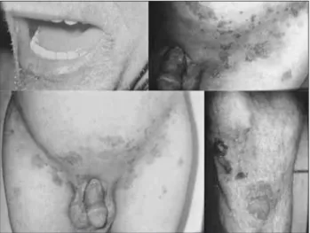
ASE REPORTA 56-year-old male patient was referred to the general sur-gery service of a hospital in Ceará to perform skin biopsy. He had diffuse and recurrent erythematous, erosive lesions, mainly in lower trunk, inguinal region (Figure 1) and lo-wer limbs for nine years without remission with medical treatment. Patient had little glycemic control with oral hypoglycemic agents and diet. He denied alcohol consump-tion or smoking habit. On examinaconsump-tion, the patient was in good general condition, thin, with reddish-crusted lesions, mainly in lower trunk, inguinal region and lower limbs; erosive and blistering lesions in the perioral area. Abdomen without visceromegaly or palpable masses. Laboratory tests: presence of mild normocytic and normochromic anemia, abnormal glucose tolerance test, reduced albumin and to-tal protein levels, and amino acids quite diminished.
Skin biopsy was performed, with histopathology com-patible with epidermal necrosis observed in the upper third of the epidermis with superficial peeling and crac-king in the stratum corneum, hyperkeratosis with
neutro-phils in the superficial layers.
In the investigation, abdominal ultrasound (US) was requested, evidencing a mass in the epigastric area on pan-creatic topography of approximately 9.0 x 6.0cm. Compu-ted tomography (CT) of the abdomen was performed to characterize more precisely the pancreatic tumor; thus, a hyperdense mass was confirmed on body-tail pancreatic topography of approximately 10 x 9.0cm with mild degree of contrast enhancement (Figure 2). Upper GI endoscopy (EGD) showed diffuse gastritis and slight bulging of the posterior gastric wall, suggesting extrinsic compression.
FIGURE 1 Erythematous, crusted plaques in perioral, inguinal and genital areas, and lower limbs.
FIGURE 2 Computed tomography (CT) of the abdomen showing a hyperdense mass on body-tail pancreatic topography measuring approximately 10 x 9 cm with mild degree of contrast enhancement. There was no evidence of metastases.
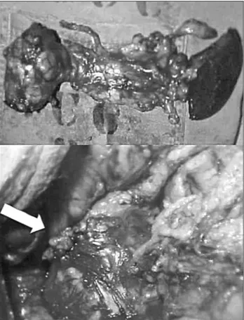
Preoperative enteral and parenteral nutrition were intro-duced. Body-caudal pancreatectomy (approximately 80% of the pancreas) and total splenectomy (Figure 3) were performed with spleen autotransplantation in greater
omentum, due to tumor sized 10 x 8.0cm, encapsulated
and with fibroelastic consistency. The histopathologic re-port indicated a tumor in the pancreatic alpha cells. Im-munohistochemistry showed expression of glucagon and chromogranin A in most tumor cells (consistent with the diagnosis of glucagonoma).
In the postoperative period, the patient progressed with surgical wound infection, left subphrenic abscess and distal pancreatic fistula. Complications were resol-ved 30 days after surgery, using surgical approach and nutritional supplementation. Improvement of glucose tolerance curve and normalization of serum levels of
glu-cagon (postoperative gluglu-cagon 66pg/mL) were observed, as well as evidence of splenic metabolism in the scintigra-phy with 400MBq of Tc-99m-colloid.
Patient received outpatient follow-up for 3 years, wi-thout clinical complications.
D
ISCUSSIONBecker et al.,4 in 1942, were the first to describe the
asso-ciation of a rash, later called necrolytic migratory erythe-ma (NME) by Wilkinson5 in 1973, to pancreatic cancer.
But it was only in 1966 that McGavran et al.6 organized
these conditions as a glucagonoma syndrome.
GLUCAGONOMASYNDROMEASSOCIATEDWITHNECROLYTICMIGRATORYERYTHEMA
REV ASSOC MED BRAS 2015; 61(3):203-206 205
neuroendocrine tumors in a period of 28 years, with glu-cagonomas in only 1.3% of these neoplasms.7
NME is present in almost 70% of patients with gluca-gonoma.8 The lesions consist of erythematous crusted
pla-ques, seen most often in the groin, intergluteal and geni-tal area. It is also found in the lower limbs, perioral region, trunk or even in a pattern of widespread distribution. It is characterized by spontaneous exacerbation and remission periods without the identification of a trigger.9
Histologically, NME can be characterized by paleness and spongiosis of the upper layer of the epidermis. A pe-rivascular lymphocytic and histiocytic infiltrate is also frequent. Necrotic keratinocytes are common and can lead to erosions, crusting and scaling.10
Pathogenesis has not been fully clarified, but is attri-buted to zinc deficiency and hypoaminoacidemia.11 It is
believed that an increase in glucagon levels can lead to loss of amino acids, secondary to increased gluconeoge-nesis.12 In the reported case, hypoaminoacidemia and
rai-sed glucagon levels were present before resection. After
the surgery, there was a significant decrease in the levels of glucagon (postoperative glucagon 66pg/mL), confir-ming the diagnosis of glucagonoma and the association between hyperglucagonemia and hypoaminoacidemia.
Clinically, glucagonoma can manifest with symptoms such as: weight loss, diabetes, diarrhea, and stomatitis. However, patients often seek medical attention for the first time due to skin changes,13 many times attributed
to eczema or psoriasis. Due to the difficulty of recogni-zing the NME and its association with glucagonoma syn-drome, patients remain without a correct diagnosis for a long time.14 In the present case, there was a delay
diagno-sis attributable to loss of clinical follow-up of the patient and the difficulty in recognizing the skin lesion – which at first was the only complaint of the patient.
Additional investigation can be made by angiogra-phy, computed tomography (CT) or magnetic resonan-ce imaging (MRI). These last two enable a better study of the pancreas, helping to characterize the tumor site accurately, which in 86-88% of cases is in the tail of the pancreas.15 Selective visceral angiography is considered
the gold standard for diagnosis and localization of the-se tumors, and its superiority is related to the hypervas-cularization of these tumors,16 despite our case report
showing a lesion with mild degree of contrast enhance-ment. Another advantage of this method is the possibi-lity to demonstrate liver metastasis even in cases where these implants are not seen with CT.16 Positron
Emis-sion tomography (PET) and octreotide scintigraphy have a prominent role, as almost all studied glucagonomas have receptors for somatostatin.17 However, methods
such as abdominal US or CT with contrast is often suf-ficient to diagnose because the tumors are often charac-terized by single and large tumor masses (ranging from 1 to 30 cm3).
Currently, the only curative therapy is surgical remo-val of the mass, which can be done either by open surgery (conventional) or laparoscopically. For those with metas-tatic liver disease, there are studies showing that these pa-tients may benefit from therapies that reduce the hepatic arterial circulation of metastases by hepatic artery embo-lization, either using chemoembolization or microsphere radioembolization.18 Other possible treatments for
disse-minated disease in the liver would be liver transplanta-tion,19 cryoablation or metastasectomy.16 For patients with
contraindications to surgery, chemotherapy with doxoru-bicin and streptozotocin can be performed, leading to a more selective damage of pancreatic islet cells.20
CARDOSO FILHO FA ETAL.
206 REV ASSOC MED BRAS 2015; 61(3):203-206
C
ONCLUSIONNME is crucial for the clinical recognition of glucagono-ma, and early diagnosis is very important for a curative therapy.
R
ESUMOSíndrome do glucagonoma associado a eritema necrolí-tico migratório.
Introdução: o glucagonoma é um tumor neuroendócri-no do pâncreas derivado das células alfa das ilhotas de Langerhans. É marcado pela produção tumoral autôno-ma de glucagon e caracterizado, dentre outros sintoautôno-mas, por eritema necrolítico migratório (ENM), uma lesão eri-tematosa circinada com áreas de necrose e descamação. Trata-se de uma doença rara com incidência mundial es-timada em 1 caso para cada 20 milhões pessoas. Relato de caso: apresentamos um caso de glucagonoma associado a ENM em um paciente de sexo masculino, 56 anos de idade, com quadro de lesões cutâneas, principal-mente em membros inferiores e região inguinal, hipergli-cemia e perda ponderal. Biópsias das lesões cutâneas foram realizadas e exames de imagem do abdome evidenciaram uma massa de 10 x 9 cm em região pancreática. O paciente foi submetido à pancreatectomia corpocaudal e esplenec-tomia total com autoimplante do baço em omento maior. O laudo histopatológico foi de tumor de células alfa pan-creáticas. Imuno-histoquímica evidenciou expressão de glucagon e cromogranina A na maioria das células tumo-rais, compatível com diagnóstico de glucagonoma. O pa-ciente apresentou seguimento de 3 anos em ambulatório sem intercorrências clínicas.
Conclusão: o ENM é importante para o reconhecimen-to clínico do glucagonoma, sendo seu diagnóstico preco-ce fundamental para uma terapia curativa de supreco-cesso.
Palavras-chave: eritema migratório necrolítico, glucago-noma, neoplasias pancreáticas, tumores neuroendócrinos.
R
EFERENCES1. Kovács RK, Korom I, Dobozy A, Farkas G, Ormos J, Kemény L. Necrolytic migratory erythema. J Cutan Pathol. 2006;33(3):242-5.
2. Wermers RA, Fatourechi V, Wynne AG, Kvols LK, Lloyd RV. The glucagonoma syndrome. Clinical and pathologic features in 21 patients. Medicine. 1996;75(2):53-63.
3. Pujol RM, Wang CY, el-Azhary RA, Su WP, Gibson LE, Schroeter AL. Necrolytic migratory erythema: Clinicopathologic study of 13 cases. Int J Dermatol. 2004;43(1):12-8.
4. Becker SW, Kahn D, Rothman S. Cutaneous manifestations of internal malignant tumors. Arch Dermatol Syphilol. 1942;45:1069–80.
5. Wilkinson DS. Necrolytic migratory erythema with carcinoma of the pancreas. Trans St John’s Hosp Dermatol Soc. 1973;59(2):244-50.
6. McGavran MH, Unger RH, Recant L. A glucagon-secreting alpha-cell carcinoma of the pancreas. N Engl J Med. 1966;274(25):1408-13. 7. Yao JC, Eisner MP, Leary C, Dagohoy C, Phan A, Rashid A, et al.
Population-based study of islet cell carcinoma. Ann Surg Oncol. 2007;14(12):3492-500. 8. Lobo I, Carvalho A, Amaral C, Machado S, Carvalho R. Glucagonoma syndrome and necrolytic migratory erythema. Int J Dermatol. 2010; 49(1):24-9.
9. Remes-Troche JM, García-de-Acevedo B, Zuniga-Varga J, Avila-Funes A, Orozco-Topete R. Necrolytic migratory erythema: A cutaneous clue to glucagonoma syndrome. J Eur Acad Dermatol Venereol. 2004;18(5):591-5. 10. Johnson SM, Smoller BR, Lamps LW, Horn TD. Necrolytic migratory erythema as the only presenting sign of a glucagonoma. J Am Acad Dermatol. 2003;49(2):325-8.
11. Shi W, Liao W, Mei X, Xiao Q, Zeng Y, Zhou Q. Necrolytic migratory erythema associated with glucagonoma syndrome. J Clin Oncol. 2010;28(20):e329-31 12. Marliss EB, Aoki TT, Unger RH, Soeldner JS, Cahill GF. Glucagon levels and
metabolic effects in fasting man. J Clin Invest. 1970;49(12):2256-70. 13. Zhang M, Xu X, Shen Y, Hu ZH, Wu LM, Zheng SS. Clinical experience in
diagnosis and treatment of glucagonoma syndrome. Hepatobiliary Pancreat Dis Int. 2004;3(3):473-5.
14. Echenique-Elizondo M, Valls AT, Orue JLE, Lizarduy IM, Aguirre JI. Glucagonoma and pseudoglucagonoma syndrome. J Pancreas. 2004; 5(1):179-85.
15. Xu Q, Chen WH, Huang QJ. Spiral CT localization of pancreatic functioning islet cell tumors. Hepatobiliary Pancreat Dis Int. 2004;3(4):616-9. 16. Castro PG, Leon AM, Trancon JG, Martínez PA, Alvarez Pérez JA, Fernández
Fernández JC, et al. Glucagonoma syndrome: a case report. J Med Case Rep. 2011;5:402.
17. Melen-Mucha G, Lawnicka H, Kierszniewska-Stepien D, Komorowski J, Stepien H. The place of somatostatin analogs in the diagnosis and treatment of the neuoroendocrine glands tumors. Recent Patents Anticancer Drug Discov. 2006;1(2):237-54.
18. King J, Quinn R, Glenn DM, Janssen J, Tong D, Liaw W, et al. Radioembolization with selective internal radiation microspheres for neuroendocrine liver metastases. Cancer. 2008;113(5):921-9.
19. Radny P, Eigentler TK, Soennichsen K, Overkamp D, Raab HR, Viebahn R, et al. Metastatic glucagonoma: treatment with liver transplantation. J Am Acad Dermatol. 2006;54(2):344-7