Autonomic nervous system monitoring in
intensive care as a prognostic tool. Systematic
review
INTRODUCTION
Since the 1970s, with the introduction of the Swan-Ganz catheter,
(1)there
has been signiicant progress in the capacity of invasive and non-invasive
hemodynamic monitoring in intensive care units (ICU) and an improved
understanding of the pathophysiological phenomena responsible for the
hemodynamic instability of critical patients.
Despite these remarkable advances, there is no unanimity as to what
therapeutic objectives should be achieved in patients with hemodynamic
instability admitted to the ICU,
(2)for the time being maintaining an individual
therapeutic attitude guided not by hemodynamic monitoring data but by
the integration of the diferent variables that can be obtained using multiple
monitoring methods.
Luis Bento1, Rui Fonseca-Pinto2,3, Pedro Póvoa4,5
1. Medical Emergency Unit, Centro Hospitalar de Lisboa Central, EPE - Lisbon, Portugal.
2. Instituto Politécnico de Leiria - Leiria, Portugal. 3. Instituto de Telecomunicações, MSP - Leiria, Portugal.
4. Multipurpose Intensive Care Unit, São Francisco Xavier Hospital - West Lisbon Hospital Center - Lisbon, Portugal.
5. NOVA Medical School, CEDOC, Universidade Nova de Lisboa - Lisbon, Portugal.
Objective:
To present a systematic
review of the use of autonomic nervous
system monitoring as a prognostic tool
in intensive care units by assessing heart
rate variability.
Methods:
Literature review of
studies published until July 2016 listed
in PubMed/Medline and conducted
in intensive care units, on autonomic
nervous system monitoring, via analysis
of heart rate variability as a prognostic
tool (mortality study). he following
English terms were entered in the search
ield: (“autonomic nervous system” OR
“heart rate variability”) AND (“intensive
care” OR “critical care” OR “emergency
care” OR “ICU”) AND (“prognosis”
OR “prognoses” OR “mortality”).
Results:
here was an increased
likelihood of death in patients who had
Conflicts of interest: None.
Submitted on April 15, 2016 Accepted on April 18, 2017
Corresponding author:
Luis Bento
Centro Hospitalar de Lisboa Central Rua José António Serrano Lisbon 1150-199 Portugal
Responsible editor: Jorge Ibrain Figueira Salluh
Monitorização do sistema nervoso autônomo em ambiente de
cuidados intensivos como ferramenta de prognóstico. Revisão
sistemática
ABSTRACT
Keywords:
Autonomic nervous
system; Heart rate variability; Intensive
care; Prognosis
a decrease in heart rate variability as
analyzed via heart rate variance, cardiac
uncoupling, heart rate volatility, integer
heart rate variability, standard deviation
of NN intervals, root mean square
of successive diferences, total power,
low frequency, very low frequency,
low frequency/high frequency ratio,
ratio of short-term to long-term fractal
exponents, Shannon entropy, multiscale
entropy and approximate entropy.
Conclusion:
In patients admitted
to intensive care units, regardless of the
pathology, heart rate variability varies
inversely with clinical severity and
prognosis.
his situation results from an overvaluation of
our view of the cardiovascular system according to
physics principles rather than a look at the capacity
and adjustment of the real-time responses of critical
patients to the pathophysiological changes induced by
the disease and imposed by our therapeutic attitudes,
either pharmacological or not. More important than
the “normalization” of a given parameter is its temporal
adjustment.
Recent studies
(3-5)have described several hemodynamic
monitoring methods, from the most invasive, such
as the Swan-Ganz catheter, to the less invasive, such as
bioimpedance and bioreactance methods. However,
although the autonomic nervous system (ANS) is
responsible for the homeostasis of the cardiocirculatory
system through the balance between the activity of the
sympathetic and parasympathetic ANS, no reference is
made to the monitoring of its activity and/or its balance
in ICU patients.
Heart rate variability (HRV) translates the oscillations
in the duration of intervals between consecutive heart
beats (NN intervals) (Figure 1) and is related to the
inluences of the ANS on the sinus node, translating the
heart’s capacity to respond to multiple physiological and
environmental stimuli, such as breathing, physical exercise,
hemodynamic and metabolic changes, orthostatism and
responses to stress induced by diseases. Moreover, the
study of HRV of the ANS is only possible in the presence
of sinus rhythm.
he objective of this article is to present a systematic
review of studies involving autonomic nervous system
monitoring of adult patients admitted to the intensive care
units by analyzing the association of multiple heart rate
variability assessment measures with the hospitalization
outcome. Prospective and retrospective randomized
controlled or cohort
studies were included.
METHODS
In this systematic review, we used the checklist
Preferred Reporting Items for Systematic Reviews and
Meta-Analyses (PRISMA)
(6)as a guide to reach the
standards accepted in systematic reviews.
he literature review of studies conducted in ICUs on
ANS monitoring was conducted by searching all of the
measures described for HRV analysis methods (Tables 1
and 2) as a prognostic tool (mortality study), published
in or before July 2016 (inclusive) using the PubMed/
MEDLINE database. he following English terms
were entered in the search ield, yielding 421 articles:
(“autonomic nervous system” OR “heart rate variability”)
AND (“intensive care” OR “critical care” OR “emergency
care” OR “ICU”) AND (“prognosis” OR “prognoses” OR
“mortality”).
After applying the ilters to limit the studies to those
involving humans aged over 19 years, without language
restriction, 193 articles were excluded.
After reading the abstracts of the 228 selected studies,
180 articles were excluded: 11 reported the monitoring of
pediatric patients, 16 were conducted outside the intensive
care setting, 119 were not related to ANS monitoring,
four did not analyze HRV, 28 did not focus on prognosis
and two were review studies.
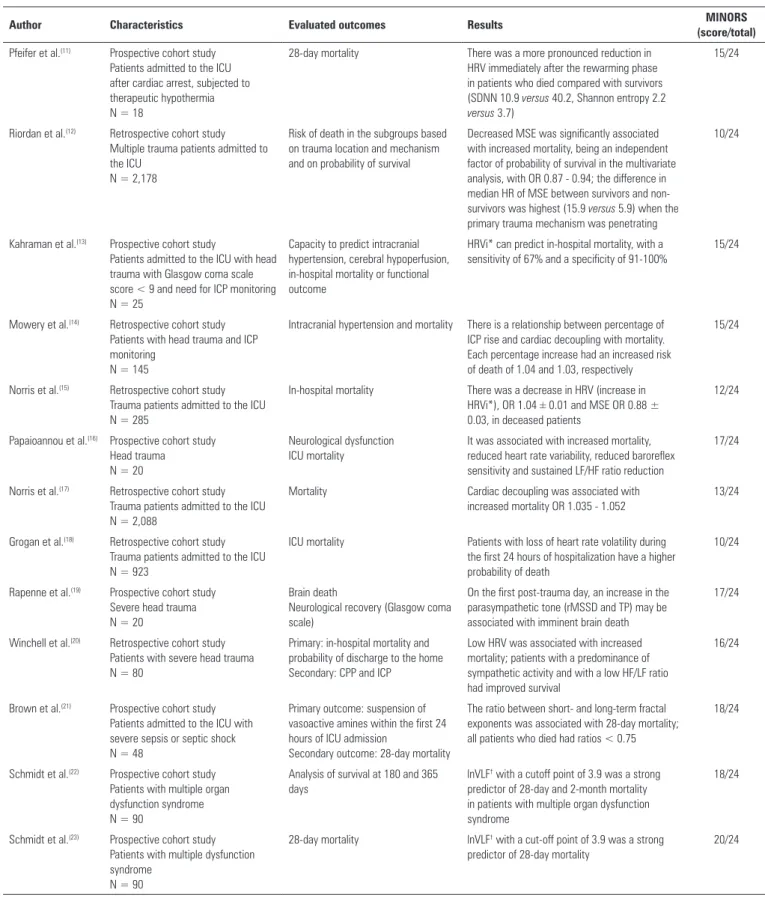
he 48 articles selected were grouped and cataloged
in EndNote
®and were read in full. Afterwards, 32
articles were excluded: 21 because they were not studies
of ICU patients (11 were performed in the Emergency
Department, ive in the prehospital setting, two in
the Cardiothoracic Surgery Service and two in the
Table 1 - Methods for the study of heart rate variability(7-9)
1. Linear methods - time domain a. Statistical measures
i. SDNN - Standard deviation of all normal NN intervals
ii. SDANN - Standard deviation of the average normal NN interval calculated over 5-minute intervals iii. SDNNi - Mean of the standard deviations of all normal NN calculated over 5-minute intervals iv. rMSSD - Square root of the mean squared differences of successive normal NN intervals v. SDSD - Standard deviation of differences between adjacent normal NN intervals
vi. NN50 - Number of pairs of adjacent normal NN intervals differing by more than 50 milliseconds
vii. pNN50 - Percentage of normal NN intervals differing by more than 50 milliseconds from the adjacent interval b. Geometric measures
i. Triangular index
ii. TINN - Triangular interpolation of normal NN intervals histogram iii. Differential index
iv. Logarithmic index 2. Linear methods - frequency domain
a. Long-term analysis (5 minutes) i. Total power
ii. VLF - Very low frequency iii. LF - Low frequency
iv. LFn - Low frequency in normalized units v. HF - High frequency
vi. HFn - High frequency in normalized units vii. LF/HF - Low frequency/high frequency ratio b. Long-term analysis (24 hours)
i. Total power
ii. ULF - Ultra low frequency iii. VLF - Very low frequency iv. LF - Low frequency v. HF - High frequency
vi. α - Slope of the linear interpolation of the spectrum in a logarithmic scale 3. Time-frequency analysis methods
a. Time-varying parametric models i. Autoregression models b. Non-parametric methods
i. Short-time Fourier transform (STFT) ii. Wavelet transform (WT) iii. Hilbert-Huang transform iv. Wigner-Ville transform 4. Non-linear methods
a. Detrended fluctuation analysis (total DTA, α1, α2 and α1/α2) b. Correlation function
Cardiology Service, and one study was conducted during
the anesthetic period) and 11 because they did not report
mortality data.
he references of the 16 selected articles were reviewed,
and whenever there was reference to a new study, that
study was evaluated; at the end of the review process, 18
articles were selected (Figure 2).
Table 2 - Definition of measures for the study of heart rate variability in the time domain(7)
Measure Unit Definition
SDNN ms Standard deviation of all normal NN intervals
SDNNi ms Standard deviation of NN calculated over 5-minute intervals
SDANN ms Standard deviation of the average NN interval
rMSSD ms Root mean square of the successive NN interval difference
pNN50 % Normal-to-normal NN intervals whose difference exceeds 50 milliseconds
Figure 2 - Article selection protocol.(6)
HRV - heart rate variability; ICU - intensive care unit.
he quality of evidence for each selected study was
assessed using the
Methodological Index for
Non-Randomized Studies (MINORS) tool.
(10)he article review (data extraction and quality
of evidence) was conducted by one author, with the
information later independently veriied by two others.
Table 3 shows the characteristics of the selected
studies.
RESULTS
he 18 selected studies are presented in table 3. he
type of study, study population, number of patients
included, HRV variables studied in the ANS monitoring,
most relevant conclusions and quality of evidence were
also analyzed.
All studies reviewed were cohort, prospective
or retrospective studies. he sample size was very
heterogeneous, ranging from 18
(11)to 2,178
(12)patients;
the sample size was not previously calculated in any
study. he most studied pathology was trauma, mainly
of the head, with a total of nine studies,
(12-20)and with
the same number of studies on patients with severe sepsis
and septic shock,
(21)multiple dysfunction syndrome,
(22,23)patients undergoing therapeutic hypothermia after cardiac
arrest,
(11)with stroke
(24)and neurosurgical patients;
(25)three
studies focused on the general population admitted to the
ICU, without discriminating the reason for admission.
he conclusions of all of the studies were obtained by
comparing the groups according to the outcome evaluated,
namely, mortality.
he results presented included increases in mortality
associated with reduction in HRV (entropy 0.65 ± 0.24
versus
0.84 ± 0.26; p < 0.05), reduction in the barorelex
(transfer function 0.43 ± 29
versus
1.11 ± 0.74; p < 0.05)
and a sustained reduction of the low frequency/high
frequency ratio (LF/HF ratio 0.22 ± 0.29
versus
0.62 ± 28;
p < 0.01);
(16)reductions in HRV, with odds ratios (ORs) of
1.03
(14)and of 1.035 - 1.052;
(17)loss of heart rate volatility
during the irst 24 hours of hospitalization, translated as
a coeicient of 0.05 in the logistic regression model (95%
conidence interval [95% CI] 1.033 - 1.071);
(18)integer
heart rate variability (HRVi) with a sensitivity of 67% and
a speciicity of 91 - 100% to predict the mortality rate
(13)or OR of 1.04;
(15)and reduction in HRV in patients
admitted to the ICU after cardiac arrest and undergoing
therapeutic hypothermia, with a standard deviation of all
normal NN intervals of 10.9 ± 4.1
versus
40.2 ± 19.5 (p =
0.01) and a Shannon entropy of 2.2 ± 0.4
versus
3.7 ± 0.6
(p = 0.008) for deceased
versus
surviving patients in the
rewarming period. Concordant results were observed in
the pre-hypothermia period.
(11)here was also an increase
in the parasympathetic tone as measured by the square
root of the mean squared diferences of successive intervals
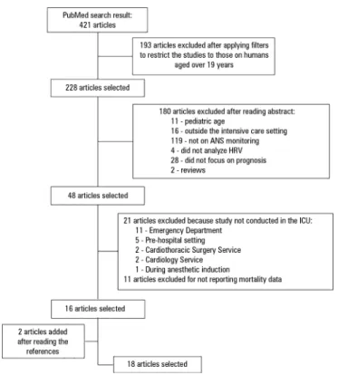
Table 3 - Characteristics of the selected studies
Author Characteristics Evaluated outcomes Results MINORS
(score/total)
Pfeifer et al.(11) Prospective cohort study Patients admitted to the ICU after cardiac arrest, subjected to therapeutic hypothermia N = 18
28-day mortality There was a more pronounced reduction in HRV immediately after the rewarming phase in patients who died compared with survivors (SDNN 10.9 versus 40.2, Shannon entropy 2.2
versus 3.7)
15/24
Riordan et al.(12) Retrospective cohort study Multiple trauma patients admitted to the ICU
N = 2,178
Risk of death in the subgroups based on trauma location and mechanism and on probability of survival
Decreased MSE was significantly associated with increased mortality, being an independent factor of probability of survival in the multivariate analysis, with OR 0.87 - 0.94; the difference in median HR of MSE between survivors and non-survivors was highest (15.9 versus 5.9) when the
primary trauma mechanism was penetrating
10/24
Kahraman et al.(13) Prospective cohort study
Patients admitted to the ICU with head trauma with Glasgow coma scale score < 9 and need for ICP monitoring N = 25
Capacity to predict intracranial hypertension, cerebral hypoperfusion, in-hospital mortality or functional outcome
HRVi* can predict in-hospital mortality, with a sensitivity of 67% and a specificity of 91-100%
15/24
Mowery et al.(14) Retrospective cohort study Patients with head trauma and ICP monitoring
N = 145
Intracranial hypertension and mortality There is a relationship between percentage of ICP rise and cardiac decoupling with mortality. Each percentage increase had an increased risk of death of 1.04 and 1.03, respectively
15/24
Norris et al.(15) Retrospective cohort study Trauma patients admitted to the ICU N = 285
In-hospital mortality There was a decrease in HRV (increase in HRVi*), OR 1.04 ± 0.01 and MSE OR 0.88 ± 0.03, in deceased patients
12/24
Papaioannou et al.(16) Prospective cohort study Head trauma N = 20
Neurological dysfunction ICU mortality
It was associated with increased mortality, reduced heart rate variability, reduced baroreflex sensitivity and sustained LF/HF ratio reduction
17/24
Norris et al.(17) Retrospective cohort study Trauma patients admitted to the ICU N = 2,088
Mortality Cardiac decoupling was associated with increased mortality OR 1.035 - 1.052
13/24
Grogan et al.(18) Retrospective cohort study Trauma patients admitted to the ICU N = 923
ICU mortality Patients with loss of heart rate volatility during the first 24 hours of hospitalization have a higher probability of death
10/24
Rapenne et al.(19) Prospective cohort study Severe head trauma N = 20
Brain death
Neurological recovery (Glasgow coma scale)
On the first post-trauma day, an increase in the parasympathetic tone (rMSSD and TP) may be associated with imminent brain death
17/24
Winchell et al.(20) Retrospective cohort study Patients with severe head trauma N = 80
Primary: in-hospital mortality and probability of discharge to the home Secondary: CPP and ICP
Low HRV was associated with increased mortality; patients with a predominance of sympathetic activity and with a low HF/LF ratio had improved survival
16/24
Brown et al.(21) Prospective cohort study Patients admitted to the ICU with severe sepsis or septic shock N = 48
Primary outcome: suspension of vasoactive amines within the first 24 hours of ICU admission
Secondary outcome: 28-day mortality
The ratio between short- and long-term fractal exponents was associated with 28-day mortality; all patients who died had ratios < 0.75
18/24
Schmidt et al.(22) Prospective cohort study Patients with multiple organ dysfunction syndrome N = 90
Analysis of survival at 180 and 365 days
lnVLF† with a cutoff point of 3.9 was a strong predictor of 28-day and 2-month mortality in patients with multiple organ dysfunction syndrome
18/24
Schmidt et al.(23) Prospective cohort study Patients with multiple dysfunction syndrome
N = 90
28-day mortality lnVLF† with a cut-off point of 3.9 was a strong predictor of 28-day mortality
20/24
Author Characteristics Evaluated outcomes Results MINORS (score/total)
Gujjar et al.(24) Prospective cohort study Acute stroke
N = 25
ICU mortality LFn was an independent predictor of survival, with a regression coefficient of -6.73 and an OR of 0.002
19/24
Haji-Michael et al.(25) Prospective cohort study
Neurosurgical patients with Glasgow coma scale score < 13
N = 29
3-month outcome Patients who died had decreased HRV, LF/HF ratio and baroreflex sensitivity
18/24
Papaioannou et al.(26) Prospective cohort study General ICU population N = 53
ICU mortality The minimum ApEn value correlated with mortality (r = 0.41; p = 0.01)
16/24
Yien et al.(27) Prospective cohort study General population admitted for noncardiac causes
N = 52
Mortality Deceased patients had decreased VLF and LF band power
16/24
Winchell et al.(28) Prospective cohort study General ICU population N = 742
Mortality The relative risk of death in patients with low HRV was 7.4, with an increased HF/LF ratio of 4.55
19/24
MINORS - Methodological Index for Non-Randomized Studies; ICU - intensive care unit; HRV - heart rate variability; MSE - multiscale entropy; OR - odds ratio; HR - hazard ratio; HRVi - integer heart rate variability; ICP - intracranial pressure; LF/HF - ratio between the low frequency component and the high frequency component; CPP - cerebral perfusion pressure; TP - total power. * Calculation of the standard deviation of the electrocardiogram signal collected every 1-4 seconds during a 5-minute interval; † natural logarithm of VLF.
... continuation
in patients with severe head injury;
(19)decreased power
in the low frequency band (low frequency in standard
units in patients with severe stroke 18.90 ± 1.36
versus
49.66 ± 2.10; p = 0.02; in the general population p < 0.05
with Schefé analysis);
(24,27)decreased natural logarithm
of the very low frequency band (lnVLF £ 3.9 with OR
2.9; in the general population p < 0.05 with Schefé
analysis);
(22,23,27,28)and decreased ratio of short- to
long-term fractal exponents; all patients admitted to the ICU
with severe sepsis or septic shock who died had a ratio
of < 0.75 (p = 0.04).
(21)he following were also found:
decreased multiscale entropy in trauma patients (8.9
versus
16.6; p < 0.0001; 7.5
versus
11.2; p < 0.001 in
patients with survival probabilities < 0.25; 7.7
versus
12.8;
p < 0.01 for patients with survival probabilities of 0.25 to
0.50; 9.4
versus
15.0; p < 0.001 for patients with survival
probabilities of 0.50 to 0.75; 9.9
versus
16.1; and p <
0.001 among those with survival probabilities ³ 0.75).
(12,15)Decreased approximate entropy (mean ApEn 0.53 ± 0.25
versus
0.62 ± 0.28; p = 0.04; minimum ApEn 0.24 ± 0.23
versus
0.48 ± 0.23; p = 0.01) with a Pearson coeicient of
0.41 (p = 0.01) was also found.
(26)hus, these studies showed that, in patients admitted
to the ICU, regardless of the pathology that led to
hospitalization, HRV varied inversely with clinical severity
and prognosis.
(29)DISCUSSION
he control of the cardiovascular system is ensured
by the balance between the activity of the sympathetic
ANS, which enervates the entire myocardium, and
the parasympathetic ANS, which enervates the sinus
node, the atrial myocardium and the atrioventricular
node.
(30)he inluence of the ANS on the heart depends
on the information it receives from the baroreceptors,
chemoreceptors, atrial receptors, ventricular receptors,
changes in the respiratory system, vasomotor system,
renin-angiotensin-aldosterone system and thermoregulatory
system.
(31)All of these inluences condition the HRV,
and the standards for its measurement, physiological
interpretation and applicability were published in 1996.
(7)he HRV can be analyzed using diferent methods,
with linear methods being the most used in clinical
practice.
he time domain is analyzed using various measures
and relects the variation in the duration of NN intervals
resulting from the depolarization of the sinus node.
which corresponds to sympathetic and parasympathetic
activity; the very low frequency band, ranging between
0.003 and 0.04 Hz, which relects the thermoregulation
cycles; and ultra low frequency components, with
variations below 0.003 Hz, modulated by the circadian
rhythm and neuroendocrine axes.
he inverse relationship enters the very low frequency
band, and the prognosis was irst described in the 1960s,
(32)when it was observed that NN interval reduction preceded
fetal distress.
he irst study conducted in the ICU was published in
1996 and concluded that HRV reduction was related to
increased mortality.
(28)Since then, all studies conducted in
the ICU have almost exclusively focused on the evaluation
of HRV, which varies inversely with clinical severity and
prognosis.
(29)Examples of clinical conditions in which HRV is
predictive of patient survival include diabetes,
(33)cancer,
(34)heart failure,
(35)acute myocardial infarction,
(36)stroke,
(37)epilepsy,
(38)Parkinson’s disease
(39)and kidney failure,
(40)among others.
In patients admitted to the ICU, in addition to being
used as a prognostic tool, HRV has also been described
as a screening tool for multiple trauma patients,
(41)as a
tool for individual monitoring of organ dysfunction,
(42)as a non-invasive tool for pain monitoring
(43)and as an
independent predictor factor for the prolongation of
hospital stay in patients undergoing heart surgery
(44)and
has been used as a tool for successful extubation
decision-making.
(45,46)Some limitations were identiied in the studies
reviewed. here is no uniformity in the variables studied
for HRV assessment, although the studies are concordant
in the conclusions presented; furthermore, the quality of
the evidence is low, due mainly to the sampled studies
being cohort studies.
CONCLUSION
Heart rate variability occurs inversely to clinical
severity and prognosis. he diiculty of introducing
autonomic nervous system monitoring in the daily
practice of intensive care units is due to the limitation of
its use as a prognostic tool and, above all, to the diiculties
involved in continuous and dynamic monitoring and in
the interpretation and applicability of its results.
Successful implementation depends on heart rate
variability monitoring going from a prognostic tool to
a real-time monitoring instrument in order to be useful
in therapeutic guidance; for example, as a guide for luid
therapy through analysis of the high frequency component
and for treatment with vasoactive amines through analysis
of the low frequency/high frequency ratio.
Objetivo:
Apresentar uma revisão sistemática do uso da
monitorização do sistema nervoso autônomo como ferramenta
de prognóstico, veriicando a variabilidade da frequência
cardíaca nas unidades de cuidados intensivos.
Métodos:
Revisão de literatura publicada até julho de 2016
na PubMed/MEDLINE de estudos realizados em unidades
de cuidados intensivos, sobre a monitorização do sistema
nervoso autônomo, por meio da análise da variabilidade da
frequência cardíaca, como ferramenta de prognóstico - estudo
da mortalidade. Foram utilizados os seguintes termos em inglês
no campo de pesquisa: (“
autonomic nervous system
” OR “
heart
rate variability
”) AND (“
intensive care
” OR “
critical care
” OR
“
emergency care
” OR “ICU”) AND (“
prognosis
” OR “
prognoses
”
OR “
mortality
”).
Resultados:
A probabilidade de morte nos doentes
aumentou com a diminuição da variabilidade da frequência
cardíaca, estudada por meio da variância da frequência cardíaca,
desacoplamento cardíaco, volatilidade da frequência cardíaca,
integer heart rate variability
, desvio padrão de todos os intervalos
RR normais, raiz quadrada da média do quadrado das diferenças
entre intervalos RR adjacentes, poder total, componente de
baixa frequência, componente de muito baixa frequência, razão
entre o componente de baixa frequência e o componente de
alta frequência), razão entre expoentes fractais de curto e longo
prazo, entropia de Shannon, entropia multiescalar e entropia
aproximada.
Conclusão:
Nos doentes internados em unidades de cuidados
intensivos, independentemente da patologia que motivou o
internamento, a variabilidade da frequência cardíaca varia de
forma inversa com a gravidade clínica e com o prognóstico.
RESUMO
REFERENCES
1. Swan HJ, Ganz W. Hemodynamic monitoring: a personal and historical perspective. Can Med Assoc J. 1979;121(7):868-71.
2. Joosten A, Alexander B, Cannesson M. Defining goals of resuscitation in the critically ill patient. Crit Care Clin. 2015;31(1):113-32.
3. Vincent JL, Rhodes A, Perel A, Martin GS, Della Rocca G, Vallet B, et al. Clinical review: Update on hemodynamic monitoring--a consensus of 16. Crit Care. 2011;15(4):229.
4. Ramsingh D, Alexander B, Cannesson M. Clinical review: Does it matter which hemodynamic monitoring system is used? Crit Care. 2013;17(2):208. 5. Kenaan M, Gajera M, Goonewardena SN. Hemodynamic assessment in
the contemporary intensive care unit: a review of circulatory monitoring devices. Crit Care Clin. 2014;30(3):413-45.
6. Liberati A, Altman DG, Tetzlaff J, Mulrow C, Gotzsche PC, Ioannidis JP, et al. The PRISMA statement for reporting systematic reviews and meta-analyses of studies that evaluate healthcare interventions: explanation and elaboration. BMJ. 2009;339:b2700.
7. Heart rate variability. Standards of measurement, physiological interpretation, and clinical use. Task Force of The European Society of Cardiology and The North American Society of Pacing and Electrophysiology. Eur Heart J. 1996;17(3):354-81.
8. Rajendra Acharya U, Paul Joseph K, Kannathal N, Lim CM, Suri JS. Heart rate variability: a review. Med Biol Eng Comput. 2006;44(12):1031-51. 9. Bailon R, Laguna P, Mainardi L, Sornmo L. Analysis of heart rate variability
using time-varying frequency bands based on respiratory frequency. Conf Proc IEEE Eng Med Biol Soc. 2007;2007:6675-8.
10. Slim K, Nini E, Forestier D, Kwiatkowski F, Panis Y, Chipponi J. Methodological index for non-randomized studies (minors): development and validation of a new instrument. ANZ J Surg. 2003;73(9):712-6. 11. Pfeifer R, Hopfe J, Ehrhardt C, Goernig M, Figulla HR, Voss A. Autonomic
regulation during mild therapeutic hypothermia in cardiopulmonary resuscitated patients. Clin Res Cardiol. 2011;100(9):797-805.
12. Riordan WP Jr., Norris PR, Jenkins JM, Morris JA Jr. Early loss of heart rate complexity predicts mortality regardless of mechanism, anatomic location, or severity of injury in 2178 trauma patients. J Surg Res. 2009;156(2):283-9. 13. Kahraman S, Dutton RP, Hu P, Stansbury L, Xiao Y, Stein DM, et al. Heart rate and pulse pressure variability are associated with intractable intracranial hypertension after severe traumatic brain injury. J Neurosurg Anesthesiol. 2010;22(4):296-302.
14. Mowery NT, Norris PR, Riordan W, Jenkins JM, Williams AE, Morris JA Jr. Cardiac uncoupling and heart rate variability are associated with intracranial hypertension and mortality: a study of 145 trauma patients with continuous monitoring. J Trauma. 2008;65(3):621-7.
15. Norris PR, Stein PK, Morris JA Jr. Reduced heart rate multiscale entropy predicts death in critical illness: a study of physiologic complexity in 285 trauma patients. J Crit Care. 2008;23(3):399-405.
16. Papaioannou V, Giannakou M, Maglaveras N, Sofianos E, Giala M. Investigation of heart rate and blood pressure variability, baroreflex sensitivity, and approximate entropy in acute brain injury patients. J Crit Care. 2008;23(3):380-6.
17. Norris PR, Ozdas A, Cao H, Williams AE, Harrell FE, Jenkins JM, et al. Cardiac uncoupling and heart rate variability stratify ICU patients by mortality: a study of 2088 trauma patients. Ann Surg. 2006;243(6):804-12; discussion 812-4.
18. Grogan EL, Morris JA Jr., Norris PR, France DJ, Ozdas A, Stiles RA, et al. Reduced heart rate volatility: an early predictor of death in trauma patients. Ann Surg. 2004;240(3):547-54; discussion 554-6.
19. Rapenne T, Moreau D, Lenfant F, Vernet M, Boggio V, Cottin Y, et al. Could heart rate variability predict outcome in patients with severe head injury? A pilot study. J Neurosurg Anesthesiol. 2001;13(3):260-8.
20. Winchell RJ, Hoyt DB. Analysis of heart-rate variability: a noninvasive predictor of death and poor outcome in patients with severe head injury. J Trauma. 1997;43(6):927-33.
21. Brown SM, Tate Q, Jones JP, Knox DB, Kuttler KG, Lanspa M, et al. Initial fractal exponent of heart rate variability is associated with success of early resuscitation in patients with severe sepsis or septic shock: a prospective cohort study. J Crit Care. 2013;28(6):959-63.
22. Schmidt H, Hoyer D, Hennen R, Heinroth K, Rauchhaus M, Prondzinsky R, et al. Autonomic dysfunction predicts both 1- and 2-month mortality in middle-aged patients with multiple organ dysfunction syndrome. Crit Care Med. 2008;36(3):967-70.
23. Schmidt H, Müller-Werdan U, Hoffmann T, Francis DP, Piepoli MF, Rauchhaus M, et al. Autonomic dysfunction predicts mortality in patients with multiple organ dysfunction syndrome of different age groups. Crit Care Med. 2005;33(9):1994-2002.
24. Gujjar AR, Sathyaprabha TN, Nagaraja D, Thennarasu K, Pradhan N. Heart rate variability and outcome in acute severe stroke: role of power spectral analysis. Neurocrit Care. 2004;1(3):347-53.
25. Haji-Michael PG, Vincent JL, Degaute JP, van de Borne P. Power spectral analysis of cardiovascular variability in critically ill neurosurgical patients. Crit Care Med. 2000;28(7):2578-83.
26. Papaioannou VE, Maglaveras N, Houvarda I, Antoniadou E, Vretzakis G. Investigation of altered heart rate variability, nonlinear properties of heart rate signals, and organ dysfunction longitudinally over time in intensive care unit patients. J Crit Care. 2006;21(1):95-103; discussion 103-4. 27. Yien HW, Hseu SS, Lee LC, Kuo TB, Lee TY, Chan SH. Spectral analysis of
systemic arterial pressure and heart rate signals as a prognostic tool for the prediction of patient outcome in the intensive care unit. Crit Care Med. 1997;25(2):258-66.
28. Winchell RJ, Hoyt DB. Spectral analysis of heart rate variability in the ICU: a measure of autonomic function. J Surg Res. 1996;63(1):11-6. 29. Gang Y, Malik M. Heart rate variability in critical care medicine. Curr Opin
Crit Care. 2002;8(5):371-5.
30. Brodde OE, Bruck H, Leineweber K, Seyfarth T. Presence, distribution and physiological function of adrenergic and muscarinic receptor subtypes in the human heart. Basic Res Cardiol. 2001;96(6):528-38.
31. Berntson GG, Bigger JT Jr., Eckberg DL, Grossman P, Kaufmann PG, Malik M, et al. Heart rate variability: origins, methods, and interpretive caveats. Psychophysiology. 1997;34(6):623-48.
32. Hon EH, Lee ST. Electronic evaluation of the fetal heart rate. VIII. Patterns preceding fetal death, further observations. Am J Obstet Gynecol. 1963;87:814-26.
33. França da Silva AK, Penachini da Costa de Rezende Barbosa M, Marques Vanderlei F, Destro Christofaro DG, Marques Vanderlei LC. Application of heart rate variability in diagnosis and prognosis of individuals with diabetes mellitus: systematic review. Ann Noninvasive Electrocardiol. 2016;21(3):223-35.
34. Zhou X, Ma Z, Zhang L, Zhou S, Wang J, Wang B, et al. Heart rate variability in the prediction of survival in patients with cancer: A systematic review and meta-analysis. J Psychosom Res. 2016;89:20-5.
35. Wu L, Jiang Z, Li C, Shu M. Prediction of heart rate variability on cardiac sudden death in heart failure patients: a systematic review. Int J Cardiol. 2014;174(3):857-60.
36. Huikuri HV, Stein PK. Heart rate variability in risk stratification of cardiac patients. Prog Cardiovasc Dis. 2013;56(2):153-9.
37. Yperzeele L, van Hooff RJ, Nagels G, De Smedt A, De Keyser J, Brouns R. Heart rate variability and baroreceptor sensitivity in acute stroke: a systematic review. Int J Stroke. 2015;10(6):796-800.
38. Lotufo PA, Valiengo L, Benseñor IM, Brunoni AR. A systematic review and meta-analysis of heart rate variability in epilepsy and antiepileptic drugs. Epilepsia. 2012;53(2):272-82.
39. Maetzler W, Liepelt I, Berg D. Progression of Parkinson’s disease in the clinical phase: potential markers. Lancet Neurol. 2009;8(12):1158-71. 40. Zhang J, Wang N. Prognostic significance and therapeutic option
41. Ryan ML, Thorson CM, Otero CA, Vu T, Proctor KG. Clinical applications of heart rate variability in the triage and assessment of traumatically injured patients. Anesthesiol Res Pract. 2011;2011:416590.
42. Green GC, Bradley B, Bravi A, Seely AJ. Continuous multiorgan variability analysis to track severity of organ failure in critically ill patients. J Crit Care. 2013;28(5):879.e1-11.
43. Boselli E, Daniela-Ionescu M, Bégou G, Bouvet L, Dabouz R, Magnin C, et al. Prospective observational study of the non-invasive assessment of immediate postoperative pain using the analgesia/nociception index (ANI). Br J Anaesth. 2013;111(3):453-9.
44. Stein PK, Schmieg RE Jr., El-Fouly A, Domitrovich PP, Buchman TG. Association between heart rate variability recorded on postoperative day 1 and length of stay in abdominal aortic surgery patients. Crit Care Med. 2001;29(9):1738-43.
45. Arcentales A, Caminal P, Diaz I, Benito S, Giraldo BF. Classification of patients undergoing weaning from mechanical ventilation using the coherence between heart rate variability and respiratory flow signal. Physiol Meas. 2015;36(7):1439-52.