Reference values and factors related to thoracic mobility
in Brazilian children
Valores de referência e fatores relacionados à mobilidade torácica em crianças brasileiras
Valores de referencia y factores relacionados con la movilidad torácica en niños brasileños
Raphaella Oliveira E. da Silva1, Tania Fernandes Campos2, Raíssa de Oliveira Borja3, Thalita Medeiros F. de Macêdo3, Juliana Souza de Oliveira1, Karla Morganna P. P. de Mendoça4
Instituição: Universidade Federal do Rio Grande do Norte (UFRN), Natal, RN, Brasil
1Mestranda em Fisioterapia pelo Programa de Pós-Graduação em Fisioterapia da UFRN, Natal, RN, Brasil
2Doutora em Psicobiologia pela UFRN; Professora Adjunta do Departamento de Fisioterapia da UFRN, Natal, RN, Brasil
3Mestre em Fisioterapia pelo Programa de Pós-Graduação em Fisioterapia da UFRN, Natal, RN, Brasil
4Doutora em Ciências da Saúde pela UFRN; Professora Adjunta do Departamento de Fisioterapia da UFRN, Natal, RN, Brasil
ABSTRACT
Objective: To provide reference values and to evaluate
the factors influencing thoracic mobility in children aged 7 to 11 years old.
Methods: A total of 166 children were assessed from
public and private schools (90 girls and 76 boys) in the city of Natal (Northeast Brazil). Demographic and anthropometric data were collected, and the thoracic perimeter was assessed by cirtometry. Non-paired Stu-dent’s t-test and variance analysis compared xiphoid respiratory coefficient between sex and ages, respectively. Axillary respiratory coefficient differences between sex and ages were tested by Mann-Whitney and Kruskal-Wallis tests, respectively, with differences located by Duncan post-hoc test. Spearman and Pearson correlation coefficients were used to verify the association between independent variables with the assessed coefficients.
Results: Xiphoid and axillary perimetry means were
5.00±1.59 and 4.75±1.56cm, respectively. There was a low correlation, without statistical significance, between xiphoid respiratory coefficient and age, sex, weight, height, and body mass index. The axillary respiratory coefficient was correlated with weight and height. Differences were found in the axil-lary respiratory coefficient in the age groups between 8–10 (p=0.03) and 10–11 years old (p=0.02).
Conclusions: Reference values for thoracic cirtometry were
provided for children aged between seven and 11 years old. Sex, age, weight, height, and body mass index did not influence
xi-phoid respiratory coefficient. The axillary respiratory coefficient was different between ages, from eight years onwards, being significantly influenced by height and weight regardless of sex.
Key-words: respiratory muscles; evaluation methods;
thorax; reference values.
RESUMO
Objetivo: Fornecer valores de referência e avaliar os
fatores que influenciam a mobilidade torácica de crianças entre sete e 11 anos.
Métodos: Foram avaliadas 166 crianças de escolas
pú-blicas e privadas (90 meninas e 76 meninos) da cidade de Natal, no estado do Rio Grande do Norte. Foram coletados dados pessoais, antropométricos e perímetros torácicos por cirtometria. O teste t de Student não pareado e a análise de variância compararam o coeficiente respiratório xifoidiano entre os sexos e as idades, respectivamente. Diferenças no coe-ficiente respiratório axilar entre os sexos e as idades foram ve-rificadas com os testes de Mann-Whitney e Kruskal-Wallis, respectivamente, com diferenças localizadas pelo teste post-hoc de Duncan. Coeficientes de correlação de Spearman e Pearson relacionaram variáveis independentes com os co-eficientes avaliados.
Resultados: As médias das perimetrias axilar e xifoidiana
foram 5,00±1,59 e 4,75±1,56cm, respectivamente. Observou-se baixa correlação, sem significância estatística, entre o coeficiente respiratório xifoidiano e as variáveis idade, sexo, peso, altura
Endereço para correspondência Karla Morganna P. P. de Mendonça
Avenida Senador Salgado Filho, 3.000 – Lagoa Nova CEP 59078-970 – Natal/RN
E-mail: [email protected]
Conflito de interesse: nada a declarar
e índice de massa corpórea. O coeficiente respiratório axilar correlacionou-se com peso e altura. Foram encontradas diferen-ças no coeficiente respiratório axilar nas faixas etárias entre oito e dez anos (p=0,03) e 10 e 11 anos (p=0,02).
Conclusões: Foram disponibilizados valores de referência
de cirtometria torácica para crianças entre sete e 11 anos. Sexo, idade, peso, altura e índice de massa corpórea não influenciaram o coeficiente respiratório xifoidiano. O coe-ficiente respiratório axilar diferiu-se entre idades, a partir dos oito anos, sendo influenciado pelo peso e pela altura, independentemente do sexo.
Palavras-chave: músculos respiratórios; métodos de
avaliação; caixa torácica; valores de referência.
RESUMEN
Objetivo: Suministrar valores de referencia y evaluar los
factores que influencian la movilidad torácica de niños de 7 a 11 años.
Métodos: Se evaluaron 166 niños de escuelas públicas y
privadas (90 muchachas y 76 muchachos) de la ciudad de Natal (Rio Grande do Norte). Se recogieron datos personales, antropométricos y los perímetros torácicos por cirtometría. Las pruebas t de Student no pareada y ANOVA compararon el coeficiente respiratorio xifoideo entre los sexos y edades, respectivamente. Diferencias en el Coeficiente respiratorio axilar entre los sexos y edades fueron probadas con Mann-Shitney y Kruskal-Wallis, respectivamente, con diferencias localizadas por la prueba post-hoc de Ducan. Coeficientes de Correlación de Spearman y Pearson correlacionaron variables independientes con los coeficientes evaluados.
Resultados: Los promedios de la perimetría axilar y
xifoi-dea fueron 5,00±1,59 y 4,75±1,56cm, respectivamente. Se observó baja correlación, sin significancia estadística, entre el coeficiente respiratorio xifoideo y las variables edad, sexo, peso, altura e índice de masa corporal. El Coeficiente Respiratorio Axilar se correlacionó con el peso y la altura. Se encontraron diferencias en el coeficiente respiratorio axilar en las franjas de edad entre 8 y 10 años (p=0,03) y 10 y 11 años (p=0,02).
Conclusiones: Se pusieron a disposición varios factores
de referencia de cirtometría torácica para niños entre 7 y 11 años. El sexo, la edad, el peso, la altura y el índice de masa corporal no influenciaron el Coeficiente Respiratorio Xifoideo. El Coeficiente Respiratorio Axilar difirió entre edades, a partir de los ocho años, siendo influenciado por el peso y la altura, independientemente del sexo.
Palabras clave: músculos respiratorios, métodos de
eva-luación, caja torácica, valores de referencia.
Introduction
Evidence-based practice is currently a reality in health-care. It consists of developing a clinical assessment through quality tests and measurements, which enables the identiica-tion of the problem. In Physical Therapy, this scientiic basis is the responsible for guiding the choice of interventions(1).
Thoracic mobility is related to the integrity of respiratory muscles that assist in thoracic expansion and retraction(2). In
clinical practice, this measurement is also used with the aim of evaluating parameters such as chest width, lung volumes and capacities, pulmonary compliance, thoracoab dominal me-chanics, diaphragmatic function, muscle work, and dyspnea(1).
Thoracoabdominal mobility and/or expansibility also provide information on the presence or absence of thoraco-pulmonary stiffness, which is often related to respiratory diseases(3). This
method has been used in individuals with respiratory diseases, in the postoperative period, and before and after therapeutic interventions(4), since thoracic mobility, among other
evalua-tion criteria of lung funcevalua-tion, can be considered an important parameter for diagnosis, follow-up of disease evolution, and assessment of the eficacy of the treatment proposed for different clinical conditions that present with respiratory impairment(5) .
There are several methods to assess thoracic mobility, mainly non-invasive, since invasive methods affect respi-ratory movements(6). Some authors(7,8) state that the most
effective way of measuring thoracic mobility is cirtometry, or dynamic thoracic perimeter measurement, because it is a simple, accessible and low cost technique(9). This evaluation
method consists of a set of measurements of the circumfe-rence of the chest and abdomen taken during respiratory movements. Thus, it is possible to evaluate lung expansion on an estimate basis(9). Currently, the applicability of the
model has increased, and it is capable of providing parame-ters referring to lung expansion.
Thoracic mobility and lung function may be altered not only due to growth and the onset of respiratory diseases, but also due to other factors, such as body composition, sex, age, height and ethnicity(7). During children’s growth,
several changes are observed in the respiratory system(1),
which makes the knowledge on thoracic mobility relevant in physical therapy practice(10).
According to Carvalho(6), in 1994 the values
centimeters, and values lower than those corresponded to reduced lung capacity. In 2001, Barros(11) showed values
higher than 3cm as normal for the mammillary region. In 2004, after evaluating healthy children and adolescents aged 8 to 14 years living in South Brazil, with no stratiication by age, Panizzi(12) proposed normal values for respiratory
coeficients in both sexes. In 2011, Oliveira et al(2) assessed
cirtometry values and the inluence of muscle training on them in children between 5 and 14 years of age diagnosed with acute leukemia, using coeficients for axillary and xi-phoid expansion and retraction.
Chest variations in dimension and proportion are partially individual and are also related to age and sex, which becomes more evident in childhood, a period of great body changes. The use of reference values to assist and guide health care professionals provides more favorable conditions for accurate diagnosis and for the adequate evaluation of the care provi-ded. However, although thoracic cirtometry is widely used, references for normal values are still scarce for the assessment of thoracic mobility in a population comprising exclusively children. Thus, the present study aims to provide reference values for thoracic mobility using thoracic cirtometry, and to evaluate the factors correlated with chest mobility in healthy Brazilian children aged 7 to 11 years from the city of Natal, state of Rio Grande do Norte, Brazil (Natal-RN).
Methods
This project was submitted to the Research Ethics Committee of Universidade Federal do Rio Grande do Norte (UFRN) and was approved under protocol number 278/2009, in accordance with Resolution 196/96 of the National Health Council. Data were collected after parents and children signed the Free and Informed Consent (FIC).
This is an observational descriptive, cross-sectional study(13).
The sample comprised children of both sexes, between seven and 11 years, enrolled in state and private schools of the city of Natal-RN. Sample size was calculated using a formula to estimate mean values(14), considering a conidence level of 95%,
for which z value is equal to 1.96. Standard deviation values and error estimate were those found by Wilson etal(15). Error estimate
was calculated by the difference in mean maximal inspiratory pressures among the groups of boys and girls. The calculation was performed by sex, resulting in 14 boys and 12 girls for each age group, totaling a minimum sample of 130 children.
Study inclusion criteria were the following: children who were not underweight or overweight/obese(16,17), it means,
those who were between the 5th and 85th percentiles in the
charts of body mass index (BMI) for age and for-sex of chil-dren/adolescents proposed by the National Center for Health Statistics(18) (NCHS); children with no evident deformity in
the thorax and/or diagnosis of acute or chronic lung disease or neuromuscular disease, no history of previous thoracic surgery or recent trauma of the upper airways, thorax or abdomen, no history of smoking nor the presence of neuro-logical and/or cognitive impairment; as well as those who were not using medication affecting muscular strength.
The following children were excluded from the sample: those who refused to complete the evaluation during the procedure, those who did not understand the necessary com-mands to perform the thoracic cirtometry, those who were absent on the evaluation day or that presented some acute respiratory disease or fever, those children who performed strenuous physical activity on the previous day or on a few moments before collection, children who were not wearing comfortable clothes and those who had a large meal until three hours before the procedures.
The schools participating in the study were selected throu-gh a randomized draw of schools in the city of Natal/RN, based on the list provided by the State Department of Education. In each of the 27 schools drawn, 50 children were selected. These children were listed by age group, and then ive boys and ive girls for each studied age were drawn. After a previous contact with these students, the following material was handed in: 1) A Free and Informed Consent for parents/legal guardian, written in an adequate language, containing explanations about the objectives, importance and procedures of the work, as well as some recommendations for the assessment day; 2) A Questionnaire that should be answered by the guardian, including questions about the general health condition of the child. Data on the eligible subjects were collected after documentation was returned. It should be emphasized that, even if the guardian had signed the consent, the child’s will was respected in case of refusal to participate.
A standardized assessment form was used to collect per-sonal and anthropometric data and information on thoracic cirtometry. Body weight was assessed using an digital scale (Douer Trading Company Limitada), with capacity of 150kg. Height was estimated with a 150cm measuring tape of, mounted on the wall 50cm above the loor. The child was positioned erect, with the head in neutral position, his/her back against the wall and heels touching the wall. The mea-surement was taken from the ground to the top of the head. The weight/height2 formula was used to calculate the Body
Thoracic perimeter was measured with a non-stretchable 150cm measuring tape, on a single occasion. With the child in an erect upright position, feet apart at shoulders’ width and arms loose besides his/her body, measurements were taken in two regions: initially in the axillary region, with the measuring tape under the axillary hollows, on the level of the third pair of costal arches, and subsequently in the xiphoid region, with the measuring tape on the xiphoid appendix, on the level of the seventh costal cartilage(20).
The examiner, positioned in front of the child, after adap-ting the measuring tape around the chest, performed three maneuvers with verbal encouragement. Initially the child was asked to breathe normally in order to determine tidal volume. Next, thoracic perimeter was measured in a maximal inhalation and subsequently in maximal exhalations. With the difference obtained between these values, the xiphoid respiratory coeficient (XRC) and the axillary respiratory coeficient (ARC) were calculated(21). To minimize possible
interferences caused by heterogeneity, data were measured by the same examiner.
The results obtained were analyzed using the SPSS 17.0 (Statistical Package for the Social Science). Descriptive sta-tistics were expressed using mean and standard deviation. The Kolmogorov-Smirnov test was used to test the norma-lity of the data. Normal distribution was found for xiphoid respiratory coeficient; therefore, the non-paired Student t test was used to the difference in XRC among the sexes. ANOVA was applied to assess the difference between ages. To evaluate the differences in the axillary respiratory coefi-cient among sexes and age groups, the Mann-Whitney and Kruskal-Wallis tests were used, respectively. The Duncan’s Post Hoc test was used to determine the differences in ARC for each age analyzed. In order to observe the existence of an association between independent variables (height, age, BMI and weight) with ARC and XRC, Spearman and Pearson correlation coeficient were applied, respectively.
Results
Among the 900 questionnaires sent to the parents of students from the 27 drawn schools, 331 were adequate-ly completed, with the free and informed consent signed by parents and/or the legal guardian. Among these, 166 school children were eligible, according to the established criteria. There were no sample losses. Therefore, the inal sample comprised 166 children, 90 girls (54.21%) and 76 boys (45.9%), with an overall mean age and for each sex of 9.0±1.2 years. Mean weight of the analyzed children was
29.6±5.6kg and mean height was 1.35±0.92m, with mean BMI of 16.18±1.46kg/m².
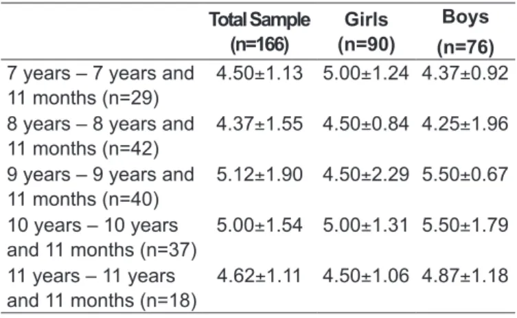
Mean xiphoid and axillary perimetries for the total sample were 5.00±1.59cm and 4.75±1.56cm, respectively. Mean values for girls were 4.87±1.57cm for axillary perimetry and 4.62±1.55cm for xiphoid perimetry. Among boys, the values found were 5.00±1.60 and 4.87±1.58cm, respectively.
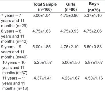
No statistical difference was observed between the xiphoid respiratory coeficient and the variables: sex, age, weight, height and BMI. The ARC showed signiicant differences in the age groups from eight years onwards (p=0.03), and these differences were found in the age groups between 8 and 10 years and 11 months (p=0.03) and betwe-en 10 years and 11 years and 11 months (p=0.02). The ARC was also signiicantly correlated with weight and height. Table 1 shows the correlations between the independent variables (sex, age, weight, height and BMI) and xiphoid and axillary respiratory coeficients. Means obtained in the thoracic perimetry of the axillary region for each age assessed
Table 1 - Correlations between respiratory coeficients and
independent variables
Sex Age Weight Height BMI
Axillary Respiratory Coeficient
r=0.13
p=0.08
r=0.09
p=0.23
r=0.16
p=0.03
r=0.20
p<0.001 r=0.01
p=0.92 Xiphoid
Respiratory Coeficient
r=0.05
p=0.45
r=0.12
p=0.11
r=0.09
p=0.24
r=0.14
p=0.05
r=0.01
p=0.80
Table 2 - Means and standard deviations of the axillary respira
-tory coeficient measured in cm, for each age and sex
Total Sample (n=166)
Girls (n=90)
Boys (n=76)
7 years – 7 years and 11 months (n=29)
5.00±1.04 4.75±0.96 5.37±1.10
8 years – 8 years and 11 months (n=42)
4.75±1.63 4.75±0.93 4.75±2.06
9 years – 9 years and 11 months (n=40)
5.00±1.85 4.75±2.10 5.50±0.88
10 years – 10 years and 11 months (n=37)
5.25±1.57 5.00±1.50 5.87±1.67
11 years – 11 years and 11 months (n=18)
are described in table 2. Table 3 displays the values obtained in the thoracic perimetry of the xiphoid region.
Discussion
The indings of the present study propose reference values for thoracic cirtometry in axillary and xiphoid regions for male and female schoolchildren aged 7 to 11 years from the city of Natal-RN. In a previous study with the same age group, Simon et al(7) suggest values for thoracic mobility
in a sample of 91 boys from South Brazil. Panizzi et al(12)
interpreted the thoracic mobility values of healthy children and adolescents and proposed reference values using axillary and xiphoid respiratory coeficients. In 2007, Caldeira et al(1)
proposed reference values for thoracic mobility, but in an adult population comprising 40 subjects.
The results found indicate that there is no association between xiphoid respiratory coeficient and the variables sex, age, weight, height and BMI. However, the association of the independent variables weight and height with the axillary respiratory coeficient was signiicant, although weak.
The lack of inluence of child’s gender on the respiratory coeficient (RC), observed in this study, corroborates the indings of the study by Kerkoski et al(22), which compared
RC values of two cirtometry techniques in children and adolescents. These authors observed that the values obtained did not differ among the sexes. On the other hand, other authors who compared thoracic cirtometry results among the sexes in child and adolescent samples(12) observed that
the values measured for males were higher compared with those of females. Recently, when assessing thoracic cirto-metry values among groups of health individuals and those
with respiratory disease, Gouilly et al(23) observed higher
values for the male sex. This contradiction is possibly due to the methodological rigor of the present study regarding the selection of the eligible children to participate in the study. The proposed methodology ensured a homogeneous sample for both sexes, especially in terms of anthropometric variables. These criteria were not described by the above mentioned authors.
The indings of this study demonstrate that axillary pe-rimetry differed among ages, between eight and 11 years, which may be explained by the predominance of the apical breathing pattern usually observed at this age(7). This
presu-mably occurs due to a better control of external intercostal muscles in comparison with diaphragmatic muscles. In this period of maturation of the respiratory system, the full control of these muscles is not obtained when a maximal inhalation is required(1,7). According to Murahovschi et al(24),
this phase leads to changes in size, shape, physiology and growth speed, indicating that this population should have a different approach than that used in adults.
The results obtained reveal that, among the study va-riables, height is the one that has a stronger inluence on thoracic mobility in children. These indings corroborate the results found by Simon et al(7), who analyzed the thoracic
mobility of 91 healthy boys aged 7 to 11 years from a private school in Itajaí, state of Santa Catarina, Brazil. The authors concluded that there is a strict relationship between thoracic mobility and the variables height and age, and boys’ height was the variable that had the most signiicant inluence on the increase in thoracic mobility. The values obtained in the present study for thoracic cirtometry, both for axillary and xiphoid coeficients, were similar to those proposed by Simon et al(7) for healthy boys.
Although some authors(1,6,7,10,25) have proposed different
methods for thoracic cirtometry, there is no consensus on its use yet. In 2005, Fregonezi et al(26) proposed that
tho-racic mobility should be measured by indices of chest wall expansion and reduction in axillary and xiphoid regions only. Later, Malagutiet al(10) recommended that the analysis
was performed using the thoracic mobility index, which consists of the difference between a maximal inhalation and total pulmonary capacity and a maximal exhalation until residual volume. Additionally, Jamami et al(27) proposed the
use of the Amplitude Index (AI), considering the highest value of three measurements, which would be included in a speciic equation, in order to take individual anthropometric differences into consideration. These authors considered as Table 3 - Means and standard deviations of the xiphoid respira
-tory coeficient measured in cm, for each age studied
Total Sample (n=166)
Girls (n=90)
Boys (n=76)
7 years – 7 years and 11 months (n=29)
4.50±1.13 5.00±1.24 4.37±0.92
8 years – 8 years and 11 months (n=42)
4.37±1.55 4.50±0.84 4.25±1.96
9 years – 9 years and 11 months (n=40)
5.12±1.90 4.50±2.29 5.50±0.67
10 years – 10 years and 11 months (n=37)
5.00±1.54 5.00±1.31 5.50±1.79
11 years – 11 years and 11 months (n=18)
normal values for AI the interval between 4 and 7cm, but for an adult population. Despite such disagreements, thoracic cirtometry is routinely used in physical therapy to quantify the measures of thoracoabdominal mobility and has been considered an accurate measure(1), with adequate intra and
interrater reliability(28).
One of the limitations of the present study could be the fact that it was conducted in a single city, which could jeo-pardize its external validity and data inference, and it is also necessary to consider the possibility of intra and interrater va-riability, an issue that can be observed in thoracic cirtometry. Moreover, the interpretation of the results was hindered by
the scarcity of previous studies with the assessed age group, the lack of standardization in the literature regarding the technical performance of thoracic cirtometry, as well by the disagreements about how to interpret the values obtained in the thoracic mobility assessment.
In conclusion, the present study provides reference values of thoracic cirtometry for children aged 7 to 11 years. Sex, age, weight, height and BMI of the children assessed did not inluence xiphoid respiratory coeficient. However, the axillary respiratory coeficient showed differences among ages, from eight years onwards, and was inluenced by children’s weight and height, regardless of sex.
References
1. Caldeira Vda S, Starling CC, Britto RR, Martins JA, Sampaio RF, Parreira VF. Reliability and accuracy of cirtometry in healthy adults. J Bras Pneumol 2007;33:519-26.
2. Oliveira KM, Macêdo TM, Borja RO, Nascimento RA, Medeiros Filho WC, Campos TF et al. Respiratory muscle strength and thoracic mobility in children
and adolescents with acute leukemia and healthy school students. Rev Bras Cancerol 2011;57:511-7.
3. Romano AM. Avaliação funcional respiratória em indivíduos com síndrome de Down [tese de mestrado]. Piracicaba (SP): Universidade Metodista de Piracicaba; 2007.
4. Basso RP, Regueiro EM, Jamami M, Di Lorenzo VA, Costa D. Relationship of the measure of the amplitude thoracoabdominal in asthmatics and healthy adolescents with the physical performance. Fisioter Mov 2011;24:107-14. 5. Melo AP, Carvalho FA. Effects of respiratory therapy in Duchenne Muscular
dystrophy – case report. Rev Neurocienc 2011;19:686-93.
6. Carvalho AA. Semiologia em reabilitação. São Paulo: Atheneu; 1994. 7. Simon KM, Carpes MF, Imhof BV, Juk DB, Souza GC, Beckert GF et al.
Avaliação da mobilidade torácica em crianças saudáveis do sexo masculino pela medição do perímetro torácico. Fisioter Pesqui 2006;13:6-12. 8. Costa D, Forti EM, Barbalho-Moulim MC, Rasera-Junior I. Study on pulmonar
volumes and thoracoabdomianl mobility in morbidity obese women undergoing bariatric surgery, treated with two different physical therapy methods. Rev Bras Fisioter 2009;13:294-300.
9. Scipioni G. Função pulmonar e perda de peso em indivíduos obesos mórbidos submetidos à gastroplastia [tese de mestrado]. Curitiba (PR): Universidade Federal do Paraná; 2010.
10. Malaguti C, Rondelli RR, de Souza LM, Dominques M, Dal Corso S. Reliability of chest wall mobility and its correlation with pulmonary function in patients with chronic obstructive pulmonary disease. Respir Care 2009;54:1703-11.
11. Barros Filho TE, Lech O. Exame físico em ortopedia. São Paulo: Manole; 2001. 12. Panizzi AC, Nunes AC, Borba C, Kerkoski E. Mobilidade torácica em
estudantes na faixa etária de 8 a 14 anos de ambos os sexos: uma análise descritiva. São José dos Campos: VIII Encontro Latino-Americano de Iniciação Cientíica e IV Encontro Latino-Americano de Pós-Graduação; 2004. 13. Burgos Rodríguez R. Metodología de investigación y escritura cientíica en
clínica. 3 ed. Granada/España: Escuela Andaluza de Salud Pública; 1998. 14. Instituto Dante Pazzanese de Cardiologia [homepage on the Internet].
Laboratório de Epidemiologia e Estatística. Tamanho de amostra para pesquisa em ciências da saúde [cited 2009 Feb 23]. Available from: http://www.lee. dante.br/pesquisa/amostragem/amostra.html
15. Wilson SH, Cooke NT, Edwards RH, Spiro SG. Predicted normal values
for maximal respiratory pressures in caucasian adults and children. Thorax 1984;39:535-8.
16. Souza RB. Pressões respiratórias estáticas máximas. J Pneumol 2002;28 (Suppl 3):S155-65.
17. Harik-Khan RI, Wise RA, Fozard JL. Determinants of maximal inspiratory pressure: the Baltimore longitudinal study of aging. Am J Respir Crit Care Med 1998;158:1459-64.
18. Centers for Disease Control and Prevention [homepage on the Internet]. United States: Body mass index-for-age. Children and adolescents, 2 to 20 years [cited 2010 Sept 9]. Available from: http://www.cdc.gov/nchs/about/ major/nhanes/growthcharts/clinical_charts.htm
19. Kuczmarski RJ, Ogden CL, Guo SS, Grummer-Strawn LM, Flegal KM, Mei Z
et al. 2000 CDC growth charts for the United States: methods and development.
Vital Health Stat 11 2002;1-190.
20. Kakizaki F, Shibuya M, Yamazaki T, Suzuki H, Homma I. Preliminary report of the effects of respiratory muscle stretch gymnastics on chest wall mobility in patients with chronic obstructive pulmonary disease. Respir Care 1999;44:409-14.
21. Trevisan ME, Soares JC, Rondinel TZ. Effects of two respiratory incentive techniques on chest wall mobility after upper abdominal surgery. Fisioter Pesq 2010;17:322-6.
22. Kerkoski E, Lenzi C, Russi ML, Chiaratti FR, Panizzi EA. Comparação entre duas técnicas de cirtometria em crianças e adolescentes. Proceeding of the VIII Encontro Americano de Iniciação Cientíica e IV Encontro Latino-Americano de Pós-Graduação. Vale do Paraíba: Universidade do Vale do Paraíba; 2004.
23. Gouilly P, Reggiori B, Gnos PL, Schuh O, Muller K, Dominguez A. À propos de la mesure de l’ampliation thoracique. Rev Kinesither 2009;9:49-55. 24. Murahovschi J. Pediatria: diagnóstico e tratamento. 5a. ed. São Paulo: Sarvier;
1995.
25. Caromano FA, Durigon OF, Landaburu C, Pardo MS. Estudo comparativo de duas técnicas de avaliação da mobilidade torácica em mulheres jovens e idosas saudáveis. Fisioter Bras 2003;4:348-52.
26. Fregonezi GA, Resqueti VR, Güell R, Pradas J, Casan P. Effects of 8-week, interval-based inspiratory muscle training and breathing retraining in patients with generalized myasthenia gravis. Chest 2005;128:1524-30.
27. Jamami M, Pires VA, Oishi J, Costa D. Efeitos da intervenção isioterápica na reabilitação pulmonar de pacientes com doença pulmonar obstrutiva crônica (DPOC). Rev Fisioter Univ São Paulo 1999;6:140-53.