ORIGIN
AL RESEAR
CH
Evaluation of respiratory
mechanics in pregnant women
Avaliação da mecânica respiratória em gestantes
Evaluación de la mecánica respiratoria en embarazadas
Anaelisa Venâncio Antunes Pinto1, Juliana Carvalho Schleder2, Christofer Penteado3, Rubneide Barreto Silva Gallo4
Mailing address: Anaelisa Venâncio Antunes Pinto – Rua Bortolo Nadal, 218 – Uvaranas – CEP: 84020-560 – Ponta Grossa (PR), Brazil
E-mail: [email protected] – Presentation: June 2014 – Accepted for publication: Dec. 2015 – Consolidated written opinion from CEP – Platform Brazil No. 503.740. Date of report: December 11, 2013.
1Undergraduate in Physiotherapy of CESCAGE.
2Student in the Master’s Program of Physiology (UFPR), Master’s Degree in Technology in Health (PUC). Specialist in Physiotherapy
in Oncology (SBFC). Physiotherapist of the Regional University Hospital of Campos Gerais (HURCG). Physiotherapist responsible for Teaching and Research in the Physiotherapy Department of the Erasto Gaertner Hospital (HEG).
3Specialist in Orthopedy, Traumatology and Sports Medicine (PUC-PR) and Manual Therapy (Inspirar). Professor of the Higher
Education Center of Campos Gerais (CESCAGE).
4PhD Candidate in Gynecology and Obstetrics at the University of São Paulo (USP). Specialist in Woman Health Physiotherapy
(UNAERP). Professor of the Higher Education Center of Campos Gerais (CESCAGE).
ABSTRACT | The respiratory function is one of the organic processes most afected by pregnancy, and it can be evaluated by cirtometry and manovacuometry. The changes in thoracic expansibility and respiratory pressures during pregnancy of healthy women were veriied. This is a prospective, descriptive and analytic study. Ninety three pregnant women who attended health unities from Ponta Grossa (Brazil) have participated. They were divided in three groups, according to the gestational period: G1 (irst trimester), G2 (second trimester) and G3 (third trimester). The following evaluations were performed with each woman: cirtometry in three points and manovacuometry. The thoracic mobility presented reduction while the pregnancy progressed (respiratory coeicient: armpit line – G1>G2>G3, middle line – G1>G2>G3, xiphoid appendix – G1>G2>G3; inspiration-rest: armpit line – G1>G2>G3, middle line – G1>G2>G3, xiphoid appendix – G1>G2>G3; rest-expiration: armpit line – G1>G2>G3, middle line – G1>G2>G3, xiphoid appendix – G1>G2>G3), in all the cases p<0.01. The maximum inspiratory and expiratory pressures decreased from G1 to G2 (p<0.01 in both cases) and to G3 (p<0.01 in both cases). We concluded that the respiratory muscular strength and the thoracic mobility present reduction with pregnancy progression.
Keywords | Respiratory Function Tests; Respiratory Mechanics; Physical Therapy Modalities.
348
RESUMO | A função respiratória é um dos processos do organismo mais afetados pela gravidez, podendo ser avaliada pela cirtometria e manovacuometria. Veriicaram-se as alterações na expansibilidade torácica e nas pressões respiratórias geradas ao longo do período gestacional de mulheres sadias. Trata-se de uma pesquisa prospectiva, descritiva e analítica. Participaram deste estudo 93 gestantes acompanhadas pelas unidades de saúde de Ponta Grossa (PR). As gestantes foram divididas em três grupos: G1 (primeiro trimestre), G2 (segundo trimestre) e G3 (terceiro trimestre). Foram realizadas as seguintes avaliações em cada gestante: cirtometria em três pontos e manovacuometria. A mobilidade torácica apresentou diminuição com a progressão da gestação (coeiciente respiratório: linha axilar – G1>G2>G3, linha média – G1>G2>G3, apêndice xifoide – G1>G2>G3; inspiração-repouso: linha axilar – G1>G2>G3, linha média – G1>G2>G3, apêndice xifoide – G1>G2>G3; repouso-expiração: linha axilar – G1>G2>G3, linha média – G1>G2>G3, apêndice xifoide – G1>G2>G3), em todos os casos p<0,01. As pressões inspiratória e expiratória máximas diminuíram do G1 para o G2 (p<0,01 nos dois casos) e para o G3 (p<0,01 nos dois casos). Concluiu-se que a força muscular respiratória e a mobilidade torácica reduzem com a progressão da gestação.
RESUMEN | Uno de los procesos más afectados del organismo por el embarazo es la función respiratoria, que se puede evaluar mediante la cirtometría y la manovacuometría. Se investigaron las alteraciones en la expansión torácica y en las presiones respiratorias durante el periodo de embarazo en mujeres sanas. Se trata de una investigación prospectiva, descriptiva y analítica. Participaron del estudio 93 embarazadas bajo supervisión de las unidades de salud en la ciudad brasileña de Ponta Grossa (PR). Se distribuyeron a las embarazadas en tres grupos: E1 (primer trimestre del embarazo), E2 (segundo trimestre del embarazo) y E3 (tercer trimestre del embarazo). Se realizaron las siguientes evaluaciones en cada una: cirtometría en tres puntos y manovacuometría. La movilidad torácica disminuyó con
la progresión del embarazo (coeficiente respiratorio: línea axilar – E1>E2>E3, línea media – E1>E2>E3, apéndice xifoides – E1>E2>E3; inspiración-reposo: línea axilar – E1>E2>E3, línea media – E1>E2>E3, apéndice xifoides – E1>E2>E3; reposo-espiración: línea axilar – E1>E2>E3, línea media – E1>E2>E3, apéndice xifoides – E1>E2>E3), en todos los casos p<0,01. Las presiones inspiratoria y espiratoria máximas disminuyeron del E1 para el E2 (p<0,01 para los dos casos) y para el E3 (p<0,01 para los dos casos). Se concluye que la fuerza muscular respiratoria y la movilidad torácica reducen con la progresión del embarazo.
Palabras clave | Embarazo; Pruebas de Función Respiratoria; Mecánica Respiratoria; Modalidades de Fisioterapia.
INTRODUCTION
Pregnancy is a special health condition that causes several adaptations in the mother’s body, generating both emotional and physical modiications. Such changes are due mainly to the result of the interaction of some hormones, which aim to promote readjustments in the woman’s body and to prepare her for this moment that is so peculiar in her life, which is pregnancy1.
hese adjustments make the body and mind of women sufer profound transformations, which may cause some discomfort, such as diicult breathing,
fatigue, dizziness, among others2. herefore, monitoring
from health care professionals during this period is essential for the balance of these discomforts, since pregnancy afects practically all systems of the human body, including the respiratory system3.
Respiratory function is signiicantly afected during pregnancy. Growth of uterus results in an elevation in the resting position of the diaphragm and a change in the coniguration of the thorax, which extends in the anteroposterior diameter. Subcostal angle increases and consequently the thoracic circumference as well. In addition, abdominal muscles are subjected to extreme stretch. During the irst gestational trimester, respiratory minute volume increases due to raised tidal volume. his hyperventilation may, therefore, explain the number of subjective complaints of dyspnea
during gestation4. Other changes can also occur, such
as increased respiratory rate, tiredness, and even more severe situations such as respiratory failure, having
serious consequences. hus, in addition to serious risk to the pregnant woman’s health, the fetus may also be negatively afected5.
here are several ways to evaluate respiratory changes, among them, by cirtometry and manovacuometry.
horacoabdominal mobility, i.e., how much the thorax and the abdomen expand during respiratory movements, is evaluated by a method called cirtometry. he instrument for conducting it is a measuring tape with scale in centimeters. With the measuring tape ixed at a certain point, values for thoracic and abdominal circumferences at diferent points are determined,
during diferent moments of the respiratory cycle6.
Manovacuometry is also a method widely used in respiratory evaluation, whose objective is to measure positive pressures (pressure gauge) and negative pressures (vacuum gauge). his allows for the measurement of inspiratory muscle strength (negative pressure) and expiratory muscle strength (positive pressure), which assists in evaluating respiratory mechanics, useful in diagnosing breathing disorders, and in determining parameters for starting and discontinuing mechanical ventilation in intensive care7.
Finally, participation in a research of this nature serves as a source of information for pregnant women about their physiological condition during this period of life. And, to the community in general, it demonstrates the importance of the physical therapist in the full care of pregnant women, reinforcing the need for the presence of this professional in prenatal care.
It is known that there are changes in the respiratory system of pregnant women; however, how these changes interfere with the respiratory mechanics and the extent of such changes over the three trimesters are not yet very well elucidated. herefore, the aim of this study was to determine the changes in thoracic expandability and respiratory pressures generated during the gestational period.
METHODOLOGY
his study was a prospective, descriptive, and analytical research. he research project was submitted to assessment by the Research Ethics Committee (CEP) of CESCAGE, and approved by written opinion No. 503.740 in December 11, 2013.
he selection of pregnant women was conducted in health units of the ive regions of the municipality of Ponta Grossa (North, South, East, West, and Center).
he procedures were performed in the health unit on the day of prenatal appointment with proper authorization from the municipal authority of Ponta Grossa.
his study included 98 pregnant women aged between 18 and 35 years, who attended prenatal care on the public health units. hree women were excluded from the study due to lack of understanding of the procedures performed and other two who reported chronic disease (asthma), which could further alter ventilatory mechanics, in addition to pregnancy.
he pregnant women were divided into three groups according to the gestational trimester: G1: irst trimester of pregnancy; G2: second trimester of pregnancy; G3: third trimester of pregnancy.
In the initial evaluation, after signature of the terms of free and informed consent and authorization for image use, personal and physical data were collected from the pregnant woman through a form prepared by the researchers.
Quantitative variables of this study were determined by conducting cirtometry (thoracic expandability) and
manovacuometry (MIPmax, MEPmax).
Pneumofunctional evaluation consisted in determining some respiratory mechanics parameters by means of two procedures on a single day, on the same day the pregnant woman had her prenatal appointment in the health unit. hese two procedures were:
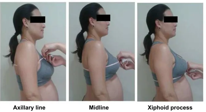
Figure 1. Reference anatomical points for performing cirtometry
Cirtometry, used to analyze thoracoabdominal mobility, is performed with a common measuring tape, considering three reference anatomical points: axillary line, xiphoid process and umbilical line9. However, in the
case of pregnant women, due to the likely displacement of the umbilical line, we considered the axillary line, xiphoid process, and a midline between the axillary line and the xiphoid process.
To position the measuring tape, we marked points with a ballpoint pen on the patient’s body surface area, and the measuring tape was placed just below
each marked point 8. Figure 1 illustrates the points of
measurement for cirtometry.
For the cirtometric evaluation, the pregnant women remained in the standing position, upright column, looking at the horizon with upper limbs relaxed along the trunk and lower limbs parallel.
Cirtometry measurements were carried out at three moments: at rest; after a deep inspiration, slowly and up to the total lung capacity; and after a maximum expiration, slowly, until the residual volume9. Later, the Respiratory
Coeicient (inspiration-expiration), Inspiration-Rest, and Rest-expiration indexes were calculated.
Manovacuometry is a method able to assess respiratory muscle strength. hrough the maximum inspiratory pressure (MIPmax), it is possible to determine inspiratory muscle strength, the normal value for a young
adult being between -90 and -120cmH2O. Expiratory
muscle strength is determined through maximum
expiratory pressure (MEPmax), the normal value for a
young adult being between +100 and +150cmH2O9.
To determine inspiratory and expiratory pressures, we used a pressure gauge manufactured by Comercial
Médica®, and irst the pregnant woman was positioned
comfortably, seated on a chair with backrest, feet on the loor, and upper limbs relaxed on the sides of the body, and then we placed a nasal clip and a
mouthpiece, which had an oriice of approximately 2 mm in diameter, to prevent the elevation of intraoral pressure because of air10.
To measure MEPmax, expiration started at the level of total lung capacity (TLC), i.e., after a deep inspiration. For this purpose, the researcher asked the pregnant woman to inlate her lungs up to TLC and shortly after perform a forced expiration sustaining pressure for 2 seconds. his was performed 3 times, and the inal result
was the highest value obtained11.
To measure the MIPmax, ideally, inspiration should
start from the residual volume (RV), i.e., after a deep expiration. hus, the researcher asked the pregnant woman to exhale all the lung volume including the residual volume, sustaining the pressure for about 2 seconds. he highest value after the three trials was considered11.
To test the normality of the sample, we applied the Kolmogorov-Smirnov test; for the variables that followed Normal distribution, the mean and standard deviation were calculated, and we applied the parametric test one-way ANOVA (ANalysis Of VAriance) with post-hoc Tukey HSD (Honest Signiicant Diferences); for the variables whose Normal distribution was not veriied, we calculated the median, the irst and
third quartile. Tests were processed by the BioEstat®
5.0statistical program.
RESULTS
After applying the inclusion and exclusion criteria, the total number of pregnant women included in the study was n=93.
Table 1 presents the characteristics of each group, emphasizing that there was signiicant diference between the groups for the variables “gestational age” and “sedentary lifestyle”:
Table 1. Characterization of the sample
Variables Group 1 (n = 31) Group 2 (n = 30) Group 3 (n = 32) p-Value
Age (years)* 24.13+4.29 24.63+4.99 24.56+5.20 >0.05
Gestational age (weeks)* 10.48+1.52 20.83+2.76 33.59+4.13 <0.01
Number of pregnancies* 2.13+1.09 1.97+1.25 1.91+1.30 >0.05
Sedentary lifestyle* 83.87% 83.33% 68.75% <0.01
Current weight (kg)¬ 63 (57; 78.5) 63.5 (59.25; 70) 67.5 (63; 71.62) >0.05
Previous weight (kg)¬ 65 (55; 76.5) 59 (53; 65.5) 56.5 (54; 66.25) >0.05
Height (cm)¬ 162 (155; 165) 160.5 (157.5; 165) 159.5 (154; 165) >0.05
n = number of the sample. We considered as sedentary the pregnant women who declared not practicing any type of physical activity at least three times a week for one hour a day
* Values are expressed as mean and standard deviation and percentage. The test used to determine the diference between groups was the one-way ANOVA (ANalysis Of VAriance) with post-hoc Tukey HSD (Honest Signiicant Diferences), adopting the signiicance value of p≤0.05
Figure 2 illustrates the evolution of thoracic mobility of the three groups under study.
Figure 2. Evolution of thoracic mobility over the gestational period
Figure 3 illustrates the evolution of respiratory pressures for the three groups under study.
Figure 3. Evolution of respiratory pressures during the gestational period
he means obtained with cirtometry and manovacuometry are presented in Table 2, emphasizing that there was signiicant diference between the groups for all variables:
Table 2. Thoracoabdominal mobility and respiratory muscle strength of the three groups assessed
Anatomical reference Variables G1 (n = 31) G2 (n = 30) G3 (n = 32) p-Value
Axillary line (cm)
Respiratory coeicient 6.84±1.98 4.53±0.94 3.16±0.81 <0.01
Inspiration-rest 3.65±1.45 2.63±0.76 1.91±0.59 <0.01
Rest-expiration 3.19±1.14 1.90±0.71 1.31±0.54 <0.01
Midline (cm)
Respiratory coeicient 6.74±2.08 4.00±1.60 3.00±0.92 <0.01
Inspiration-rest 3.71±1.42 2.50±0.86 1.94±0.50 <0.01
Rest-expiration 3.03±1.25 2.03±0.61 1.13±0.61 <0.01
Xiphoid process (cm)
Respiratory coeicient 6.74±2.08 4.00±1.60 3.00±0.92 <0.01
Inspiration-rest 3.71±1.42 2.50±0.86 1.94±0.50 <0.01
Rest-expiration 3.03±1.25 2.03±0.61 1.13±0.61 <0.01
MIPmax (cmH2O) -98.39±14.63 -74.00±16.32 -69.06±19.07 <0.01
MEPmax (cmH2O) 100.32±13.78 76.00±14.53 72.19±19.13 <0.01
n = number of the sample. Values are expressed as mean and standard deviation. The test used to determine the diference between groups was the one-way ANOVA (ANalysis Of VAriance) with post-hoc Tukey HSD (Honest Signiicant Diferences), adopting the signiicance value of p≤0.05
In comparing the three measures (respiratory coeicient, inspiration-rest diference and rest-expiration diference), for the three reference anatomical points, the same situation was observed: there was a decrease in values, that is, decreased thoracic mobility, from G1 to G2, from G1 to G3, and from G2 to G3.
he same can be observed regarding respiratory muscle strengths.
DISCUSSION
he fact that the variables of characterization of the samples “age”, “number of pregnancies”, “current
weight”, “previous weight” and “height” have no signiicant diference among the three groups (p>0.05) indicates that they are similar, favoring the comparison between them. As expected, the variable “gestational age” showed signiicant diference (p<0.05) among the groups, as it was the criterion adopted to divide them.
Cirtometry has been pointed out by many authors as a simple and accessible measure to assess thoracic
mobility8. It was possible to observe, in studies that
focus on the technique of cirtometry as a means of evaluation, a lack of standardization and diferent
manners of describing how it should be conducted12.
addition to the basal region (12th rib) or the umbilical
region; however, some use only two points of reference13.
In this study, we considered the axillary line, xiphoid process, and the midline between the axillary line and the xiphoid process, because of the displacement of the umbilical region due to the gestational period.
he results of cirtometry, in this study, point to a decrease in thoracic mobility with the progression of pregnancy. his fact occurred when comparing the groups, in the three anatomical points considered. Similar results were found in a study of 150 pregnant women monitored in the Hospital das Clínicas of the University of São Paulo14.
he assessment of respiratory pressures through manovacuometry in this study pointed to a decrease in both maximum inspiratory and maximum expiratory pressures as pregnancy progressed. A study involving 150 pregnant women in São Paulo showed signiicant decrease in comparing the three trimesters
of gestation14, and the greatest diference was observed
in the comparison between the irst and the second trimester of pregnancy, as in the case of this research. Initially, a marked decrease in the values for respiratory pressures could be expected for the third trimester, the period in which uterine volume is higher. It was probably not observed because, according to Lemos (2011), there is an increase in abdominal pressure at the end of expiration due to the greater volume of the uterus associated with an increase in compliance of the thoracic wall, resulting in decreased FRC and alteration in the rest position of the respiratory system; with the elevation of the diaphragm, there is an increase in the area of apposition in relation to the rib cage, thus raising the ability to generate tension. Diaphragm muscle ibers are in an optimal position of length-tension, and diaphragmatic descent control is favored by decreased abdominal compliance; in addition, there are factors such as no change in transdiaphragmatic pressure and an equitable contribution of intercostal and diaphragmatic muscles to tidal volume, resulting in facilitation to maintain respiratory muscle strength14.
A research shows that, in addition to decrease in respiratory pressures with the progression of pregnancy, there is also a decrease when comparing the values found for pregnant women in relation to non-pregnant
women15. However, there are analyses that suggest that
respiratory pressures do not change signiicantly during pregnancy, such as the one conducted in Recife in 2010, with 120 low-risk pregnant women aged between 20
and 29 years16. Nevertheless, the same study reported
a trend toward signiicance for decrease in MEPmax in
the beginning of pregnancy, which is inconsistent with the data collected by this research. Another study also suggested no change in respiratory pressures; however, this study was conducted only with pregnant women in the last trimester of gestation17. A study that evaluated
37 primigravidae aged between 18 and 30 years with gestational age of ≥24 weeks with preeclampsia stated no diference between respiratory pressures in relation to control group (same characteristics, except for diagnosis of preeclampsia), in addition to presenting values for
MIPmax (-107cmH20) and MEP (95cmH20) which
were higher than the values of all three groups in this study. his may be due to diference in the equipment, since the technique used to measure was the same, or alterations in life habits such as smoking and physical activity, not speciied in the studies18.
CONCLUSION
We observed that thoracic mobility decreases with the progression of pregnancy in the three anatomical points considered, and respiratory muscle strength also decreases during the gestational trimesters; a decrease which, as observed, has greater intensity at the beginning of pregnancy.
Finally, we suggest that future studies monitor pregnant women throughout the gestational period, i.e., evaluate them in the irst, second and third trimester of pregnancy.
REFERENCES
1. Constantine MM. Physiologic and pharmacokinetic changes in pregnancy. Front Pharmacol. 2014;5:65.
2. Jensen D, Webb KA, Davies GAL, O’DonnellInda DE. Mechanical ventilatory constraints during incremental cycle exercise in human pregnancy: implications for respiratory sensation. J Physiol. 2008; 586(19):4735-50.
3. Marcos IACG. Pulmão e gravidez. Rev Port Pneumol. 2007; 13(2):213-37.
4. Lemos A, Souza AI, Andrade AD, Figueiroa JN, Cabral-Filho JE. Força muscular respiratória: comparação entre primigestas e nuligestas. J Bras Pneumol. 2011. 37(2):193-9. 5. Surita FG, Nascimento SL, Pinto e Silva JL. Exercício físico e
gestação. Rev Bras Ginecol Obstet. 2014; 36(12):531-4. 6. Costa D, Forti EMP, Barbalho-Moulim MC, Rasera-Junior
toracoabdominal de portadoras de obesidade mórbida, submetidas à cirurgia bariátrica, tratadas com duas diferentes técnicas de isioterapia. RBF. 2009;13(4):294-300. 7. Onaga Fi, Jamami m, Ruas g, Di Lorenzo vap, Jamami lk.
Inluência de diferentes tipos de bocais e diâmetros de traqueias na manovacuometria. Fisioter Mov. 2010;23(2):211-9. 8. Caldeira VS, Starling CCD, Britto RR, Martins JÁ, Sampaio RF,
Parreira VF. Precisão e acurácia da cirtometria em adultos saudáveis. J Bras Pneumol. 2007; 33(5):519-26.
9. Forti E, Ike D, Barbalho-Moulim M, Rasera Jr I, Costa D. Efects of chest physiotherapy on the respiratory function of postoperative gastroplasty patients. Clinics, 2009; 64(7):683-9.
10. Teixeira CA, Santos JE, Silva GA, Souza EST, Mart JAB. Prevalência de dispnéia e possíveis mecanismos isiopatológicos envolvidos em indivíduos com obesidade graus 2 e 3. J Bras Pneumol. 2007;33(1):28-35.
11. Almeida IP, Bertucci NR, Lima VP. Variações da pressão inspiratória máxima e pressão expiratória máxima a partir da capacidade residual funcional ou da capacidade pulmonar total e volume residual em indivíduos normais. Mundo Saúde, 2008;176-82.
12. Pedrini A, Gonçalves MA, Leal BE, Yamaguti WPS, Paulin E. Comparação entre as medidas de cirtometria
tóraco-abdominal realizadas em decúbito dorsal e em ortostatismo. Fisioter Pesqui. 2013;20(4):373-8.
13. Archija LRF, Selman JPR, Dal Corso S, Lanza FC. Cirtometria torácica está relacionada à força dos músculos respiratórios e aos volumes pulmonares em indivíduos saudáveis. RBF. 2012;16(Supl 1):168.
14. Caromano F, Sayuri E, Cruz CMV, Candeloro JM, Burti JS, Andrade LZ. Mobilidade torácica e pressões respiratórias máximas durante a gestação. Fisioter Bras. 2006;7(1):5-7.
15. Almeida LGD, Constâncio JF, Santos CVS, Silva TG, Raposo MT. Análise comparativa das PE e PI máximas entre mulheres grávidas e não-grávidas e entre grávidas de diferentes períodos gestacionais. Rev Saúde Com. 2005;1(1):9-17.
16. Lemos A, Souza AI, Figueroa JN, Cabral-Filho JE, Andrade AD. Respiratory muscle strength in pregnancy. Respir Med. 2010;4:1638-44.
17. Lemos A, Caminha MA, Melo Jr EF, Andrade AD. Avaliação da força muscular respiratória no terceiro trimestre de gestação. RBF. 2005;9(2):151-6.