SUMMARY
Colorectal cancer is the 3rd most common malignant neoplasm in the West. About 50% of patients develop liver metastases throughout the course of the disease. hose are res ponsible for at least twothirds of deaths. Advances in surgical techniques and improve ment in chemotherapy regimens have allowed ofering treatment with curative intent to an increasing number of patients. his article reviews recent advances in the treatment of liver metastases, including strategies to increase resection (e.g., portal vein emboliza tion, radiofrequency ablation, twostage hepatectomy, conversion therapy and reverse treatment strategy) and hepatectomy in the presence of extrahepatic disease. Finally, the results of surgical treatment of liver metastases at the Hospital A.C. Camargo are briely shown.
Keywords: Abdominal neoplasms; colorectal neoplasms; digestive system neoplasms; liver neoplasms; hepatectomy; colorectal surgery.
Advances in the surgical treatment of colorectal liver metastases
FELIPE JOSÉ FERNÁNDEZ COIMBRA1, THIAGO COSTA PIRES2, WILSON LUIZDA COSTA JUNIOR3, ALESSANDRO LANDSKRON DINIZ4,
HÉBER SALVADORDE CASTRO RIBEIRO3
1 Surgical Oncology; M.Sc. in Oncology, IHPBA, AHPBA, CBC, TECA; Director of the Department of Abdominal Surgery of Hospital AC Camargo, São Paulo, SP 2 Surgical Oncology; Department of Abdominal Surgery of Hospital AC Camargo, São Paulo, SP
3 Surgical Oncology; TCBC, TECA; Department of Abdominal Surgery of Hospital AC Camargo, São Paulo, SP 4 Surgical Oncology; TCBC, IHPBA, TECA; Department of Abdominal Surgery of Hospital AC Camargo, São Paulo, SP
Study performed at Department of Abdominal Surgery of Hospital AC Camargo - Surgical Oncology of Digestive System, São Paulo, SP
Submitted on: 09/14/2010 Approved on: 01/25/2011
Correspondence to: Felipe José Fernández Coimbra Rua José Getúlio, 579, conj 42 –
Aclimação São Paulo – SP CEP: 01509-001 [email protected]
INTRODUCTION
Colorectal cancer is the 3rd most common malignant neo plasm in the West. Approximately 50% of the patients de velop liver metastasis during the disease evolution, which are responsible for at least twothirds of the deaths16. To date, the only potentially curative therapy for these pa tients is the surgical treatment. However, only 10% to 20% are candidates to resection. When patients are submitted to complete resection, the iveyear survival can range from 37% to 58% in the most recent series68.
Although only some of these patients are candidates to surgical treatment, the absolute number of resectable individuals is signiicant. If a free estimate is made for the Brazilian population, based on the incidence rates sup plied by the National Cancer Institute (Instituto Nacional do Cancer – INCA) for colorectal cancer in 2009/2010, which is 27,000 new cases/year, one can suppose that around 13,500 (50%) patients have or will have colorec tal liver metastases (CRLM), of which 2,700 to 4,050 pa tients/year (20% to 30%) will be potential candidates for liver resections.
he increase in the number of surgical indications, with the inclusion of bilateral metastases, with no lim it for the number and size of nodules, associated with the improvement in systemic treatment outcomes with the use of new regimens with high response rates (going from less than 20% to approximately 50%), can change patients that were initially unresectable into resectable ones and treatments that were initially palliative into curative ones911.
he present article reviews the recent advances in the treatment of liver metastases, including strategies to increase resection and hepatectomies in the presence of extrahepatic disease. Finally, we briely show the results of the surgical treatment of liver metastases in Hospital A.C. Camargo.
RESECTABILITYCRITERIA
he capacity to remove all liver metastases with free margins and preserve a future remnant liver (FRL) of at least 20% of the total liver volume (TLV) in patients with a healthy liver, in the absence of unresectable extrahepatic disease, deines most cases regarding liver resectability. Moreover, it is nec essary to guarantee adequate arterial and portal inlow, as well as biliary drainage and venous return (outlow). Some patients might need a FRL volume > 20%.
Patients that have been submitted to many chemother apy cycles (intensive chemotherapy) need FRL of at least 30%, whereas for patients with chronic hepatopathy one can estimate 40%. here is still a great deal of controversy regarding what is considered intensive chemotherapy. In Hospital A. C. Camargo, that is considered as more than six cycles of the usual regimen carried out currently, such as FOLFOX (5Fluorouracil and oxaliplatin), FOLFIRI (5Fluorouracil and irinotecan) or FOLFOXIRI (5Fluoro uracil, oxaliplatin and irinotecan).
In patients candidate to extensive resections, it is nec essary to calculate with higher accuracy the FRL volume. For that purpose, it is necessary to perform liver volum etry. he direct measurement of the FRL is performed by computed tomography (CT). Among the existing formu las, we used the one described by Vauthey et al. to calculate the standardized total liver volume1213 (Figure 1).
As for the margin, diferently from what was believed in the past, a margin of at least 1 cm is not mandatory. Busquets et al.14 in a multicentric study with 557 patients, compared the resection margins from 1 mm to 1 cm and observed that there was no signiicant diference in global and freeofdisease survival. herefore, the main objective is to achieve free margins, even though the goal is a 1cm margin14.
PREOPERATIVEEVALUATIONANDSTAGINGEXAMINATION
Initially, the sequelae of previous treatments (for in stance, previous hepatectomy and chemotherapy) must be considered as well as patients’ comorbidities (obesity, diabetes mellitus, alcohol consumption, liver cirrhosis). he morbidity associated with liver steatosis, very oten a consequence of the systemic treatment, is a controversial issue, as although there is a histological liver lesion, its inluence on mortality remains controversial15.
he main examination to be carried out for the stag ing is the CT with a protocol for liver, where thinsection CT images are acquired (preferably in equipment with multidetectors) in four phases: precontrast, arterial, portal and equilibrium or late phase. It is considered the goldstandard by most specialized centers, as it allows the accurate assessment of resectability, the number of nodules and their association with liver structures and adjacent organs, in addition to performing the liver volu metry.
Other examinations can also be efectively per formed, especially the magnetic resonance (MR), which allows the acquisition of images that are equivalent to the tomography in terms of quality. Some believe that at this time of preoperative chemotherapy and obesity, the MR can be very important, due to the higher capacity of dif ferentiating areas of steatosis from secondary nodules, which has yet to be deinitively demonstrated.
he colonoscopy must be always used to rule out the possibility of primary tumor recidivism. Chest images by xrays or CT are also mandatory to assess the presence of lung metastases.
better patient selection for surgery, with a 5year survival of 58% being observed in this group of patients; however, the great criticism faced by this study is not comparing PETCT with the currently available highdeinition to mographic images. One must be aware of falsenegative results ater the chemotherapy. hus, even when there is no uptake of a nodule visualized before the chemo therapy, the indication for resection is maintained, as the decreased sensitivity of PETCT in detecting metastases postchemotherapy is wellknown17, mainly before two weeks ater its completion.
he PETCT can also predict the response to chemo therapy when 18FFU is used instead of 18FFDG18.
One of the most important questions regarding the restaging postchemotherapy is the discrepancy be tween imaging study results and surgical indings19,20. Angliviel et al.20 showed that there is more than 50% of result discrepancy in CT indings postchemotherapy at the restaging when compared with the surgical indings. Carnaghi et al.19 pointed out that both PETCT and CT have limited sensitivity (60%) for restaging of CRLM postchemotherapy, especially for lesions < 1 cm.
Benoist et al.21 evaluated 66 patients that had com plete response at the imaging examinations ater “neo adjuvant” chemotherapy and were submitted to surgical exploration and systematic clinical followup. Of these, 32 had lesions identiied at the surgery and 23 were iden tiied at the clinical followup in the same sites of the pre vious lesion. he conclusion is that 83% of the patients that had a complete response at the imaging examina tions have macro or microscopic residual disease or early recidivism. From our point of view, this information is of utmost importance for the indication of surgical explora tion and resection of previously compromised areas, even in patients in whom complete radiological response was observed and for whom a curative treatment is intended.
SURGICALTREATMENT
The surgical procedure must be initiated with the sys tematic exploration of the abdominal cavity, with spe cial attention when assessing the presence of extra hepatic disease. Colon, peritoneum, retroperitoneal lymph nodes, celiac trunk and hepatic hilum are evalu ated and biopsies and microscopic examination of fro zen samples are carried out in all suspected sites.
All assessment modalities are important during liver evaluation. The presence of nodules, postchemothera py scars, retractions or areas suggestive of fibrosis must be observed. At palpation, the presence of hardened, round, firm, or fibroelastic areas that can be superfi cial, easily palpable or deep can be noticed. These must be assessed carefully, as the presence of the liver paren chyma between the tumor and the examiner’s hand can make the evaluation difficult. The examination must
be carried out by surgeon by sliding the hands over the entire liver surface and it must always be bimanual, in creasing the sensitivity to identify deep lesions, espe cially in the left lobe.
The intraoperative ultrasonography is currently an essential tool for staging and surgical planning and therefore, a mandatory examination in any liver sur gery. It can identify 20% to 30% of the nodules that were not detected at the conventional examinations. In our country, Cohen MP et al.22 demonstrated that the in traoperative ultrasonography in surgeries performed to resect liver metastases changes the surgical strategy in 25.7% of the cases and is extremely useful in identifying lesions < 1 cm.
The type of resection must guided by the number and location of lesions and by the need to attain tumor free margins. The anatomic resections, that is, exeresis of liver segments or lobes, respecting the regions de limited by venous and arterial vascularization, in addi tion to the biliary drainage, are preferable, as they allow lower blood loss and carry a lower risk of compromised margins. However, there is no difference in survival re garding the nonanatomical resections, as long as the margins are free23,24. The types of resection are: segmen tectomies, bisegmentectomies, central hepatectomies, lobectomies, trisegmentectomies, enucleations and combinations of these forms. Resections that are con comitant to the primary tumor are safe and feasible, as long as they are carried out by an experienced tem and follow the oncologic principles.
STRATEGIESTO INCREASERESECTABILITY
As previously described, resectability is currently de fined by a new paradigm, where the possibility of re section of liver lesions must be considered, as well as the complete resection of extrahepatic lesions and the quality (inflowoutflow) and quantity of remnant liver after the surgery and not exclusively by the tumor clini copathological factors. Therefore, previously used cri teria, such as number of nodules, size of lesions, bilat eralism and presence of extrahepatic disease (as long as resectable) must be considered prognostic factors and not a contraindication for resection.
BEFORE: based on what was resected.
CURRENTLY: based on what will remain ater resection.
PORTAL VEINEMBOLIZATION
In general, 20% of the normal FRL is considered safe ater an extensive resection. However, the sectioning volume for FRL in patients with livers presenting steatosis, steato hepatitis (30% RLV) or cirrhosis (> 40%) must be higher. In general, the right lobe represents twothirds of the liver volume and the let only onethird. Frequently, patients with multiple liver lesions are submitted to right hepatectomy ex tended to segment IV (or right trisegmentectomy).
On average, these surgeries remove around 84% of the liver volume in the absence of compensatory hypertrophy of the remnant liver25. However, a high degree of indi vidual variation can be observed in the volumes of liver segments and lobes. To prevent surgeries in patients with FRL lower than the desired volume, the portal vein embo lization must be carried out to induce contralateral lobe hypertrophy25.
he idea came from the observation that when there is invasion of a portal vein branch by the tumor, there is hypertrophy of the contralateral lobe. Technically it is performed through catheterization by radioscopy of the lobe or segmental vein, followed by vessel embolization by embolic material (coils, thrombin, cyanoacrylate, mi crospheres, etc). It is a relatively safe procedure; its rate of complications varies from 5% to 8%. hen expected vol ume growth of the FRL is of 8% to 16%2628.
he portal vein embolization is more frequently used as part of the multimodal treatment regimens, which in clude preoperative chemotherapy and hepatectomy, as most part of these patients already presents with more than one factor of poor prognosis, such as multiple lesions, bilobar lesions, compromised lymph nodes at the primary and extrahepatic metastases.
Some authors evaluated whether the use of CT before or ater the portal embolization could impair liver hyper trophy; however, the results showed no impairment in liver hypertrophy when volume increase is desired29,30.
TWO-STAGEHEPATECTOMY
In extreme situations, in which there are multiple metas tases in both hepatic lobes, twostage resections can be the best therapeutic option and the only chance of cure, pre serving an adequate volume of FRL.
he initial results had a high rate of liver failure and postoperative mortality > 10%31, very diferent from what is currently observed with the routine use portal vein em bolization in specialized centers.
he recommendation is that at the irst intervention, the removal of the liver metastasis be carried out in the liver parenchyma that one wishes to preserve (FRL), to prevent the excessive growth of metastases ater the portal low deviation by embolization. It is usually a parenchyma sparing resection carried out in the lobe or segments that exhibit less disease damage (usually the let lobe) attaining
tumorfree margins, and allowing the preservation of most of the lobe or segments in question. here is a 4to6 week interval to surgery and volumetry control is always per formed before and ater this period, to ensure that there is FRL with an adequate volume.
At the second stage of the procedure, a more extensive resection is performed, most oten from the right lobe, which extends to the IV segment. It is seldom necessary to perform let portal vein embolization for right lobe hyper trophy, as the volume of the latter is hardly ever lower than the desired volume. As it is a complex procedure, it must be performed only in curative situations.
In several situations, CT is indicated during the time between the embolization and surgery, without the use of monoclonal antibody (Bevacizumab), when it is being used in the CT regimen. he objective is to prevent tumor growth during the period when waiting for the second phase of the surgery.
RADIOFREQUENCY
Another alternative to the twostage surgery is the asso ciation of radiofrequency (RF) ablation with liver resec tion, which in some situations can expand the number of patients eligible for surgery. However, the RF has a higher risk of recidivism in comparison with the resection, main ly in lesions > 3 cm. It use must be restricted to cases in which the resection is not possible due to lack of adequate FRL volume7.
CONVERSION THERAPY
Many patients have such extensive liver disease at diag nosis that they cannot be candidates to liver resection through any of the strategies mentioned before. However, there are cases in which the reduction of hepatic lesions through CT can enable the surgical treatment, transform ing an initially unresectable disease into a resectable one. When the CT is used for that purpose, it is called conver sion chemotherapy.
he conversion CT consists of administration of thera peutic regimens with a high rate of response, aiming at the decrease in tumor volume to allow the resection of metas tases, while obtaining an adequate liver volume.
he main therapeutic options are FOLFOX or FOL FIRI, or a combination of both (FOLFOXIRI), with re sponse rates of 4866%3235, 3962%3638 and 5671,4%39,40, respectively. According to some authors, there is a “con version” in around 10% to 20% of individuals initially con sidered to unresectable9, with survival rates similar to that observed in patients that are initially resectable41. Higher response rates can be obtained by adding targettherapy with cetuximab or bevacizumab.
as the lesions are resectable, preventing unnecessary liver toxicity that result from CT excess and also an eventual progression of the disease ater a long period of treatment. hus, the patients must be followed together with the surgeon, through control image assessment every two months, aiming at detecting lesion response and identify ing, as soon as possible, the moment when the metastases become resectable. If there is no adequate radiological re sponse, a new CT scheme can be attempted, always aiming at conversion.
EXTRAHEPATIC DISEASE
Traditionally, the presence of extrahepatic metastases of colorectal origin was considered an absolute contraindi cation for hepatectomy42. As a consequence of safer sur gical procedures and the evolution of the efectiveness of CT schemes, hepatic resections started to be performed in association with extrahepatic metastasis resection, for selected groups of patients. he main sites of extrahepatic disease to be considered are: portal lymph nodes, perito neum and lungs.
he metastases for portal lymph nodes in the context of CRLM result from the lymphatic drainage of the liver and thus, represent the localregional dissemination of liver metastases43. Patients with macroscopic metastases for por tal lymph nodes have an unfavorable evolution, with little chance of iveyear survival44,45. However, it is possible to select patients with a better prognosis based on the loca tion of the afected lymph nodes. Jaeck et al.46 demonstrated that whereas patients with lymph node metastases along the common hepatic artery and celiac trunk have 0% 1year survival, those with lymph node metastases located in the hepatoduodenal ligament had a 38% 3year survival. hese indings were conirmed by Adam et al.47, who showed a 5year survival of 25% in the analysis of 47 patients with perihepatic lymph node metastases in the hepatoduode nal ligament, whereas there were no survivors among those with metastases in the celiac or paraaortic trunk.
herefore, only patients with hepatoduodenal ligament lymph node metastasis must be considered for liver resec tion. hose with retroperitoneal lymph node disease must receive palliative treatment.
he lungs, together with the liver, are the most com mon sites of metastases in colorectal tumors. Several stud ies have demonstrated that the resection of the lung dis ease can lead to longterm survival48. However, little has been studied on the presence of synchronic lung and liver metastases. Six studies addressed this question, showing that although it is oten necessary to perform new resec tions per early recidivism, the global 5year survival var ies from 2774%49. he main factors that seem to inluence prognosis are: number of pulmonary lesions, number of hepatic lesions and the synchronic versus metachronic presentation.
Peritoneal carcinomatosis occurs in 1325% of pa tients with colorectal tumors. If treated only with sys temic CT, this condition leads to death in less than one year, with a median survival ranging from 5.2 to 6.9 months50. However, similarly to hepatic metastases, it is believed that it does not always represent disseminated systemic disease, but a localregional form of dissemi nation (transmural deposit of tumor cells), which can be treated by peritonectomy and hyperthermic intra peritoneal chemotherapy (HIPEC)51. hus, patients with restricted peritoneal disease can beneit from this treat ment52. Two studies speciically addressed the association between peritoneal carcinomatosis and liver metastases. Carmignani et al.53 evaluated 27 patients with peritoneal carcinomatosis, of which 16 had liver metastases as the only additional site of the disease and 4 other had liver and lung metastases. he procedures aiming at complete cytoreduction had a morbidity of 14.8%, with no deaths and a median survival of 15.2%. Elias et al. reported on the treatment of 27 patients with CRLM and synchronic peritoneal carcinomatosis, of which 14 patients had car cinomatosis detected preoperatively and 13, intraopera tively54. here was a postoperative death (4%) on 14th day due to undiagnosed peritonitis and the morbidity was 58%. With a median followup of 6.1 years, the global 5year survival was 26.5%, with seven patients being diseasefree; as for the cases of recidivism, only three had been located in the peritoneum. he only prognos tic factor with statistical signiicance was the number of liver nodules > 2. However, these indings still need to be corroborated by other randomized studies with larger samples. hus, wellselected patients, as long as they are submitted to treatment in specialized centers, can un dergo the simultaneous treatment of liver metastases and peritoneal carcinomatosis.
REVERSE TREATMENTSTRATEGY
Patients with synchronic liver metastases are classically submitted to primary tumor resection, followed by long CT periods and subsequently, if there is no disease pro gression during this period, they are referred to liver resection55,56. However, patients with advanced liver dis ease can have metastasis progression during the primary tumor treatment, making the lesions unresectable. his problem becomes especially important in patients with rectal tumors (who oten necessary need to undergo neoadjuvant RT, in which the concomitant CT has only a radiosensitizing function) and in those with surgical complications caused by the primary tumor treatment.
An attempt to overcome the problem has been the use of a new treatment strategy, called the reverse treatment strategy, where there is an inversion of the classic treat ment sequence59,60.
Hence, liver metastases – the main determinants for the deinition of the treatment curative characteristic – are treated before the primary tumor. Patients with asymp tomatic colorectal tumor with large, but resectable liver metastases or patients with initially unresectable metasta ses that achieved conversion ater chemotherapy are can didates to this type of treatment.
In our service, we recommend starting these patients’ management with chemotherapy, aiming at the immediate treatment of both the liver metastases and the micrometa static systemic disease. he main concerns regarding this approach are the possibility of complications related to the primary tumor (pain, bleeding or obstruction) or the pro gression of liver metastases during the CT period. How ever, the irst is a rare event, not diferent from the rates of complications or bridle obstructions in patients submitted to surgery61,62, whereas the latter represents such a poor prognosis that these patients would hardly have beneited from any initial surgical treatment63.
TREATMENTOUTCOMES
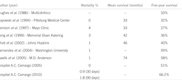
Even considering the increase in surgical indications for larger tumors, multiple nodules, synchronous bilobar lesions and extrahepatic disease, one can observe an in crease in survival throughout the last decades, going in a period of ive years from 30% in the oldest series to more than 50% in the current ones (Table 1).
A published analysis of 70 patients submitted to sur gery in our institution between January 1999 and June 2005 showed a iveyearsurvival of 51%64. A more recent reassessment of our series, taking into account 142 surger
ies in 121 patients in recent years, showed a global survival of 66.2% in ive years and 54.9% in seven years (data not published).
CONCLUSION
he perfecting of surgical techniques together with safer procedures, as well as the improvement in chemotherapy regimens have allowed doctors to ofer patients with liver metastasis the possibility of curative treatment or long term survival. Factors that were previously considered contraindications for the surgery, such as number of me tastases, synchronous metastases and even the presence of extrahepatic disease, must be considered only as prognos tic factors and must not prevent the patient from having the opportunity of being treated.
REFERENCES
1. Bouvier AM, Remontet L, Jougla E, Launay G, Grosclaude P, Velten M et al. Incidence of gastrointestinal cancers in France. Gastroen terol Clin Biol. 2004;28:87781.
2. Faivre J, Manfredi S, Bouvier AM. Epidemiology of colorectal cancer liver metastases. Bull Acad Natl Med. 2003;187:81522; discussion 223.
3. Geoghegan JG, Scheele J. Treatment of colorectal liver metastases. Br J Surg. 1999;86:15869.
4. Rastogi T, Hildesheim A, Sinha R. Opportunities for cancer epidemi ology in developing countries. Nat Rev Cancer 2004;4:90917. 5. Welch JP, Donaldson GA. he clinical correlation of an autopsy
study of recurrent colorectal cancer. Ann Surg. 1979;189:496502. 6. Yamamoto J, Shimada K, Kosuge T, Yamasaki S, Sakamoto M, Fu
kuda H. Factors inluencing survival of patients undergoing hepa tectomy for colorectal metastases. Br J Surg. 1999;86:3327. 7. Abdalla EK, Vauthey JN, Ellis LM, Ellis V, Pollock R, Broglio KR
et al. Recurrence and outcomes following hepatic resection, radio frequency ablation, and combined resection/ablation for colorectal liver metastases. Ann Surg. 2004;239:81825; discussion 257. 8. Ercolani G, Grazi GL, Ravaioli M, Cescon M Gardini A, Varotti G
et al. Liver resection for multiple colorectal metastases: inluence of parenchymal involvement and total tumor volume, vs number or lo cation, on longterm survival. Arch Surg. 2002;137:118792.
Table 1 – Outcomes of liver resection for metastatic colorectal cancer
Author (year) Mortality % Mean survival (months) Five-year survival
Hughes et al. (1986) - Multicêntrico – – 33%
Gayowski et al. (1994) - Pittsburg Medical Center 0 33 32%
Jamison et al. (1997) - Mayo Clinic 4 33 27%
Fong et al. (1999) - Memorial Sloan Katering 3 42 36%
Choti et al. (2002) - Johns Hopkins 1 46 40%
Fernandez et al. (2004) - Washington University 1 – 59%
Pawlik et al. (2005) - M.D. Anderson 1 74 58%
Hospital A.C. Camargo (2005) 0 – 51%
Hospital A.C. Camargo (2010) 0.9 (30 days)
9. Adam R, Delvart V, Pascal G, Vallanu A, Castaing D, Azoulay D et al. Rescue surgery for unresectable colorectal liver metastases down staged by chemotherapy: a model to predict longterm survival. Ann Surg. 2004;240:64457; discussion 578.
10. Giacchetti S, Itzhaki M, Gruia G. Longterm survival of patients with unresectable colorectal cancer liver metastases following infusional chemotherapy with 5luorouracil, leucovorin, oxaliplatin and sur gery. Ann Oncol. 1999;10:6639.
11. Pozzo C, Basso M, Cassano A, Quirino M, Schinzari G, Frigilia N et al. Neoadjuvant treatment of unresectable liver disease with irinote can and 5luorouracil plus folinic acid in colorectal cancer patients. Ann Oncol. 2004;15:9339.
12. Vauthey JN, Abdalla EK, Doherty DA, Gertsch P, Loyer R, Ellis LM et al. Body surface area and body weight predict total liver volume in Western adults. Liver Transpl. 2002;8:23340.
13. Mosteller RD. Simpliied calculation of bodysurface area. N Engl J Med. 1987;317:1098.
14. Busquets J, Pelaez N, Alonso S, Grande L. he study of cavitation al ultrasonically aspirated material during surgery for colorectal liver metastases as a new concept in resection margin. Ann Surg. 2006;244:6345.
15. Choti MA. Chemotherapyassociated hepatotoxicity: do we need to be concerned? Ann Surg Oncol. 2009;16:23914.
16. Fernandez FG, Drebin JA, Linehan DC, Dehdashti F, Siegel BA, Strasberg SM. Fiveyear survival ater resection of hepatic metas tases from colorectal cancer in patients screened by positron emis sion tomography with F18 luorodeoxyglucose (FDGPET). Ann Surg. 2004;240:43847; discussion 4750.
17. Lubezky N, Metser U, Geva R, Nakache R, Shmuele E, Klausner JM et al. he role and limitations of 18luoro2deoxyDglucose positron emission tomography (FDGPET) scan and computerized tomography (CT) in restaging patients with hepatic colorectal me tastases following neoadjuvant chemotherapy: comparison with op erative and pathological indings. J Gastrointest Surg. 2007;11:4728. 18. DimitrakopoulouStrauss A, Strauss LG, Schlag P, Hohenberger
P, Irnagartinger G, Oberdorfer F et al. Fluorine18luorouracil to predict therapy response in liver metastases from colorectal carci noma. J Nucl Med. 1998;39:1197202.
19. Carnaghi C, Tronconi MC, Rimassa L, Tondulli L, Zuradelli M, Ro dari M et al. Utility of 18FFDG PET and contrastenhanced CT scan in the assessment of residual liver metastasis from colorectal cancer following adjuvant chemotherapy. Nucl Med Rev Cent East Eur. 2007;10:125.
20. Angliviel B, Benoist S, Penna C, El Hajjam M, Chagnon S, Julie C et al. Impact of chemotherapy on the accuracy of computed tomogra phy scan for the evaluation of colorectal liver metastases. Ann Surg Oncol. 2009;16:124753.
21. Benoist S, Brouquet A, Penna C, Angliviel B, Benoist S. Complete response of colorectal liver metastases ater chemotherapy: does it mean cure? J Clin Oncol. 2006;24:393945.
22. Cohen MP, Machado MA, Herman P. Imapcto da ultrasonograia intraoperatória nas cirurgias para ressecção de metástases hepáti cas. Arq Gastroenterol. 2005;42:20612.
23. Lee WS, Kim MJ, Yun SH, Chung HK, Lee WY, Yun HR et al. Risk factor stratiication ater simultaneous liver and colorectal resec tion for synchronous colorectal metastasis. Langenbecks Arch Surg. 2008;393:139.
24. Zorzi D, Mullen JT, Abdalla EK, Pawlik TM, Adres A, Muratore A et al. Comparison between hepatic wedge resection and ana tomic resection for colorectal liver metastases. J Gastrointest Surg. 2006;10:8694.
25. Abdalla EK, Denys A, Chevalier P, Nemr RA, Vauthey JN. Total and segmental liver volume variations: implications for liver surgery. Surgery 2004;135:40410.
26. Vauthey JN, Chaoui A, Do KA, Bilimori MM, Hicks M, Alsfassie G et al. Standardized measurement of the future liver remnant prior to extended liver resection: methodology and clinical associations. Surgery 2000;127:5129.
27. Farges O, Belghiti J, Kianmanesh R, Regimbeau JM. Portal vein em bolization before right hepatectomy: prospective clinical trial. Ann Surg. 2003;237:20817.
28. Madoff DC, Hicks ME, Abdalla EK, Morris JS, Vauthey JN. Por tal vein embolization with polyvinyl alcohol particles and coils in preparation for major liver resection for hepatobiliary malig nancy: safety and effectivenessstudy in 26 patients. Radiology. 2003;227:25160.
29. Elias D, Lasser P, Rougier P, Ducreux M, Bognel C, Roche A. Frequency, technical aspects, results, and indications of major hepatectomy after prolonged intraarterial hepatic chemother apy for initially unresectable hepatic tumors. J Am Coll Surg. 1995;180:2139.
30. Bismuth H, Adam R, Levi F, Farabos C, Waltcher F, Castaing D et al. Resection of nonresectable liver metastases from colorectal cancer after neoadjuvant chemotherapy. Ann Surg. 1996;224:509 20; discussion 202.
31. Elias D, De Baere T, Roche A, Mducreux, Leclere J, Lasser P. Dur ing liver regeneration following right portal embolization the growth rate of liver metastases is more rapid than that of the liver parenchyma. Br J Surg. 1999;86:7848.
32. Giacchetti S, Perpoint B, Zidani R, Le Bain N, Fagguiolo R, Focan C et al. Phase III multicenter randomized trial of oxaliplatin add ed to chronomodulated fluorouracilleucovorin as firstline treat ment of metastatic colorectal cancer. J Clin Oncol. 2000;18:136 47.
33. Levi F, Zidani R, Brienza S, Dogliotti L, Perpoint B, Rotarski M. A multicenter evaluation of intensified, ambulatory, chronomodu lated chemotherapy with oxaliplatin, 5fluorouracil, and leucov orin as initial treatment of patients with metastatic colorectal car cinoma. International Organization for Cancer Chronotherapy. Cancer 1999;85:253240.
34. BertheaultCvitkovic F, Jami A, Ithzaki M, Brummer PD, Brienza A, Adam R et al. Biweekly intensified ambulatory chronomodu lated chemotherapy with oxaliplatin, fluorouracil, and leucov orin in patients with metastatic colorectal cancer. J Clin Oncol. 1996;14:29508.
35. Levi F, Misset JL, Brienza S, Metzger G, Itzakhi M, Caussanel JP, et al. A chronopharmacologic phase II clinical trial with 5fluo rouracil, folinic acid, and oxaliplatin using an ambulatory mul tichannel programmable pump. High antitumor effectiveness against metastatic colorectal cancer. Cancer 1992;69:893900. 36. Kohne CH, Van Cutsem E, Wils J, Bokemeyer C, El Serafi M, Lutz
MP et al. Phase III study of weekly highdose infusional fluoro uracil plus folinic acid with or without irinotecan in patients with metastatic colorectal cancer: European Organisation for Research and Treatment of Cancer Gastrointestinal Group Study 40986. J Clin Oncol. 2005;23:485665.
37. Douillard JY, Cunningham D, Roth AD, Navarro M, James RD, Kar asek P et al. Irinotecan combined with luorouracil compared with luorouracil alone as irstline treatment for metastatic colorectal can cer: a multicentre randomised trial. Lancet 2000;355:10417. 38. Saltz LB, Cox JV, Blanke C, Rosen LS, Moore MH, Maroun JA et al.
Irinotecan plus luorouracil and leucovorin for metastatic colorectal cancer. Irinotecan Study Group. N Engl J Med. 2000;343:90514. 39. Tournigand C, Andre T, Achille E, Lledo G, Fresh M, MeryMignaro
D et al. FOLFIRI followed by FOLFOX6 or the reverse sequence in ad vanced colorectal cancer: a randomized GERCOR study. J Clin Oncol. 2004;22:22937.
40. Falcone A, Masi G, Allegrini G, Danesi R, Pfanner E, Brunetti IM et al. Biweekly chemotherapy with oxaliplatin, irinotecan, infusional Fluorouracil, and leucovorin: a pilot study in patients with metastatic colorectal cancer. J Clin Oncol. 2002;20:400614.
41. Adam R, Avisar E, Ariche A. Fiveyear survival following hepatic re section ater neoadjuvant therapy for nonresectable colorectal. Ann Surg. Oncol. 2001;8:34753.
42. Scheele J, Stang R, AltendorfHofmann A, Paul M. Resection of colorectal liver metastases. World J Surg. 1995;19:5971.
43. August DA, Sugarbaker PH, Schneider PD. Lymphatic dissemination of hepatic metastases. Implications for the followup and treatment of patients with colorectal cancer. Cancer 1985;55:14904.
45. Ekberg H, Tranberg KG, Andersson R, Lundstedt C, Hägerstrand I, Ranstam J et al. Determinants of survival in liver resection for colorectal secondaries. Br J Surg. 1986;73:72731.
46. Jaeck D, Nakano H, Bachellier P, Inoue K, Weber C, Oussoultzoglou E et al. Signiicance of hepatic pedicle lymph node involvement in patients with colorectal liver metastases: a prospective study. Ann Surg Oncol. 2002;9:4308.
47. Adam R, de Haas RJ, Wicherts DA, Aloia TA, Delvart V, Azoulay D et al. Is hepatic resection justiied ater chemotherapy in patients with colorectal liver metastases and lymph node involvement? J Clin Oncol. 2008;26:367280.
48. Ashley AC, Deschamps C, Alberts SR. Impact of prognostic factors on clinical outcome ater resection of colorectal pulmonary metasta ses. Clin Colorectal Cancer 2006;6:327.
49. Carpizo DR, DAngelica M. Liver resection for metastatic colorec tal cancer in the presence of extrahepatic disease. Lancet Oncol. 2009;10:8019.
50. Sadeghi B, Arvieux C, Glehen O, Beaujard AC, Rivoire M, Baulieux J et al. Peritoneal carcinomatosis from nongynecologic malignancies: results of the EVOCAPE 1 multicentric prospective study. Cancer. 2000;88:35863.
51. Carpizo DR, DAngelica M. Liver resection for metastatic colorec tal cancer in the presence of extrahepatic disease. Ann Surg Oncol. 2009;16:241121.
52. Yan TD, Black D, Savady R, Sugarbaker PH. Systematic review on the eicacy of cytoreductive surgery combined with perioperative intraperitoneal chemotherapy for peritoneal carcinomatosis from colorectal carcinoma. J Clin Oncol. 2006;24:40119.
53. Carmignani CP, OrtegaPerez G, Sugarbaker PH. he management of synchronous peritoneal carcinomatosis and hematogenous metas tasis from colorectal cancer. Eur J Surg Oncol. 2004;30:3918. 54. Elias D, Benizri E, Pocard M, Ducreux M, Boige V, Lasser P. Treat
ment of synchronous peritoneal carcinomatosis and liver metastases from colorectal cancer. Eur J Surg Oncol. 2006;32:6326.
55. Choti MA, Sitzmann JV, Tiburi MF, Sumetchotimetha W, Rangsin R, Schulik RD et al. Trends in longterm survival following liver resec tion for hepatic colorectal metastases. Ann Surg. 2002;235:75966.
56. Wicherts DA, Miller R, de Haas RJ, Bitsakou G, Vibert E, Veihan LA et al. Longterm results of twostage hepatectomy for irresectable colorectal cancer liver metastases. Ann Surg. 2008;248:9941005. 57. Martin R, Paty P, Fong Y, Grace A, Cohen A, De Matteo R et al. Simulta
neous liver and colorectal resections are safe for synchronous colorectal liver metastasis. J Am Coll Surg. 2003;197:23341; discussion 412. 58. Reddy SK, Pawlik TM, Zorzi D, Gleisner AL, Ribeiro D, Assumpção
L et al. Simultaneous resections of colorectal cancer and synchro nous liver metastases: a multiinstitutional analysis. Ann Surg On col. 2007;14:348191.
59. Brouquet A, Mortenson MM, Vauthey JN, Abdalla EK. Surgical strategies for synchronous colorectal liver metastases in 156 consec utive patients: classic, combined or reverse strategy? J Am Coll Surg. 2010;210:93441.
60. Mentha G, Majno PE, Andres A, RubbiaBrandt L, Morel P, Roth AD. Neoadjuvant chemotherapy and resection of advanced synchro nous liver metastases before treatment of the colorectal primary. Br J Surg. 2006;93:8728.
61. Poultsides GA, Servais EL, Saltz LB, Patil S, Kameny NE, Guillem JG et al. Outcome of primary tumor in patients with synchronous stage IV colorectal cancer receiving combination chemotherapy without surgery as initial treatment. J Clin Oncol. 2009;27:337984. 62. Tebbutt NC, Norman AR, Cunningham D, Andreyev J. Intesti
nal complications ater chemotherapy for patients with unresect ed primary colorectal cancer and synchronous metastases. Gut. 2003;52:56873.
63. Adam R, Pascal G, Castaing D, Azoulay D, Delvart V, Paule B et al. Tumor progression while on chemotherapy: a contraindication to liver resection for multiple colorectal metastases? Ann Surg. 2004;240:105261; discussion 614.