SUMMARY
Objective: he aim of this study was to evaluate the prevalence of hemodynamically signiicant infrain-guinal bypasses stenosis using reverse great saphenous vein grat. Methods: From March of 2008 to March of 2009, 56 infrainguinal bypasses were performed with reverse great saphenous vein grat in 56 patients. On the 30th post-operative day, 32 out of 56 patients were submitted to vascular ultrasonogra-phy. he prevalence of signiicant grat stenosis was determined. In addition, the diagnosis of stenosis was related to the clinical and surgical characteristics of the patients. he variables analyzed at the moment of diagnosis were the localization of the grat stenosis, the risk factors associated with stenosis and the association of vascular ultrasonography indings with ankle-brachial pressure index (ABI). Results: he overall prevalence of signiicant grat stenosis was 48.4%. Out of the total number of observed stenosis, 19.4% were considered severe, and 29% mild or moderate. here was no signiicant association between the presence of signiicant stenosis and the following variables: gender, diabetes, hypertension, smoking, hipercholesterolemia, grat diameter, site of the distal anastomosis, and grat composition. here was a weak agreement between ABI and vascular ultrasonography in detecting stenosis in general (K = 0.30; CL95% 0.232 - 0.473; p = 0.018). However, there was a substantial agreement in detecting severe stenosis (K = 0.75; CL95% 0.655 - 0.811; p = 0.0001). Conclusion: here was a high prevalence of stenosis on the 30th post-operative day, mostly localized in the proximal half of the vein grat. here was no signiicant association of stenosis with clinical and surgical factors analyzed. ABI and vascular ultrasonography had weak agreement with the diagnosis of stenosis in general and an important agreement for the diagnosis of severe stenosis.
Keywords: Color Doppler ultrasonography; Pulsed Doppler ultrasound; vascular grat occlusion; angi-ography; vascular patency.
Trabalho realizado no Programa de Pós-graduação em Ciências Aplicadas à Cirurgia e à Oftalmologia da Faculdade de Medicina da UFMG,
Belo Horizonte, MG
Submitted on: 10/19/ 2010
Approved on: 1/25/2011
Correspondence to:
Francesco Evangelista Botelho Rua Professor Antonio Aleixo,
760/1401 Bairro Lourdes Belo Horizonte – MG – Brazil CEP: 30180-150 [email protected]
Conlict of interest: None.
Stenosis of reverse great saphenous vein graft in infrainguinal arterial
revascularization
FRANCESCO EVANGELISTA BOTELHO1, TARCIZO AFONSO NUNES2, TÚLIO PINHO NAVARRO3, BRUNO LIMADE CASTRO4, DANIEL LOPES PINHEIRO5, JOSE OYAMA MOURA LEITE6, PETRONIO GENEROSO THOMAZ7,RENATO SAMY ASSAD8
1 M.Sc. in Science and Vascular Surgeon, Universidade Federal de Minas Gerais – UFMG, Belo Horizonte, MG, Brazil 2 Associate Professor; General Surgeon at UFMG, Belo Horizonte, MG, Brazil
3 Assistant Professor; Practitioner Vascular Surgeon; UFMG, Belo Horizonte, MG, Brazil 4 Registered Vascular Surgeon; Vascular Surgeon at UFMG, Belo Horizonte, MG, Brazil 5 Undergraduate Student at Medical College, UFMG, Belo Horizonte, MG, Brazil
INTRODUCTION
he bypass using the great saphenous vein to treat critical lower limb ischemia is a time-honored therapeutic op-tion, showing reduced surgical mortality ratios and good success rates in preserving the ischemic limb1. his oper-ation, however, is subject to frequent grat stenosis both in anastomoses and in the great saphenous vein body resulting from myointimal hyperplasia and/or technical matters, mainly over the irst postoperative year. hese stenoses can progress to a great saphenous vein throm-bosis and cause the procedure failure2.
he development of the vascular ultrasonography en-abled noninvasive hemodynamic monitoring ater lower limb bypasses. Many studies3 showed the method accura-cy in diagnosing signiicant stenoses and thus surgical in-terventions to treat stenoses threatening the vein patency could be carried out before thrombosis occurrence. Vas-cular ultrasonography also enabled prevalence and natu-ral history of venous grat stenoses to be known. The ste-noses occurring earlier progress more rapidly to critical stenosis, compared with later stenoses, thus stressing the ultrasonography importance in early postoperative period4. The best way to follow-up the patients postop-eratively remains controversial, and routine ultrasound surveillance has not been recommended by the TASC II consensus, based on an European multicenter study results5.
International reports have showed ultrasonography beneit in the early diagnosis of stenoses, but there are no similar studies in Brazil. hus, this study purpose is to know the prevalence of stenoses in great saphenous vein infrainguinal bypasses, their distribution along the grat, as well as the associated risk factors. An additional pur-pose was to study the concurrence between the ABI6 and vascular ultrasonography in diagnosing stenoses.
METHODS
his study was approved by the Ethics Research Commit-tee at Universidade Federal de Minas Gerais (protocol ETIC
339/07). In this study, patients undergoing infrainguinal bypass with reverse great saphenous vein grat at the Hos-pital Tolentino Neves (Belo Horizonte, MG) from March 2008 to March 2009 were included. All patients had lower limb obstructive arterial disease that was atherosclerotic in origin and showed severe ischemia grade (trophic lesion or pain at rest). Patients who died over the postoperative period, who had saphenous vein thrombosis before the ul-trasonography, those undergoing lower limb bypass using conduits other than the great saphenous vein, those who did not attend for the postoperative ultrasonography and those who did not adhere to the protocol were excluded from the study.
Data regarding the surgical procedure, such as grat extension, diameter of the vein and the venous segments,
and ABI measurement at the hospital discharge, were col-lected and commented about at the questionnaire used in this study.
Ater hospital discharge, the patients were instructed to come to a follow-up visit on the postoperative 30th day for clinical revaluation, ABI measurement and a revascu-larized limb ultrasonography. he patients were examined in the supine position with light hip joint abduction and light knee lexion. Ultrasound examinations were carried out using the ultrasound machine GE Vivid 6 (GE, USA), by a single examiner and using a linear transducer with the frequency ranging from 5 MHz to 10 MHz. he color Doppler box was angulated 60 degrees. he ultrasound ex-amination followed the sequence below:
• Initially, the whole venous grat track was covered by the ultrasound linear transducer in the color mode, with increased focal velocity being observed.
• he peak systolic velocity (PSV) recorded in the donor artery, in the proximal anastomosis, in proximal, mid-dle, and distal thirds of the venous grat, in the distal anastomosis and in the recipient artery was obtained. he anastomoses and the grat were characterized for the presence of stenoses7 by using the arterial blood low velocity criteria for stenosis classiication. he peak sys-tolic velocity (PSV) and the velocity ratio (VR) were con-sidered. he severe stenoses were classiied as those with a PSV higher than 300 cm/s and VR higher than 3.5; moder-ate stenoses were those with PSV 180 to 300 cm/s e VR 2 to 3.5; absent stenosis was shown when PVS was lower than 180 cm/s and VR was lower than 2.
he patients with mild to moderate stenosis were as-signed to clinical examination and serial ultrasonogra-phies. hose with severe stenosis or a grat body velocity lower than 45 cm/s and/or ABI fall higher than 0.15, com-pared to the hospital charge examination, were assigned to an arteriography and a new surgical procedure, in case the stenosis was conirmed8.
STATISTICAL ANALYSIS
he qualitative variables analyzed were clinical features, gender, diabetes, smoking, hypertension, great saphenous vein diameter, distal anastomosis site, ABI variation.
he quantitative variables analyzed were age and blood low velocity in bypass segments. he analysis was done by minimum and maximum value observed and by calculating the means and standard deviations. For qualitative analysis, absolute and relative frequencies were calculated.
Fisher’s exact test was used to test the association be-tween qualitative variables and stenosis presence for ex-pected frequencies lower than 5 or the chi-square test was used when frequencies were expected to be higher. he signiicance level used for the tests was 5%.
RESULTS
Out of 56 infrainguinal bypasses using reverse great saphe-nous vein, ive patients experienced early occlusion from bypass thrombosis, three died, and 16 did not show up on the 30th day to undergo an ultrasonography.
he group who came to the follow-up consisted of 32 patients (21 males, 61.6%), representing 57.1% of oper-ated patients. he clinical features of patients are listed in
Table 1. In one patient, the ultrasonography could not be
performed because of a technical diiculty. He was obese and had an infected dehiscent wound, precluding the grat full extension view. hus, in 31 patients (55.4%), the ul-trasonography and the achievement of the used data were possible in this study analysis.
Ater the vascular ultrasonography, the patients were classiied into three groups, according to stenoses found in the grats: no stenosis, 16 patients (51.6%); mild/moderate stenosis, nine patients (29%); marked stenosis, six patients (19.4%).
he patients with severe stenosis were assigned to arte-riography and underwent stenosis repair ater the diagnosis was conirmed. Out of six patients assigned to arteriogra-phy, one had a grat occlusion and ive patients had steno-ses repaired by endovascular treatment. All these stenosteno-ses were found at the grat proximal segment. he patients with a mild to moderate stenosis were instructed on the need to undergo serial sonographic assessments and those without a stenosis were recommended a quarterly sonographic as-sessment over the irst year. he association among
clini-cal and demographic variables, grat composition and the presence of stenoses was studied. In the current study, there was no signiicant association between the variables studied and the presence of bypass stenoses (Table 2).
Table 3 shows there was an agreement between the ABI and vascular ultrasonography in 67.74% of patients whose outcome was signiicant (p = 0.018). Upon assessing the agreement grade by the Kappa index, a weak agreement was observed (Kappa index < 0.45). Upon diagnosing se-vere stenoses, an agreement between the ABI and vascular ultrasonography was found in 91.30% of patients, a sig-niicant result (p = 0.0001).Upon assessing the agreement grade by the Kappa index, the agreement was fair (Kappa index 0.75).
DISCUSSION
he current study found a high rate of stenoses in grats10.
All patients studied had critical ischemia as a surgery indication and 2/3 bypasses were infrapopliteal. his inding is substantially diferent from the cited papers reporting signiicant intermittent claudication and a lower obstructive arterial disease intensity in lower limbs, re-lected by the high percentage of femoropopliteal revas-cularizations. It must be underscored that six patients with a severe stenosis were referred to surgical treatment, stressing the need of an earlier sonography. he treatment of lower limb critical ischemia by revascularizations using the great saphenous vein needs a peroperative planning to select the bypass coniguration and the postoperative clinical and sonographic surveillance strategy to reach the long-term required patency rates.
In 1973, Szilagyi et al.11 demonstrated by postoperative
follow-up arteriography that one-third of infrainguinal bypasses with great saphenous vein developed stenoses re-quiring surgical repair within the irst postoperative year.
More recent studies conirmed prior indings12 and
identi-ied myointimal hyperplasia as the mechanism responsible
for stenosis appearance13, making postoperative follow-up
of patients a requirement.
he ultrasonography on the 30th day aims at identifying
severe stenoses that should be repaired and mild to mod-erate stenoses that need continued surveillance, as their natural history is unfavorable compared with stenosis-free bypasses. By ultrasonography, it is possible to assess the venous grat adaptation to the arterial low and the myo-intimal hyperplasia pattern developed by each saphenous bypass, while predicting the grat biological behavior, their trend to stenosis appearing and consequent occlusion risk.
Stenoses in venous grats in early postoperative
pe-riod have a poor clinical course. Wilson et al.14 assessed
123 patients undergoing lower limb revascularization by means of vascular ultrasonography performed at the hos-pital discharge and found a 37% stenosis prevalence. In 26 (57%) patients with stenoses, there was a worsening,
Clinical characteristics Total (%)
Males
Diabetes mellitus Kidney failure Hypertension Hypercholesterolemia Smoking
Intermittent claudication Pain at rest
Trophic lesion
Supragenicular femoropopliteal bypass Infragenicular femoropopliteal bypass Distal femoral
Great saphenous vein diameter > 3 mm Composite graft*
21 (61.6%) 9 (29%) 1 (3.2%) 18 (58.1%) 21 (67.7%) 18 (58.1%) 0 (0.0%) 4(12.9%) 27 (87.1%)
5 (16.1%) 5 (16.1%) 21 (67.7%) 27 (87.1%) 05 (16.1%) *Graft consisting of 2 great saphenous vein segments joined by an end-to-end anastomosis.
with impaired hemodynamics requiring a surgical inter-vention. Nielsen15, by following 42 patients, demonstrated poor clinical course in early stenoses compared with late stenoses (one-year patency 51% vs. 92%, p = 0.03).
Fer-ris et al.16 reviewed 224 lower limb revascularizations and identiied a 26% stenosis prevalence in ultrasonography imaging performed within the irst six weeks. he group of patients with stenosis showed a ive-year patency lower than the stenosis-free group (77 vs. 83, p = 0.05), even with
sonographic surveillance, having demonstrated partially limited interventions in extending the grat survival.
he quality of the venous grat used in bypasses is a determinant factor for stenosis development; among the grat characteristics, its diameter is increasingly consid-ered by surgeons. In the current study, the inluence of the grat diameter on stenosis appearing is reviewed. he majority of grats (27 grats/87.1%) reviewed had a diam-eter > 3 mm, a fact that can be explained by the preopera-tive selection or by the higher early thrombosis incidence in reduced diameter grats which would not be a part of the sample studied on the 30th postoperative day. In this sample, a signiicant relationship between grat diameter and stenosis could not be identiied. Idu et al.17 reviewed 300 bypasses with autologous grats to identify risk fac-tors associated with stenoses. Ater multiple regression analysis in which several factors were considered, such as grat extension, proximal and distal anastomosis sites, venovenous anastomoses at the grat body, technique used (in situ or reverse) and atherosclerosis risk factors,
the only independent factor predicting stenosis was a grat diameter lower than 3.5 mm. he great saphenous
vein grats with a diameter < 3.5 mm showed lower pa-tency compared with alternative grats constructed from arm veins or the small saphenous vein and, according to the authors, the former should not be used when the lat-ter is available.
Another aspect to be considered when reviewing the venous grat quality and its proneness to developing steno-ses is the venovenous anastomosteno-ses at the grat body joining non-adjacent segments of the great saphenous vein. his technique is used when great saphenous vein segments show macroscopic changes indicating that segment is in-appropriate, such as no dilatation when prepared or wall thickening secondarily to prior inlammatory processes. In settings with insuicient extension of the available great saphenous vein, the construction of the venovenous anastomosis with a contralateral great saphenous vein seg-ment or alternative venous grats is required. he steno-ses could appear in anastomosis sites or in vein segments with microscopic changes. In the current study, ive saphe-nous bypasses (16.1%) were constructed from composite grats: two (40%) with stenoses on the early sonographic imaging and a prevalence similar to that found in other bypasses (odds ratio [OR]: 0.36; p = 0.4). A multicenter study with patients from the PREVENT III study pointed out technical aspects associated with the grat thrombosis. One of the aspects studied was the composite grat paten-cy (with venovenous anastomoses)18. In that study, 15% of grats were composite grats, with a reduced one-year pri-mary patency compared with grats consisting of a single great saphenous vein segment (OR: 1.47; 95% conidence interval [95%CI]: 1.18-1.84).
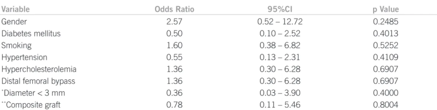
Table 2 – Statistical analysis of the variables associated with stenosis prevalence in 31 infrainguinal bypasses using the great saphenous vein
Variable Odds Ratio 95%CI p Value
Gender
Diabetes mellitus Smoking Hypertension Hypercholesterolemia Distal femoral bypass
*Diameter < 3 mm
**Composite graft
2.57 0.50 1.60 0.55 1.36 1.36 0.36 0.78
0.52 – 12.72 0.10 – 2.52 0.38 – 6.82 0.13 – 2.31 0.30 – 6.28 0.30 – 6.28 0.03 – 3.90 0.11 – 5.46
0.2485 0.4013 0.5252 0.4109 0.6907 0.6907 0.4000 0.8004
*Great saphenous vein graft diameter assessed by vascular ultrasonography. **Graft consisting of ³ 2 great saphenous vein segments.
Methods compared N concurrent cases (%) K 95%CI p
Ankle-brachial index with vascular
ultrasonography in overall stenoses 67.74 0.305 0.232 a 0.473 0.018
Ankle-brachial index with vascular
ultrasonography in severe stenoses 91.30 0.7473 0.655 a 0.811 —0.0001
he grat extension and the distal anastomosis site can determine the stenosis arising, depending on the blood low velocity, as reduced velocities stimulate myointimal hyperplasia19. he bypasses with distal anastomoses locat-ed at the popliteal artery have higher low velocity com-pared with infrapopliteal bypasses because of the difer-ence in blood outlow found20. In the present paper, 67.7% of reviewed bypasses had a distal anastomosis at infrapop-liteal arteries, relecting clinical picture severity in these patients. here was no signiicant diferences in prevalence for stenoses between femoropopliteal and infrapopliteal bypasses (40% vs. 47.6%, p = 0.445).
In this series, most stenoses were at the bypass proxi-mal segment (proxiproxi-mal anastomosis, proxiproxi-mal and middle thirds in the venous grat). One of the explanations for this fact is the reverse great saphenous vein use, whose seg-ment with the largest diameter and the least sensitive to stenoses in the great saphenous vein corresponds to the bypass distal third. A similar distribution is found in the literature21. No papers reviewing the diferent steno-sis locations when the bypass was constructed with the great saphenous vein in a in situ position were found; if
it is adopted, the anastomosis diameter will be gradually reduced from proximal to distal.
he clinical signiicance of stenoses located at the proximal artery and at the proximal anastomosis and their prognostic importance has been debated. Some authors22 demonstrated the stenoses located at that segment are poorly symptomatic, compared with those located at the bypass distal segments. Others debate the prognostic sig-niicance of stenosis located at the proximal anastomosis, as well as their repair beneit23. he increased low velocity at the proximal anastomosis is frequently found on rou-tine sonographic imaging and may not be related to the stenosis. At this location, the blood low undergoes he-modynamic changes secondary to a caliber and thickness mismatching between the artery and vein anastomosed, which is not observed when the blood low is assessed at the venous grat body. Maybe velocity criteria are not either valid as an indication of grat body stenosis or they do not have the same prognostic value when applied to low analy-sis at the proximal anastomoanaly-sis.
he main reason for using vascular ultrasonography in postoperative follow-up in lower limb revascularizations is the feasibility of identifying stenoses that would increase the grat occlusion risk even if they are not identiied by a clinical evaluation. he clinical evaluation is based on a reduced ABI. he ABI measurement and its comparison with the prior examination can indicate grat stenoses when there is a fall higher than 0.15. In the investigation sample, four patients with an ABI fall were identiied, all of them with a severe stenosis found by ultrasonography. De-spite the good agreement observed between the ABI and ultrasonography to diagnose severe stenoses (k = 0.7473),
the ABI did not identify two patients with severe stenoses and nine with mild to moderate stenoses, all of them de-tected by ultrasonography. hus, the postoperative surveil-lance strategy based only on the ABI measurement may not screen patients with severe stenoses requiring imme-diate interventions. In addition, it would not identify mild to moderate stenosis grats requiring continued surveil-lance in view of a possible stenosis progression.
Bandyk et al.24 stressed that about 20% to 40% of pa-tients having a stenosis with hemodynamic impact iden-tiied by vascular ultrasonography did not experience symptoms or a ABI fall. Papanicolaou et al.25 pointed out there is no correlation between the ABI variations and peak systolic velocity variations in stenoses measured by vascular ultrasonography. Green et al.26, in a prospective study, compared both postoperative grat surveillance methods. Patients with ABI fall and with ultrasound evi-dence of mild to moderate stenosis along the grat had 4% of acute grat thrombosis. In those with an ABI fall higher than 0.10 associated with ultrasound diagnosis of hemo-dynamically signiicant stenosis, the grat thrombosis risk was 66%, showing the importance of the ABI fall in identi-fying thrombosis risk in grats.
STUDYLIMITATIONS
he current study is limited by the reduced sample size, thus precluding stenosis-related risk factors to be identi-ied. Developing a logistic regression model with several risk factors and a larger sample of patients is desirable for future associations. Another limitation regards the lost to follow-up patients compared to the early cases. his fact occurred because of the early grat occlusion and because of patients’ non-attendance to postoperative surveillance examination. hese latter patients could have grats with a diferent stenosis prevalence compared to the sample studied, changing the outcome. A hypothesis that would warrant non-attendance could be attributed to a course with no stenosis and consequently a successful operation. Several measures are required to reduce the grat early oc-clusion: observation of the operation quality during sur-gery via imaging; identiication of patients with thrombo-philias to start systemic anticoagulation early and surgical team training to reduce occlusion resulting from technical failures. he patients’ adherence to regular postoperative surveillance should be encouraged by the health team and those who drop out should be encouraged to return.
CONCLUSION
estab-lished for patients with early grat stenoses. hese patients could be beneited by ultrasound surveillance added to the clinical examination. However, the ultrasound surveil-lance strategy restricted only to early stenosis patients was not tested in the current study. Further prospective studies are required to prove this approach relevance and eicacy.
REFERENCES
1. Tunis SR, Bass EB, Steinberg EP he use of angioplasty, bypass sur-gery, and amputation in the management of peripheral vascular dis-ease. N Engl J Med. 1991;325(8):556-62.
2. Davies MG, Hagen PO. Pathophysiology of vein grat failure: a re-view. Eur J Vasc Endovasc Surg. 1995;9(1):7-18.
3. Pilcher DB, Ricci MA. Vascular ultrasound. Review. Surg Clin North Am. 1998;78(2):273-93.
4. Moidi R, Kelman J, Berry O, Bennett S, Murie JA, Dawson AR. Signiicance of the early postoperative duplex result in infrain-guinal vein bypass surveillance. Eur J Vasc Endovasc Surg. 2007 Sep;34(3):327-32.
5. Davies AH, Hawdon AJ, Sydes MR, hompson SG. Is duplex sur-veillance of value ater leg vein bypass grating? Principal results of the Vein Grat Surveillance Randomised Trial (VGST).Circulation 2005;112(13):1985-91.
6. Rutherford RB, Baker JD, Ernst C, Johnston KW, Porter JM, Ahn S et al. DN. Recommended standards for reports dealing with lower extremity ischemia: revised version. J Vasc Surg. 1997;26(3):517-38. 7. Buth J, Disselhof B, Sommeling C, Stam L. Color-low duplex cri-teria for grading stenosis in infrainguinal vein grats. J Vasc Surg. 1991;14(6):716-26.
8. Westerband A, Mills JL, Kistler S, Berman SS, Hunter GC, Marek JM. Prospective validation of threshold criteria for intervention in infrainguinal vein grats undergoing duplex surveillance. Ann Vasc Surg. 1997;11(1):44-8.
9. Bland JM, Altman DG. Statistical methods for assessing agree-ment between two methods of clinical measureagree-ment. Lancet 1986;1(8476):307-10.
10. Caps MT, Cantwell-Gab K, Bergelin RO, Strandness DE Jr. Vein grat lesions: time of onset and rate of progression. J Vasc Surg. 1995;22(4):466-74.
11. Szilagyi DE, Elliott JP, Hageman JH, Smith RF, Dallolmo CA. Bio-logic fate of autogenous vein implants as arterial substitutes: clinical, angiographic and histopathologic observations in femoro-popliteal operations for atherosclerosis. Ann Surg. 1973;178(3):232-46. 12. Mills JL, Bandyk DF, Gahtan V, Esses GE. he origin of infrainguinal
vein grat stenosis: a prospective study based on duplex surveillance. J Vasc Surg. 1995;21(1):16-22.
13. Sayers RD, Jones L, Varty K, Allen K, Morgan JD, Bell PR, London NJ. he histopathology of infrainguinal vein grat stenoses. Eur J Vasc Surg. 1993;7(1):16-20.
14. Wilson YG, Davies AH, Currie IC, McGrath C, Morgan M, Baird RN, Lamont PM. he value of pre-discharge Duplex scanning in infrainguinal grat surveillance. Eur J Vasc Endovasc Surg. 1995;10(2):237-42.
15. Nielsen TG. Natural history of infrainguinal vein bypass stenoses: early lesions increase the risk of thrombosis. Eur J Vasc Endovasc Surg. 1996;12(1):60-4.
16. Ferris BL, Mills JL Sr, Hughes JD, Durrani T, Knox R. Is early post-operative duplex scan surveillance of leg bypass grats clinically im-portant? J Vasc Surg. 2003;37(3):495-500.
17. Idu MM, Buth J, Hop WC, Cuypers P, van de Pavoordt ED, Tordoir JM. Factors inluencing the development of vein-grat stenosis and their signiicance for clinical management. Eur J Vasc Endovasc Surg. 1999;17(1):15-21.
18. Schanzer A, Hevelone N, Owens CD, Belkin M, Bandyk DF, Clowes AW, Moneta GL, Conte MS. Technical factors afecting autogenous vein grat failure: observations from a large multicenter trial. J Vasc Surg. 2007;46(6):1180-90.
19. Schwartz LB, ODonohoe MK, Purut CM, Mikat EM, Hagen PO, McCann RL. Myointimal thickening in experimental vein grats is dependent on wall tension. J Vasc Surg. 1992;15(1):176-86. 20. Belkin M, Ratery KB, Mackey WC, McLaughlin RL, Umphrey SE,
Kunkemueller A, ODonnell TF. A prospective study of the determi-nants of vein grat low velocity: implications for grat surveillance. J Vasc Surg. 1994;19(2):259-65.
21. Landry GJ, Moneta GL, Taylor LM Jr, Edwards JM, Yeager RA, Porter JM. Long-term outcome of revised lower-extremity bypass grats. J Vasc Surg. 2002;35(1):56-62.
22. Landry GJ, Liem TK, Mitchell EL, Edwards JM, Moneta GL. Factors afecting symptomatic vs asymptomatic vein grat stenoses in lower extremity bypass grats. Arch Surg. 2007;142(9):848-53.
23. Ryan SV, Dougherty MJ, Chang M, Lombardi J, Raviola C, Cal-ligaro K. Abnormal duplex indings at the proximal anastomosis of infrainguinal bypass grats: does revision enhance patency? Ann Vasc Surg. 2001;15(1):98-103.
24. Bandyk DF, Johnson BL, Gupta AK, Esses GE. Nature and man-agement of duplex abnormalities encountered during infrainguinal vein bypass grating. J Vasc Surg. 1996;24(3):430-6.
25. Papanicolaou G, Beach KW, Zierler RE, Strandness DE Jr. he re-lationship between arm-ankle pressure diference and peak systolic velocity in patients with stenotic lower extremity vein grats. Ann Vasc Surg. 1995;9(6):554-60.