Objective: To describe mechanisms by which glucocorticoids cause osteoporosis, with fracture risk, combining this learning with a possible professional behavior change.
Data sources: A systematic search on SciELO, PubMed, Scopus, and Medline databases was carried out for consensus, review articles, including systematic reviews and meta-analysis, which were published in English, between 2000 and 2016. Keywords used on the search were the following: glucocorticoids, fractures, osteoporosis, bone health, vitamin D, children, and adolescents.
Data synthesis: The review was divided into four topics: 1) introduction, with a brief focus on pediatric fractures; 2) osteoporosis in children and adolescents, highlighting it as a silent cause of fractures; 3) glucocorticoids and secondary bone disease, describing deleterious mechanisms of this steroids group on bone structure; 4) molecular efects of glucocorticoids excess on bone, with details about the harmful mechanisms on bone molecular level.
Conclusions: Glucocorticoids excess determines early bone disease, favoring the occurrence of fractures. Thus, a child or an adolescent who uses glucocorticoids, especially systemically and chronically, but also repeats cycles at high cumulative doses of the medication, needs care and guidance related to bone health at the onset of treatment. On the other hand, the presence of fractures, even if related to trauma, can be a sign of underlying and unknown bone fragility, which may be secondary to the use of glucocorticoids and/ or vitamin D deficiency.
Keywords: glucocorticoids; osteoporosis; fractures; children; adolescents.
Objetivo: Descrever os mecanismos pelos quais os glicocorticoides provocam osteoporose, com risco consequente de fraturas, integrando esse conhecimento a uma possível mudança de conduta dos proissionais de saúde.
Fontes de dados: Foi realizada pesquisa aprofundada nas bases de dados (SciELO, PubMed, Medline e Scopus), buscando consensos, artigos de revisão, incluindo revisões sistemáticas e meta-análises, publicados em inglês, entre 2000 e 2016. As palavras-chaves utilizadas na busca foram glicocorticoides, esteroides, fraturas, osteoporose, saúde óssea, crianças e adolescentes.
Síntese dos dados: A revisão foi dividida em quatro tópicos principais: 1) introdução, com breve enfoque nas fraturas em pediatria; 2) osteoporose em crianças e adolescentes, destacando-a como causa silenciosa de fraturas; 3) glicocorticoides e doença óssea secundária, com a descrição dos mecanismos deletérios desse grupo de esteroides na estrutura óssea; 4) efeitos moleculares do excesso de glicocorticoides no osso, com o detalhamento dos mecanismos nocivos a nível molecular do tecido ósseo.
Conclusões: Os glicocorticoides em excesso determinam doença óssea precoce, favorecendo a ocorrência de fraturas. Dessa forma, uma criança ou adolescente que requer corticoterapia, sobretudo crônica e sistêmica, mas também em ciclos repetidos com doses cumulativas altas, necessita de cuidados e orientações relacionados à saúde óssea logo ao início do tratamento. Por outro lado, aqueles com fratura, mesmo entrelaçada a um trauma, podem sinalizar fragilidade óssea subjacente e desconhecida, incluindo a secundária ao uso de glicocorticoides e à deiciência de vitamina D. Palavras-chave: glicocorticoides; osteoporose; fraturas; crianças; adolescentes.
ABSTRACT
RESUMO
*Corresponding author. E-mail: [email protected] (V.M.P.P. Melo).
aUniversidade Federal de Pernambuco (UFPE), Recife, PE, Brazil.
Received on May 24, 2016; approved on September 27, 2016, available online on May 15, 2017.
Glucocorticoid-induced
bone disease: mechanisms and
importance in pediatric practice
Doença óssea induzida pelos glicocorticoides:
mecanismos e importância na prática pediátrica
INTRODUCTION
European studies show that 30–50% of young people will suf-fer at least one fracture up to 17 years of age, with an annual incidence of approximately 103–257 per 10,000 individuals. Fractures occur most frequently among boys (61–64%) in the age range of 13–14 years. Among girls, the predominant age range is 11–12 years. he bones of the forearm are the most frequently afected.1-5 Fractures occurred in 12.1% of accidents
involving Brazilian children aged 0–9 years, mostly caused by falls at home.6
In addition to the intensity of the trauma, a variety of intrin-sic and extrinintrin-sic factors – including genetics, puberty, adequate intake of calcium, protein, and vitamin D, obesity, physical activity, chronic inlammatory morbid conditions, and use of medications – afect the occurrence of a fracture. Genetic inheritance, which accounts for 60–80% of bone mass, is the most important factor.7
OSTEOPOROSIS IN
CHILDREN AND ADOLESCENTS
A fracture may indicate underlying bone fragility, that is, oste-oporosis, which is not restricted to the elderly. he National Institute of Health (NIH), an American organization, estab-lished the current deinition of osteoporosis in 2001 after a consensus: “a skeletal disorder characterized by compromised bone strength predisposing to an increased risk of fracture.”8
Osteoporosis is silent, prevalent worldwide, and its morbid-ity and mortalmorbid-ity are associated with the fractures it causes.9
Bone, which is apparently inert, has protective and support-ive functions, and actsupport-ively participates in the mineral metab-olism. herefore, maintaining bone health is essential as the bone communicates with the immune system, and it is con-sidered an endocrine organ, since it produces hormones such as ibroblast growth factor 23 (FGF23) and osteocalcin.10,11
Two types of bones are identiied in human beings: corti-cal bone, also known as compact bone, which represents 85% of the skeleton, and has protective and mechanical function; and trabecular, also known as spongy bone, which provides strength and mostly has metabolic functions.12,13 Bone tissue
is composed of a matrix (mainly collagen), minerals (especially calcium, phosphorus, and magnesium), and cells (osteoblasts, osteocytes, and osteoclasts). Bone mineral is accrued through-out childhood and adolescence, and reaches a plateau at around the end of the second decade, which is named peak bone mass (PBM) and will determine bone mass throughout life.14
It is estimated that 10% increase in PMO can delay senile osteoporosis in 13 years.15 Approximately 40–60% of adult
bone mass is accrued in adolescence, and 25% of the PBM
is accrued during the period of two years around the growth spurt.15,16 Even without increased bone loss, an adult can develop
osteoporosis because he or she has not reached their PBM in childhood and adolescence.17 As mentioned earlier, the genetic
inheritance accounts for 75% of the variance in the PBM.7,18
Bone mineral density is the amount of mineral mass per area or bone volume.18 However, the concept of bone health
is more comprehensive and is not only related to bone mass. he latter is considered the most important and more easily measurable parameter.17 Morphology (size and proportion of
cortical and trabecular compartments) and quality (microar-chitecture, turnover, and mineralization) also contribute to the bone strength.19
hree major techniques are used to assess bone mineral density: dual-energy X-ray absorptiometry (DXA), quantitative computed tomography (QCT), and quantitative ultrasound.20
DXA, whose results are expressed in grams/cm2, uses
paral-lel beam radiation that pass through the soft tissue and bone and are captured by a sensor located oppositely.21 herefore,
it depicts the bone in two-dimensional form. Despite being the most used technique, DXA does not assess bone geome-try, does not distinguish trabecular bone from cortical bone, and in pediatrics, it requires comparison with healthy indi-viduals of the same age, sex, and ethnicity, analyzed by similar equipment and software.22
Peripheral quantitative computed tomography (pQCT) and high-resolution peripheral quantitative computed tomography (HR-pQCT), which are techniques restricted to research using low radiation, assess the bone in three-dimensional form, quan-tify the contribution of geometry and mass to bone strength, and diferentiate the trabecular bone from the cortical bone.22,23
Quantitative ultrasound is a quick, inexpensive, and simple technique. It provides information on the elasticity and bone structure, but the quality of the results, which are dependent on the operator, still does not allow to replace DXA.20 However,
training and the development of professionals can be valuable considering the advantages of this examination.
Osteoporosis has diagnostic peculiarities among children and adolescents. Biochemical markers of bone turnover may be afected by many factors, and there are diiculties in the interpretation of densitometry related to growth and puberty, preventing the isolated use of this tool for diagnostic deini-tion.20,22 herefore, experts gathered in a worldwide consensus
established the diagnosis of osteoporosis in children, in addition to the indications, interpretation, and the use of bone densi-tometry in this age group.22,24 he isolated inding of one or
associated with a clinically signiicant history of fracture (two or more long bone fractures up to 10 years of age, or three or more fractures of long bones below 19 years of age), are diag-nosis of osteoporosis in this age group.22,25
he actual incidence and prevalence of osteoporosis in the pediatric population are not well deined; however, it is known that osteoporosis may compromise both sexes and occur at any age, being classiied as primary or genetic (whose main represen-tative is osteogenesis imperfecta) and secondary to conditions generally associated with each other (chronic inlammation, neoplasms, prolonged immobilization, use of certain medica-tions, hormonal and nutritional disorders).10,26 Anticonvulsants,
anticancer (especially methotrexate), calcineurin inhibitors, anticoagulants, and glucocorticoids (GC) are drugs associ-ated with the development of osteoporosis,10 with emphasis to
the last ones, which are widely used in pediatric prescription.
GLUCOCORTICOIDS AND
SECONDARY BONE DISEASE
GC are steroid hormones physiologically produced by the adrenal glands and synthesized for intermittent or continuous systemic and topical use. GC are recognized by the signiicant anti-inlammatory and immunomodulatory activities, being used to control the pathogenesis of autoimmunity and/or inlammation in a variety of diseases. hey have been increas-ingly used in the pediatric age group.27 A study carried out in
the UK found that 1.2% of children received at least one pre-scription of oral corticosteroids within one year, mostly for treating asthma attacks.28
his is the group most assisted by the general pediatrician. Asthma is a chronic, obstructive, and inlammatory pulmo-nary disease, with diferences in clinical presentation, severity, and response to treatment, which is usually based on the use of GC.29 he Global Initiative for Asthma (GINA), an
organi-zation that brings together pulmonologists, and annually pre-pares guidelines for asthma management, has recommended therapy in stages or “steps” for the control of asthma, whose severity classiication (mild, moderate, and severe), is established on the basis of the response to treatment, and may change over time.30 he initial treatment for most patients with asthma
(except mild cases in preschool children) includes inhaled GC for several months and with the lowest efective daily dose. On the other hand, oral systemic GC is part of rescue therapy in asthma attacks (cycles of 1 and 2 mg/kg/day of predniso-lone for three to ive days) and continuous treatment of the most severe cases.30
Chronic inlammatory diseases of the connective tissue, kid-neys, nervous system, and digestive tract, in addition to cancer
and transplants, also require therapy with oral systemic GC, but continuously and for more than three months. Juvenile idiopathic arthritis, systemic lupus erythematosus, juvenile dermatomyositis, Crohn’s disease, and nephrotic syndrome are the main examples.31-33 he chronic inlammatory disease
itself promotes the release of cytokines, malnutrition, pro-longed immobilization, loss of muscle mass, delayed puberty, and reduced sun exposure, afecting negatively bone health.31,33
However, clinical trials on volunteers showed that there is a direct corticoid action in decreased bone formation, indepen-dent of inlammation.34 In any case, the sum of the harmful
actions of the disease and steroid therapy converges to com-promise bone health.
If used chronically, GC are considered the main cause of secondary and iatrogenic osteoporosis.27,35 However, the
poten-tial for fractures is often disregarded by the professional who prescribes the GC and ignored by the patient and the family. he relative risk increases with dose and duration of steroid therapy, whereas the absolute risk is determined by a variety of associated clinical conditions. However, low daily doses of 2.5 mg of prednisolone, in intermittent and repeated cycles, may have cumulative and also harmful efect.36 he risk is higher in the
irst three months of continuous therapy and slowly decreases after its conclusion; however, it does not seem to return to nor-mality.37 In addition, this rapid increase in the risk of fracture
is not registered by densitometry tests, which suggests a more signiicant change in bone quality over quantity.35 herefore,
the absence of alterations in bone densitometry does not rule out the existence of GC-induced osteoporosis.
Extensive study of a British cohort, including individuals aged 4–17 years, revealed that the annual use of more than four cycles of oral systemic GC increased fracture risk, whereas another cohort study, with the same age range and in the same country, revealed an association of increased risk of fracture with severity of asthma, and not with the use of inhaled steroid.28,38
GC-induced bone disease mainly afects trabecular bone, justifying the higher occurrence of vertebral and rib fractures.40
However, a British study revealed that the humerus was the most fractured bone among children who used four or more short cycles of oral corticosteroids.28 Experimental, molecular,
and genetic research has been elucidating the mechanisms of action of GC excess on bones. hese steroids have a catabolic efect on the muscle and the vitamin D, which compromises bone mineralization.41,42 Bone and muscle form a unit, with
mutual trophic inluence. Moreover, chronic corticosteroid therapy promotes adipogenesis and obesity, with capture and reduction of vitamin D.43,44
Figure 1 Pathogenesis of fractures related to glucocorticoids (GC) excess.
GC excess
Bone
Osteoporosis
Fractures
↑ Catabolism
Muscle
Obesity
Vitamin D
↓25 (OH) D
Myopathy
Falls
↓ GH/alt. PTH/h.sexual
↓ Intestinal absorption Ca++
↑ Urinary elimination Ca++
GC excess increases the risk of fractures mainly by direct action on the bone, resulting in osteoporosis. Increased muscle catabolism and vitamin D, decreased GH secretion, changes in sex steroids and pulsatility of PTH, as well as reduced intestinal absorption and increased urinary elimination of Ca++, also determined by GC, contribute to the bone fragility. GC myopathy leads to muscle weakness, favoring falls, and therefore fractures. In addition, obesity, associated with chronic use of GC, captures and decreases vitamin D. GC: glucocorticoids; PTH: parathyroid hormone; Ca++: calcium.
*Figure elaborated by the authors based on the references14-22,34.
hormone (GH), and changes in the metabolism of sex ste-roids and in parathyroid hormone (PTH) pulsatility are other indirect negative efects of GC on bone health.40 However,
the bone tissue itself is a target for the GC. Research has shown that the direct action of these steroids on bone cells is the main mechanism in the genesis of the resulting osteopo-rosis.37,40 Figure 1 summarizes the pathogenesis of secondary
fracture to GC excess.
MOLECULAR EFFECTS
OF GLUCOCORTICOIDS
EXCESS ON BONE
GC’s efects occur by four mechanisms: the classic genomic (the most important), which involves the cytosolic GC receptors (cGCR), and is divided into two processes, transrepression and transactivation; nongenomic secondary, which is also initiated with cGCR; nongenomic executed by membrane receptors (mCGR); and nonspeciic nongenomic, resulting from inter-actions with cell membranes (including the organelles).45-47
he anti-inlammatory and immunomodulatory actions are
derived from these processes, as well as adverse efects, which are determined by one or more of the four mechanisms.
he polymorphism of the genes related to these receptors may contribute to diferences in the intensity of bone diseases and fractures induced by the use of GC.47 Clinical observation
and studies on cell metabolism of GC also identiied several sensitivity to these hormones, related to intracellular enzyme system 11β-hydroxysteroid dehydrogenase (11β-HSD), specif-ically to type 1 (11β-HSD1), which converts inactive forms of corticosteroids (cortisone and prednisone) in active forms (cor-tisol and prednisolone).27,48 Immunohistochemical and in situ
hybridization studies showed 11β-HSD1 expression in osteo-blasts increasing with age, which favors the greater concentra-tion of GC in these cells, and which may undergo modulaconcentra-tion by cytokines, growth factors, and other enzymes.36
Increased expression of 11β-HSD1 enzyme is considered a risk factor for GC-induced osteoporosis.12 In its active form,
the GC binds with a receiver (GRα or GRβ), a member of the nuclear receptor super family,49 and migrates from the
In the cytoplasm, after activation by HSD1 (only in the case of cortisone and prednisone) and binding to the GC receptor (GRα
or GRβ), the glucocorticoid-receptor migrates to the nucleus, where it can transactivate or transrepress gene expression, leading to a deleterious action on the bone. Inactivation of active corticosteroids by HSD2 impairs receptor binding and action at the nuclear level.
GC: glucocorticoid; HSD: 11β- hydroxysteroid dehydrogenase
enzimatic system; HSD1: type 1 11β- hydroxysteroid dehydrogenase; HSD2: type 2 11β- hydroxysteroid dehydrogenase.
*Figure elaborated by the authors based on the references15,17,22,26.
Figure 2 Genomic mechanism of action of GC and the
hydroxysteroid dehydrogenase (HSD) enzymatic system.
Corticosteroid HSD1
Inactive HSD2 Active
Corticosteroid Receptor
Nucleus
Transrepression Transactivation
Alteration in gene expression
Deleterious action on bone (activator protein 1 [AP1], nuclear factor κB [NF-kB], signal transducer, and activator of transcription 5 [STAT5]), resulting in transactivation or transrepression.36,49 herefore, the actions
of GC at the genome level, which modiies gene expression, will impact the structure of bone tissue.
he other side of the HSD system equation is composed of GC-inactivating enzyme and HSD 2. he sensitivity of difer-ent types of GC to this enzyme varies, and dexamethasone, by having a luorine atom at the 9α position of the B ring, with site of HSD2 blocked, is the steroid which is most resistant to such inactivation, and therefore is what causes osteoporosis the most.35 Figure 2 outlines the 11β-HSD enzyme system and
the connection with the genomic mechanism of GC action. he result of activation or the resistance to inactivation of GC excess in bone contributes to osteoporosis.
Owing to this nuclear action, GC interferes in the forma-tion, diferentiaforma-tion, destrucforma-tion, and survival of bone cells. hey perform a metabolic control by modulation of various regulatory factors and of matrix proteins, such as collagen, alka-line phosphatase, the receptor activator of nuclear factor kappa B – NF-kB (RANK), the receptor activator of nuclear factor
kappa B ligand (RANKL)/osteoprotegerin (OPG), osteocal-cin, osteopontin (OPN), the pro- and antiapoptotic proteins, in addition to growth factors and cytokines, which interfere signiicantly in bone metabolism.49
Experimental studies have revealed that an important bone signaling pathway, the Wnt/β-catenin pathway, is impaired by excess GC. he term Wnt derives from the merge of the name of 2 loci involved, int-1 and Wg, but also comprises
other protein signaling pathways, being the Wnt/β-catenin which is the main signaling pathway.50 he Wnt binds to a
double set of receivers (proteins 5 and 6 related to the low density lipoprotein receptor [Lrp5 and Lrp6]), and to a family member of the frizzled proteins, activating dishevelled pro-tein, and receiving the binding of Axin propro-tein, which binds to the proteins of degradation complex (especially glycogen synthase kinase-3b [GSK-3b]), preventing the phosphor-ylation and inactivation of β-catenin, which accumulates within the cell.50,51
he β-catenin accumulated and agglomerated in the cell cytoplasm translocates into the nucleus, where it binds to members of the family of transcription factors (T-cell factor/ lymphocyte elongation factor [TCF/Lef ]), regulating gene expression to favor bone homeostasis through the targeting of diferentiation of mesenchymal stem cells into osteoblasts and inhibiting apoptosis of osteoblasts and osteocytes.50,51 In
addi-tion, by promoting the expression of osteoprotegerin (OPG), competitor of RANKL, inhibits osteoclastogenesis.51 herefore,
this signaling pathway contributes to the accumulation of bone mass. Bone response to mechanical overload is also inluenced by Wnt β-catenin.50
An experimental study found that dexamethasone inhib-its all Wnt β-catenin pathway from the cell surface to nuclear translocation.52 In addition, experiments with animals showed
that the use of GC, especially for prolonged periods, increases the expression of antagonists of Wnt β-catenin pathway (scle-rostin and protein 1 of protein Dickkopf family [DKK1]), mainly produced by osteocytes.51 hese indings reveal the
complexity of the negative actions of these steroids in this protein pathway, which partly constitutes its harmful efects on the bone.
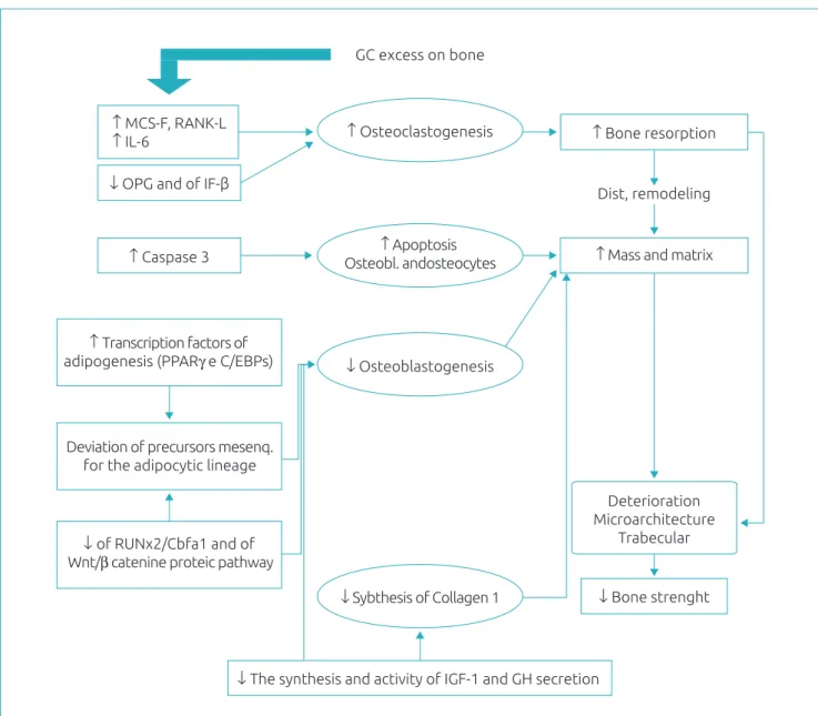
From a series of molecular alterations, GC excess results in an initial increase in osteoclastogenesis, reduction in osteoblastogenesis, increased apoptosis of osteoblasts and osteocytes, and a decrease in the synthesis of type 1 collagen. Thus, bone resorption and decrease of bone mass and matrix, that is, a remodeling disorder, with microarchitecture deterioration and bone strength impairment. MCF-S: macrophage colony-stimulating factor; RANK-L: receptor activator of nuclear factor kappa B ligand; IL-6: Interleukin-6; OPG: Osteoprotegerin; IF-β: interferon β; PPARγ (peroxisome proliferator-activated receptor-γ); and C/EBPS (CCAAT/enhancer binding proteins): family of transcription factors promoters of adipogenesis; Runx2/Cbfa1: transcription factors, members of the Runx family, promoters of osteoblastic diferentiation and migration and vascular invasion of the bone; IGF-1: insulin-like growth factor 1; GH: growth hormone.
*Figure elaborated by the authors based on the references15,16,22,30-37.
Figure 3 Synthesis of the main deleterious actions of glucocorticoids excess in the bone. GC excess on bone
↑ MCS-F, RANK-L
↑ IL-6 ↑ Osteoclastogenesis ↑ Bone resorption
Dist, remodeling ↓ OPG and of IF-β
↑ Mass and matrix
↓ Bone strenght ↑ Apoptosis
Osteobl. andosteocytes ↑ Caspase 3
↓ Osteoblastogenesis ↑ Transcription factors of
adipogenesis (PPARγ e C/EBPs)
Deterioration Microarchitecture
Trabecular
↓ Sybthesis of Collagen 1
↓ The synthesis and activity of IGF-1 and GH secretion Deviation of precursors mesenq.
for the adipocytic lineage
↓ of RUNx2/Cbfa1 and of Wnt/β catenine proteic pathway
production (main component of the bone matrix) and stimu-lating factors formation (such as the IGF-1).31,37,47 herefore,
the initial phase of resorption dominance is followed by the chronic phase of compromising the formation.
GC excess causes, in brief, decreased osteoblastogen-esis, increased apoptosis of osteoblasts and osteocytes, and temporary increase of osteoclastogenesis and osteoclast
survival.37 The favoring of apoptosis of osteocytes, which are
sensors for damage and repair support, interferes in the bone remodeling, with reduction in the replacement of the bone excavated by osteoclasts.27,31,47,53,54 A fact that stands
Experimental studies show that OPG (decreased by GC) promotes endothelial proliferation (angiogenesis), whereas the RANKL oppositely inhibits angiogenesis.55 In addition, the
osteoblasts and osteocytes produce vascular endothelial growth factor (VEGF) and the increase in its apoptosis, secondary to GC excess, would be an additional factor to promote the breakdown of bone vasculature (connected to the canalicu-lar luid low), with a loss of hydraulic support and decreased bone strength in relation to the bone mass.56 he GC excess
seems to afect the bone in many diferent ways, in a sequence of damages, related to dose and duration of exposure, but also dependent on the individual genetic characteristics.
CONCLUSIONS
he deepening of research techniques has enabled valuable indings for understanding the action of GC, especially if chronically used, at cellular bone level. his opens a pathway for more research concerning therapy and prevention for the resulting bone disease. he professional who prescribes steroid and those who assist children and adolescents with fractures should be aware of the possibility of associated bone fragility, to direct their research and improve their preventive and ther-apeutic approach.
RECOMMENDATIONS
A child or adolescent requiring chronic and systemic cortico-steroid therapy in repeated cycles, with high cumulative doses (> 1 g/year), needs care and guidance related to bone health, as soon as the therapy is initiated. he correct administration, including the time, duration, and mode of discontinuing treat-ment, should be clariied.
Nutritional follow-up is essential to prevent obesity, but also to ensure adequate intake of calcium (1,300 mg/day between
9 and 18 years) and proteins, as well as to maintain suicient levels of vitamin D, with a minimum daily intake of 600 IU, after the irst year of life.22 he recommendation for the daily
exposure to sunlight for approximately 20 minutes, must be accompanied by serum levels of 25(OH) of vitamin D, at the onset of therapy, and every three to six months, depending on the results. If serum levels are below 30 ng/mL, the oral replace-ment with medication is recommended.
he regular and supervised physical activity, although it may lead to accidents,57 should be recommended, considering
the beneicial action on bone and muscle strength, in addition to the prevention of obesity.
he suspicion of spontaneous fractures, especially inverte-brae (evidenced by pain or loss of height), must be latent and requires radiological investigation.22 he peculiarity of
verte-bral involvement highlights the importance of clinical moni-toring and routine anthropometry. he professional who cares for a child or adolescent with fracture, especially after a mild trauma, should be alert to the possibility of underlying bone fragility, which includes questioning the use of GC and inves-tigating deiciency of vitamin D.
he physician should not fear to prescribe corticosteroids for asthma and other inlammatory diseases if well indicated. He or she should only be aware that the need must be precise and that the use of repeated cycles, with cumulative doses greater than 1 g/year, may interfere with bone health. he search for synthetic forms of GC, that act only on the anti-inlammatory and immunomodulatory mechanisms, without the harmful side efects, is a present hope.49,58
Funding
his study did not receive funding.
conflict of interests
he authors declare no conlict of interests.
REFERENCES
1. Clark EM. The epidemiology of fractures in otherwise healthy children. Curr Osteoporos Rep. 2014;12:272-8.
2. Cooper C, Dennison EM, Leufkens HG, Bishop N, Staa TP. Epidemiology of childhood fractures in Britain: a study using the general practice research database. J Bone Miner Res. 2004;19:1976-81.
3. Chevalley T, Bonjour JP, Rietbergen B, Ferrari S, Rizzoli R. Fractures during childhood and adolescence in healthy boys: relation with bone mass, microstructure, and strength. J Clin Endocrinol Metab. 2011;96:3134-42.
4. Hedström EM, Svensson O, Bergström U, Michno P. Epidemiology of fractures in children and adolescents. Acta Orthop. 2010;22:148-53.
5. Ryan LM. Forearm fractures in children and bone health. Curr Opin Endocrinol Diabetes Obes. 2010;17:530-4.
7. McDevitt H, Ahmed SF. Establishing good bone health in children. Paediatr Child Health (Oxford). 2010;20:83-7.
8. Lorentzon M, Cummings SR. Osteoporosis: the evolution of a diagnosis. J Intern Med. 2015;277:650-61.
9. Sànchez-Riera L, Wilson N, Kamalaraj N, Nolla JM, Kok C, Li Y, et al. Osteoporosis and fragility fractures. Best Pract Res Clin Rheumatol. 2010;24:793-810.
10. Mäkitie O. Causes, mechanisms and management of paediatric osteoporosis. Nat Rev Rheumatol. 2013;9:465-75.
11. Stubbs J, Liu S, Quarles LD. Role of fibroblast growth factor 23 in phosphate homeostasis and pathogenesis of disordered mineral metabolism in chronic kidney disease. Semin Dial. 2007;20:302-8.
12. Feng X, McDonald JM. Disorders of bone remodeling. Annu Rev Pathol. 2011;6:121-45.
13. Rho JY, Kuhn-Spearing L, Zioupos P. Mechanical properties and the hierarchical structure of bone. Med Eng Phys. 1998;20:92-102.
14. Boot AM, Ridder MA, Sluis IM, Slobbe I, Krenning EP, Keizer-Schrama SM. Peak bone mineral density, lean body mass and fractures. Bone. 2010;46:336-41.
15. Bonjour JP, Chevalley T. Pubertal timing, bone acquisition, and risk of fracture throughout life. Endocr Rev. 2014;35:820-47.
16. Golden NH, Abrams SA, Committee on Nutrition. Optimizing bone health in children and adolescents. Pediatrics. 2014;134:1229-43.
17. Kanis JA, Johansson H, Oden A, McCloskey EV. Assessment of fracture risk. Eur J Radiol. 2009;71:392-7.
18. Sopher AB, Fennoy I, Oberield SE. An update on childhood bone health: mineral accrual, assessment and treatment. Curr Opin Endocrinol Diabetes Obes. 2015;22:35-40.
19. Ma NS, Gordon CM. Pediatric osteoporosis: where are we now? J Pediatr. 2012;161:983-90.
20. Estrada A, Ramnitz MS, Gafni RI. Bone densitometry in children and adolescents. Curr Opin Obstet Gynecol. 2014;26:339-46.
21. Chun KJ. Bone densitometry. Semin Nucl Med. 2011;41:220-8.
22. Bachrach LK. Diagnosis and treatment of pediatric osteoporosis. Curr Opin Endocrinol Diabetes Obes. 2014;21:454-60. 23. Roggen I, Roelants M, Sioen I, Vandewalle S, De Henauw
S, Goemaere S, et al. Pediatric reference values for tibial trabecular bone mineral density and bone geometry parameters using peripheral quantitative computed tomography. Calcif Tissue Int. 2015;96:527-33.
24. Bianchi ML, Leonard MB, Bechtold S, Högler W, Mughal MZ, Schönau E, et al. Bone health in children and adolescents with chronic diseases that may afect the skeleton: the 2013 ISCD Pediatric Oicial Positions. J Clin Densitom. 2014;17:281-94. 25. Gordon CM, Leonard MB, Zemel BS, International Society for Clinical Densitometry. 2013 Pediatric Position Development Conference: executive summary and reflections. J Clin Densitom. 2014;17:219-24.
26. Titmuss AT, Biggin A, Korula S, Munns CF. Diagnosis and Management of Osteoporosis in Children. Curr Pediatr Rep. 2015;3:187-99.
27. Cooper MS. Glucocorticoids in bone and joint disease: the good, the bad and the uncertain. Clin Med. 2012;12:261-5.
28. Staa TP, Cooper C, Leufkens HG, Bishop N. Children and the risk of fractures caused by oral corticosteroids. J Bone Miner Res. 2003;18:913-8.
29. National Asthma Education and Prevention Program. Expert Panel Report 3 (EPR-3): Guidelines for the Diagnosis and Management of Asthma-Summary Report 2007. J Allergy Clin Immunol. 2007;120:S94-138.
30. Horak F, Doberer D, Eber E, Horak E, Pohl W, Riedler J, et al. Diagnosis and management of asthma – Statement on the 2015 GINA Guidelines. 2016;1-14.
31. Scheven E, Corbin KJ, Stagi S, Stefano S, Cimaz R. Glucocorticoid-associated osteoporosis in chronic inlammatory diseases: epidemiology, mechanisms, diagnosis, and treatment. Curr Osteoporos Rep. 2014;12:289-99. 32. Tsampalieros A, Gupta P, Denburg MR, Shults J, Zemel BS,
Mostoui-Moab S, et al. Glucocorticoid efects on changes in bone mineral density and cortical structure in childhood nephrotic syndrome. J Bone Miner Res. 2013;28:480-8. 33. Burnham JM. Inlammatory diseases and bone health in
children. Curr Opin Rheumatol. 2012;24:548-53.
34. Briot K, Roux C. Glucocorticoid-induced osteoporosis. RMDOpen. 2015;1:e000014.
35. Weinstein RS. Glucocorticoid-induced osteoporosis and osteonecrosis. Endocrinol Metab Clin North Am. 2012;41:595-611.
36. Seibel MJ, Cooper MS, Zhou H. Glucocorticoid-induced osteoporosis: mechanisms, management, and future perspectives. Lancet Diabetes Endocrinol. 2013;1:59-70. 37. Buehring B, Viswanathan R, Binkley N, Busse W.
Glucocorticoid-induced osteoporosis: an update on efects and management. J Allergy Clin Immunol. 2013;132:1019-30.
38. Staa TP, Bishop N, Leufkens HG, Cooper C. Are inhaled corticosteroids associated with an increased risk of fracture in children? Osteoporos Int. 2004;15:785-91.
39. Hansen KE, Kleker B, Safdar N, Bartels CM. A systematic review and meta-analysis of glucocorticoid-induced osteoporosis in children. Semin Arthritis Rheum. 2014;44:47-54. 40. Henneicke H, Gasparini SJ, Brennan-Speranza TC, Zhou
H, Seibel MJ. Glucocorticoids and bone: local effects and systemic implications. Trends Endocrinol Metab. 2014;25:197-211.
41. Cianferotti L, Brandi ML. Muscle-bone interactions: basic and clinical aspects. Endocrine. 2014;45:165-77.
42. Society for Adolescent Health and Medicine. Recommended vitamin D intake and management of low vitamin D status in adolescents: a position statement of the society for adolescent health and medicine. J Adolesc Health. Society for Adolescent Health and Medicine. 2013;52:801-3. 43. Çizmecioğlu FM, Etiler N, Görmüş U, Hamzaoğlu O, Hatun Ş.
Hypovitaminosis D in obese and overweight schoolchildren. J Clin Res Pediatr Endocrinol. 2008;1:89-96.
© 2017 Sociedade de Pediatria de São Paulo. Published by Zeppelini Publishers. This is an open access article under the CC BY license (http://creativecommons.org/licenses/by/4.0/).
45. Spies CM, Strehl C, Goes MC, Bijlsma JW, Buttgereit F. Glucocorticoids. Best Pract Res Clin Rheumatol. 2011;25:891-900.
46. Stahn C, Löwenberg M, Hommes DW, Buttgereit F. Molecular mechanisms of glucocorticoid action and selective glucocorticoid receptor agonists. Mol Cell Endocrinol. 2007;275:71-8. 47. Bouvard B, Audran M, Legrand E, Chappard D. Ultrastructural
characteristics of glucocorticoid-induced osteoporosis. Osteoporos Int. 2009;20:108-92.
48. Weinstein RS, Wan C, Liu Q, Wang Y, Almeida M, O’Brien CA, et al. Endogenous glucocorticoids decrease skeletal angiogenesis, vascularity, hydration, and strength in aged mice. Aging Cell. 2010;9:147-61.
49. Moutsatsou P, Kassi E, Papavassiliou AG. Glucocorticoid receptor signaling in bone cells. Trends Mol Med. 2012;18:348-59. 50. Johnson ML, Kamel MA. The Wnt signaling pathway and
bone metabolism. Curr Opin Rheumatol. 2007;19:376-82. 51. Guañabens N, Gifre L, Peris P. The role of Wnt signaling and sclerostin in the pathogenesis of glucocorticoid-induced osteoporosis.Curr Osteoporos Rep. 2014;12:90-7.
52. Chen Z, Xue J, Shen T, Mu S, Fu Q. Curcumin alleviates glucocorticoid-induced osteoporosis through the regulation of the Wnt signaling pathway. Int J Mol Med. 2016;37:329-38.
53. Lian JB, Stein GS. Runx2/Cbfa1: a multifunctional regulator of bone formation. Curr Pharm Des. 2003;9:2677-85. 54. Ramji DP, Foka P. CCAAT/enhancer-binding proteins: structure,
function and regulation. Biochem J. 2002;365:561-75. 55. McGonigle JS, Giachelli CM, Scatena M. Osteoprotegerin and
RANKL diferentially regulate angiogenesis and endothelial cell function. Angiogenesis. 2009;12:35-46.
56. Weinstein RS. Glucocorticoids, osteocytes, and skeletal fragility: the role of bone vascularity. Bone. 2010;46:564-70. 57. Clark EM, Ness AR, Tobias JH. Vigorous physical activity increases fracture risk in children irrespective of bone mass: a prospective study of the independent risk factors for fractures in healthy children. J Bone Miner Res. 2008;23:1012-22.