C
A
SO
C
L
ÍN
IC
O
412
Revista Cientíica da Ordem dos Médicos www.actamedicaportuguesa.com ABSTRACT
Female genital tuberculosis remains a major health problem in developing countries and is an important cause of infertility. As symptoms, laboratory data and physical indings are non-speciic, its diagnosis can be dificult. We describe a case of a 39-year-old woman suffering from peri-umbilical pain and increased abdominal size for one year, anorexia, asthenia, weight loss, occasionally dysuria and dyspareunia, and four months amenorrhea. Laboratory data revealed cancer antigen 125 (CA-125) level of 132.3 U/mL, erythrocyte sedimentation rate of 42 mm/h, and gamma-globulins of 2.66 g/dL. Computer Tomography scan showed loculated ascites. It was initially suspected a carcinomatous origin, but ascites evaluation was negative for malignant cells. Magnetic Resonance Imaging from another hospital showed endometrial heterogeneity. Therefore, an endometrial biopsy was performed demonstrating an inlammatory iniltrate with giant cells of type Langhans and bacteriological culture identiied Mycobacterium tuberculosis.
Keywords: Tuberculosis, Female Genital; Endometritis; Peritonitis, Tuberculous; Ovarian Neoplasms.
Di Giovanni SE, et al. Endometrial tuberculosis, Acta Med Port 2016 Jun;29(6):412-415
CONFLITOS DE INTERESSE
Os autores declaram que não possuem conlitos de in-teresses.
FONTES DE FINANCIAMENTO
Os autores declaram não ter recebido subsídios ou bol-sas para a elaboração do artigo.
REFERÊNCIAS
1. Zaietta GA, Raval AA, Murillo L, Mehta J, Byrd RP Jr, Roy TM. Case report and review of the literature: spontaneous aortobronchial istula. Tenn Med. 2013;106:39-42.
2. Santiago S, Tobias J, Williams AJ. A reappraisal of the causes os hem-optysis. Arch Intern Med. 1991;151:2449-51.
3. Hirshberg B, Biran I, Glazer M, Kramer MR. Hemoptysis: etiology, evalu-ation, and outcome in a tertiary referral hospital. Chest. 1997;112:440-4. 4. Kazerooni EA, Williams DM, Abrams GD, Deeb GM, Weg JG. Aorto-bronchial istula 13 years following repair of aortic transection. Chest. 1994;106:1590-4.
5. Miyazaki M, Hiraga S, Kitamura M, Takamiya T, Iida T, Hida M, et al. Aor-tobronchial istula complicated with an aortic aneurysm in hemodialysis patient. Nephron.1990;56:101-2.
6. Mac Intosh EL, Parrott JC, Unruh HW. Fistulas between the aorta and tracheobronchial tree. Ann Thorac Surg. 1991;51:515-9.
7. Demeter SL, Cordasco EM. Aortobronchial istula: Keys to successful management. Angiology. 1980;31:431-5.
8. Szolar DH, Riepl T, Stiskal M, Preidler KW. Aortobronchial istula as a late complication of posttraumatic chronic aortic aneurysm. AJR Am J
Roentgenol. 1995; 164:1511-3.
9. Milano A, De Carlo M, Mussi A, Falashi F, Bortolotti U. Aortobronchial istual after coarctation repair and blunt chest trauma. Ann Thorac Surg 1999;67:539-41.
10. Coblentz CL, Sallee DS, Chiles C. Aortobronchopulmonary istula com-plicating aortic aneurysm: diagnosis in four cases. AJR. 1988;150:535−8. 11. Ono M, Takamoto S, Kawauchi M, Egami J, Kotsuka Y. Aortobron-chial istula late after transverse arch replacement. Ann Thorac Surg. 2000;70:964-6.
12. Urschel JD. The diagnostic importance of computed tomography in aor-tobronchial istula−A case report. Angiology. 1993;44:817-9.
13. Agarwal P, Chughtai A, Matzinger F, Kazerooni EA. Multidetector CT of aortic thoracic aneurisms. RadioGraphics. 2009;29:537-52.
14. Ferretti GR, Choplin RH, Haponik EF, Hudspeth AS. Case report. Aortic pseudoaneurysm with aortobronchial istula: diagnosis with CT angiog-raphy. J Comput Assist Tomogr. 1996;20:975−8.
15. Riesenman PJ, Brooks JD, Farber MA. Thoracic endovascular aortic repair of aortobronchial istulas. J Vasc Surg. 2009;50:992-8.
Endometrial Tuberculosis Simulating an Ovarian Cancer
Tuberculose Endometrial Simulando um Carcinoma do
Ovário
1. Department of Radiological Sciences. Institute of Radiology. Catholic University of Sacred Heart. A. Gemelli Hospital. Rome. Italy. 2. Department of Radiology. Instituto Português de Oncologia de Lisboa. Lisboa. Portugal.
3. Department of Radiology. Hospital do Espírito Santo. Évora. Portugal. 4. Radiology Department. Hospital Central do Funchal. Funchal. Portugal.
Autor correspondente: Silvia Eleonora Di Giovanni. [email protected]
Recebido: 07 de abril de 2016 - Aceite: 25 de maio de 2016 | Copyright © Ordem dos Médicos 2016
Silvia Eleonora DI GIOVANNI1, Teresa Margarida CUNHA2, Ana Luisa DUARTE3, Inês ALVES4
Acta Med Port 2016 Jun;29(6):412-415 ▪ http://dx.doi.org/10.20344/amp.7706
RESUMO
A tuberculose genital feminina continua a representar uma patologia importante nos países em desenvolvimento e constitui uma causa importante de infertilidade. Os seus sintomas, achados laboratoriais e exame físico não são especíicos, tornando difícil o seu dia-gnóstico. Descrevemos o caso de uma doente do sexo feminino, de 39 anos, com dor peri-umbilical e aumento do volume abdominal desde há um ano, anorexia, astenia, perda ponderal, ocasionalmente disúria e dispareunia, assim como amenorreia desde há quatro meses. Os dados laboratoriais mostraram valores de 132,3 U/mL do marcador tumoral CA-125, 42 mm/h de velocidade de sedimen-tação e 2,66 g/dL de gama-globulinas. A tomograia computadorizada mostrou ascite loculada. Inicialmente suspeitou-se de etiologia maligna, mas o exame citológico do líquido ascítico foi negativo para células malignas. Foi efectuada ressonância magnética pélvica, noutra instituição, que revelou heterogeneidade do endométrio. Foi então realizada biópsia endometrial que revelou um iniltrado inla-matório com células gigantes de Langhans e o exame bacteriológico isolou Mycobacterium tuberculosis.
C
A
SO
C
L
ÍN
IC
O
Revista Cientíica da Ordem dos Médicos www.actamedicaportuguesa.com 413
Di Giovanni SE, et al. Endometrial tuberculosis, Acta Med Port 2016 Jun;29(6):412-415
INTRODUCTION
Tuberculosis (TB) is an important health problem in
developing countries.1 Genitourinary TB accounts for about
15% of all extra-pulmonary TB and, after lung, is the most
common site.2 The endometrium is involved in approximately
50-60% of women with female genital TB (FGTB).3-8
FGTB is typically asymptomatic and is usually diagnosed incidentally during infertility evaluation. Symptomatic disease usually presents with pelvic pain, and/or menstrual irregularities.3
The disease can mimic many conditions, including bowel
disease, malignancy and other infectious diseases.2 FGTB
presenting as abdominal lump with raised Cancer Antigen 125 (CA-125) and ascites can be a differential diagnosis of advanced ovarian malignancy or disseminated peritoneal
carcinomatosis.2
No laboratory test is suficiently sensitive or speciic to allow a diagnosis; however lymphocytosis, accelerated erythrocyte sedimentation rate (ESR) and increased gamma-globulin levels in blood can be found. The imaging
indings are also not speciic.4
Hysteroscopy provides direct information about endometrial integrity and may reveal a scarred atrophic endometrial layer with adhesions varying from mild to
severe, leading to Asherman’s syndrome and secondary
amenorrhea.3 However, histopathological evidence
from biopsies of premenstrual endometrial tissue or the demonstration of tubercle bacilli in cultures of menstrual blood or endometrial curetting is necessary to provide a conclusive diagnosis of the disease.3-5,8
CASE REPORT
A 39-year-old woman from Guinea-Bissau with abdominal mass and ascites suspected to be an ovarian cancer was referred to our hospital for diagnostic and therapeutic evaluation.
She reported peri-umbilical pain and increased abdo-minal size for one year; she also reported anorexia,
asthe-nia, weight loss, occasionally dysuria and dyspareuasthe-nia, and amenorrhea for four months. The patient had no signiicant medical history. No history of TB in relatives was described. Physical examination detected ascites and left inguinal lymphadenopathy. The speculum examination was painless and unremarkable. Laboratory data revealed CA-125 serum level of 132.3 U/mL (reference range, 0-35 U/mL), ESR 42 mm/h (normal value < 30 mm/h), gamma-globulins 2.66 g/ dL (normal range, 0.5-1.5 g/dL). Human Immunodeiciency Virus test was negative. The intradermal test injection of puriied protein derivative had a maximum diameter of 15 mm. The chest X-ray (CRX) showed densiication in the right lung base, likely due to ibrotic bands, with associated bronchiectasis.
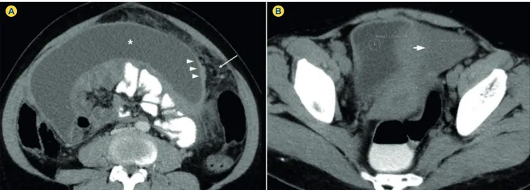
A computer tomography (CT) scan was performed and revealed the presence of pseudocystic lesion in the abdomen, associated with smooth peritoneal thickening and omental fat stranding (Fig. 1).
For the exclusion of an occult malignancy, gastrointestinal endoscopy and colonoscopy were performed and no signiicant indings were found. Drainage of the ascitic luid was also performed and no malignant cells were detected nor bacteriological growth was observed.
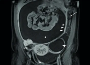
The images of a previous magnetic resonance imaging (MRI) exam from another hospital were revaluated, showing thickened and heterogeneous endometrium with an irregular surface (Fig. 2).
Therefore, hysteroscopy was performed and the endometrial biopsy revealed marked chronic necrotizing granulomatous inlammation with type Langhans giant cells. The bacteriological direct examination after Ziehl-Neelsen staining was negative, but bacteriological culture in Lowenstein-Jensen identiied Mycobacterium tuberculosis complex.
DISCUSSION
FGTB occurs secondary to primary disease in the
Figure 1 - Endometrial Tuberculosis in a 39-year-old woman. Contrast-enhanced axial computed tomography image in the portal-venous phase (A) and unenhanced axial computed tomography image (B) after oral and rectal contrast administration (A, B) show ascites (aster-isk in A) and smooth thickening of the peritoneum (arrowheads in A), associated with omental fat stranding (arrow in A). Note the slightly hight attenuation of the ascitic luid (20 Hounsield Unit) compared to urine within the bladder (1.2 Hounsield Unit) (thick arrow in B).
C
A
SO
C
L
ÍN
IC
O
414
Revista Cientíica da Ordem dos Médicos www.actamedicaportuguesa.com Di Giovanni SE, et al. Endometrial tuberculosis, Acta Med Port 2016 Jun;29(6):412-415
lung, lymph nodes, urinary tract, bones, joints, and bowel. The spread is usually by the hematogenous or lymphatic route. Sexual transmission and direct spread from other intraperitoneal foci are very rare.1,4,7,8
The fallopian tubes are involved in 90-100% of the cases, endometrium in 50-60%, ovaries in 20-30%, and cervix in 5-15%.3-9 TB of vagina and vulva is rare (1-2%).6,9 The highest prevalence (75% of all cases) is seen in the reproductive age group (20-45 years), with great impact on fertility.9,10 It is rare in postmenopausal women and
comprises 1% of postmenopausal bleeding cases.4,7,8,11
FGTB is often asymptomatic or presents with non-speciic symptoms. Therefore, it is dificult to establish the
true incidence, since many cases remain undiagnosed.4
When present, commonest initial indings are infertility (44%), pelvic pain (25%) and vaginal bleeding (18%);
less frequent symptoms are amenorrhea (5%), vaginal discharge (4%), and postmenopausal bleeding (2%). Rarely, it can manifest with ascites, abdominal mass,
tubo-ovarian abscess and vague abdominal distention.4,8,9,11
Authors suggest that when ascites is present, it is indicative of tuberculous peritonitis (TBP).1 As described in
literature, there are three types of TBP: the wet type, that presents as either free or loculated ascites, associated or not with diffuse and smooth peritoneal thickening; the dry type, characterized by peritoneal and mesenteric thickening with caseous nodules, lymph nodes enlargement and ibrinous adhesions; inally, the ibrous type, that shows remarkable omental thickening and enlargement of bowel loops clinically resembling a mass.12,13
In our case, the presence of smooth and regular peritoneal thickening and lobulated ascites can be attributed to the wet type of TBP (Fig. 3).
In presence of these indings, the differential diagnosis with peritoneal carcinomatous can be dificult. Furthermore, since the laboratory indings are non-speciic, they cannot provide an accurate diagnosis of FGTB; abdominal and gynecologic examinations may be normal, and CRX is also unremarkable in most cases, as in ours.
On hysterosalpingography, acute endometritis can be identiied as irregularity of the contour of the endometrial cavity, while the presence of ibrosis, scarring, and calciications are characteristics of the chronic form.10 However, the inal diagnosis of FGTB is obtained by the detection of Mycobacterium tuberculosis, either by direct microscopic examination or after positive cultures in
pathological samples.3-5,8 The sampling is obtained through
endometrial biopsy, laparoscopy or laparotomy or, less frequently, hysterectomy.4
After treatment, reduction of ascites and CA-125 serum levels have been described, as well as abdominal distension
and abdominopelvic mass resolution.2
In conclusion, the diagnosis of FGTB remains a challenge, due to the wide range of clinical presentations, non-speciic laboratory indings and the superimposition of
Figure 3 - Wet type tuberculous peritonitis. Axial T2-weighted magnetic resonance imaging (A) and gadolinium-enhanced fat-suppressed T1-weighted magnetic resonance imaging (B) show loculated ascites (asteriskin AandB) and thin septa within the ascitic luid (black arrow in A). Note the diffuse, smooth and regular peritoneal thickening with marked contrast uptake after intravenous contrast injection (arrowheads in B).
Figure 2 - Endometrial Tuberculosis in a 39-year-old woman. Coro-nal gadolinium-enhanced fat-suppressed T1-weighted magnetic resonance imaging shows large amounts of ascites in the perito-neal cavity (asterisk), with smooth thickening and enhancement of the peritoneum (arrowheads) and omental thickening (white arrow). Simple cyst in the left ovary (thick arrow) and right-sided hydrosal-pinx (open arrow) are also seen. Note the irregular myometrium/ endometrium interface, with thickened and heterogeneous endo-metrium (black arrows).
C
A
SO
C
L
ÍN
IC
O
Revista Cientíica da Ordem dos Médicos www.actamedicaportuguesa.com 415
Di Giovanni SE, et al. Endometrial tuberculosis, Acta Med Port 2016 Jun;29(6):412-415
imaging indings with other diseases, particularly peritoneal carcinomatosis. However, in patients in childbearing age presenting with abdominal mass and ascites, particularly if coming from a developing country, FGTB should be considered.
PROTECTION OF HUMANS AND ANIMALS
The authors declare that the procedures were followed according to the regulations established by the Clinical Research and Ethics Committee and to the Helsinki Declaration of the World Medical Association.
DATA CONFIDENTIALITY
The authors declare having followed the protocols in use at their working center regarding patient’s data publication.
CONFLICTS OF INTEREST
The authors declare that there are no conlicts of interest.
FUNDING SOURCES
No subsidies or grants contributed to this work.
REFERENCES
1. Sahin H, Isik H, Uygun IIikhan S, Tanriverdi H, Bilici M. Disseminated tuberculosis in a non immun compromised patient with a complicated diagnosis. Respir Med Case Rep. 2014;14:1-3.
2. Noor S, Nahar N, Bilkis A, Jabin T. Abdomino Pelvic Tuberculosis Versus Advanced Ovarian Malignancy: A Diagnostic Dilemma. Chatt Maa Shi Hosp Med Coll J. 2015;14:65-9.
3. Nabag WO, Nur Hassan A, Sayed DM, El Sheikh MA. Endometrial Tuberculosis and Secondary Amenorrhea: A Report of Three Cases in Sudan. J Minim Invasive Surg Sci. 2012;1:30-3.
4. Elbahraoui H, Elmazghi A, Bouziane H, Elghanmi A, Lakhdar A, Ferhati D. Post-menopausal tuberculous endometritis simulating endometrial cancer: report of a case. Pan Afr Med J. 2012;11:7.
5. Errarhay S, Hmidani N, Fatmi H, Saadi H, Bouchikhi C, Amarti A, et al. Post-menopausal endometrial tuberculosis mimicking carcinoma: An important differential diagnosis to consider. Int J Mycobacteriol. 2013;2:118-20.
6. Sharma JB. Current Diagnosis and Management of Female Genital Tuberculosis. J Obstet Gynaecol India. 2015;65:362-71.
7. Mondal SK. Histopathologic analysis of female genital tuberculosis:
a ifteen-year retrospective study of 110 cases in eastern India. Turk Patoloji Derg. 2013;29:41-5.
8. Güngördük K, Ulker V, Sahbaz A, Ark C, Tekirdag AI. Postmenopausal tuberculosis endometritis. Infect Dis Obstet Gynecol. 2007;2007:27028. 9. Ramilo I, Caeiro F, Mendinhos G, Santos AP, Pereira J. Tuberculose do
aparelho reprodutivo feminino: experiência de 10 anos. Rev Port Doenc Infec. 2014;2:62-9.
10. Shah HU, Sannananja B, Baheti AD, Udare AS, Badhe PV. Hysterosalpingography and ultrasonography indings of female genital tuberculosis. Diagn Interv Radiol. 2015;21:10-5.
11. Júlio C, Amaral N, Biscaia I, Torrezão I, Fatela A. Tuberculose genital: uma causa rara de hemorragia pós-menopausa. Acta Med Port. 2010;4:723-6.
12. Rocha EL, Pedrassa BC, Bormann RL, Kierszenbaum ML, Torres LR, D’Ippolito G. Abdominal tuberculosis: a radiological review with emphasis on computed tomography and magnetic resonance imaging indings. Radiol Bras. 2015;48:181-91.