Evaluation of EMG signals Compression by
JPEG 2000 called 1D
Ntsama Eloundou Pascal #1, Pierre Ele *2 Serfebé Zoua Dieudonné #3, Emmanuel Tonye *4 #
Electronics Laboratory, Physics Department, University of Ngaoundéré Faculty of Science, PO Box 454, Ngaoundéré, Cameroon
1
ENSP, University of Yaoundé I, Cameroon 2
Abstract—In this paper, we are conducting an evaluation the compression of electromyographic signals (EMG) through of standard modified JPEG 2000 called 1D. We illustrate that; this method can also be used to compress EMG signals. The technique consists of cutting the signal into small segments or micro vectors. The EMG signal compression through this method aims at solving the problems of transmission and optimizes storage. A comparison of the results obtained with those of the literature shows a net improvement. The results obtained on real signals are presented in terms of the objective criteria of evaluating performance.
Keyword-Compression, EMG signal, Modified JPEG 2000 1D
I. INTRODUCTION
Electromyography is very important for the diagnostic of pathologies in patients suffering from neuromuscular disorders [1]. Its registration is called electromyography [1], [2]. When this record takes enough time, then storage problem comes out and mostly transmission primarily through telecommunications networks in case of remote operations or diagnostic aid. The real-time transmission requiring a significant speed transmission, compression is the most appropriate approach to solve this problem. Therefore solving the problems of storing or transmission a large amount of data; returns to the question of data compression is important. To achieve such a result, literature offers two types of compression: lossy compression that usually include a quantification phase and lossless compression. Lossless compression is likely to achieve high compression ratio with difficulties.
Much work has been done to address this need, especially in the case of ElectroCardioGram signal (ECG). Most of these studies have resulted in compression methods related to specific characteristics of the waveform of the processed signal, thus inapplicable to the EMG signals. However, in the case of EMG, the work is negligible in number [3]-[5].
Compression methods for transformation have been applied successfully in electrophysiological signals [6]-[8]. But, the compression method by wavelet transform, the JPEG 2000 standard applied especially to image processing has shown its superiority (high compression ratio equal quality and important resolution) [9], [10] and justifies the long time taken in it. We apply on the EMG signals, the compression method of JPEG 2000 standard called 1D modified compared to the work compression of surface electromyographic signals using two-dimensional techniques [11], where the EMG signal is converted into a 2D signal, in order to increase the correlation between pixels and to allow better handling of the parameters in image mode. Thus, an evaluation model of lossy compression based on JPEG 2000 algorithm modified for 1D signal is applied to our different EMG signals measured during isometric contraction. The performances of this method under study are determined by the compression ratio, signal to noise ratio, the PRD and the subjective criteria.
The plan of this paper consists of three joints: the presentation of JPEG 2000 standard, procedure coding algorithm called modified JPEG 2000 1D, analysis and interpretation of the results.
II. JPEG2000
• The color transformation;
• The decorrelator JPEG 2000 uses wavelet transform. The wavelet transform is a way to represent a signal in time-frequency form. Wavelet transform is based on small waves, called wavelets, of varying frequencies and limited duration. Wavelet transform uses multiple resolutions where different frequencies are analyzed with different resolutions. A Discrete Wavelet Transform (DWT) is applied on the source image data. After component transform, the tile components are decomposed into different decomposition levels using a discrete wavelet transform. These decomposition levels contain a number of subbands populated with coefficients that describe the horizontal and vertical spatial frequency characteristics of the original tile component planes. The DWT is dyadic and be performed with the non-reversible Daubechies 9/7 bi-orthogonal filter, which provides for higher lossy compression;
• According to [10], quantification is the process by which the resulted transform coefficients are reduced in precision. This operation is lossy. Each of the transform coefficients y of the subband b is quantized to the value q according to the formula:
( ) y
q sign y
b
=
Δ (1)
Quantization operation is defined by the step size
Δ
b
, the selection of the step size is quite flexible, but there are a few restrictions imposed by the JPEG 2000 standard.q is the quantified value of subband b , y is the value to be quantified.
One quantization step per subband is allowed. The transform coefficients are quantized with a uniform scalar quantization with dead-zone.
For dequantification, the following formula is used:
[
]
( )
z
=
q
+
r sign q
Δ
b
(2)Where r is a value set by the codec developer.
For the entropy coding, the coding of each sub-band is performed by the bit-plane [10], where the bits encode strengths before the least significant bits.
The JPEG 2000 standard has done some compression biomedical signals work, including ECG signals [13].
III.THE MODIFICATION JPEG2000CALLED 1D
The general scheme of JPEG 2000 can be modified so as to compress the EMG signal. It consists in this case of four modules. Namely: the division into segments, the decorrelation transforms into lifting scheme 1D, the uniform scalar quantification and arithmetic coding. The compression chain implementation is illustrated by Fig. 1. In order to obtain best results, cutting of EMG signal into segments of 16384 points was chosen.
A. Wavelets and Lifting Scheme (LS)
Dyadic processing of a signal
F t
( )
is its projection on the basis of wavelet functions(
,)
,k n k n
ψ
∈ ∈ such that:
,
1 ( )
2 2
k n k k
t n t
ψ = ψ −
(3)
( )
t
ψ
is the function of wavelet mother with expansion giving rise toψ
k n,( )
t
. The functionF t
( )
can then be written:, , ;
( ) k n k n( )
k n
F t =
Cψ
t (4),
k n
C
are the coefficients.When one wants to preserve the linearity in phase or left right symmetry treatment while ensuring perfect reconstruction, wavelet bases used must be biorthogonal. A bank of filters with finite impulse response at two channels can realize wavelet transformation.
Fig. 1. Chain of compression
According to this figure, the lifting scheme basically consists of two steps: the prediction P and update U. The lifting scheme was introduced in 1994 by Win Swleden [15]. It is a method that is overcome from the notion of dilation and translation. Lifting based filtering consists of a sequence of very simple filtering operations for which alternately odd samples values of the signal are updated with a weighted sum of even sample values, and even sample values are updated with a weighted sum of odd sample value.
Fig. 2. The forward (analysis and synthesis) wavelet transform using P and U stand for prediction and update, respectively
This decomposition has the advantage of generalizing the notion of wavelets for the design of second generation wavelets. They are more flexible and efficient than the first generation wavelets because, they are designed for spaces where the notion of frequency is no longer defined.
B. Decomposition Algorithm
The algorithm for calculating the coefficients of the transformed, which is done by lifting scheme differs according to wavelet used [16]. As for our work, we have considered wavelet biorthogonal corresponding to the pair of filters 9/7 JPEG 2000. The wavelet coefficients are expressed as [16] as follows:
• Update data odd index:
(
)
'
2n 1 2n 1 2n 2n 2
x
+=
x
++ ×
a
x
+
x
+Where a is a constant that correspond to -1.586
• Updated index data pairs:
(
)
'' ' '
2n 2n 2n 1 2n 1
x
=
x
+ ×
b
x
−+
x
+Where b is a constant that correspond to -0.052
• Calculation of the coefficients from the high-pass filtering by the formula:
(
)
' '' ''
2 1 2 2 2
n n n n
H
=
x
++ ×
c
x
+
x
+Where c is a constant which is 0.883
• Calculation of the coefficients from the low-pass filtering by the formula:
(
)
''
2 1
n n n n
L
=
x
+ ×
d
H
−+
H
Where d is a constant which is 0.444.
At the end of this step, we obtain two types of coefficients: the detail coefficients (Ln) and approximation coefficients (Hn), depending on decomposition level.
For compression algorithm, the EMG signal is comprised of N samples. To facilitate the algorithm, the composed vector of the approximation and detail coefficients, from the decorrelation and classified as details are following approximations will be called "decorrelation vector." Compression is done in the following manner:
1 Cutting of EMG in tiles or windows, 16384 samples each for better compression; 2 Choose the first window;
3 Apply the wavelet transform by lifting scheme on this window;
Division into micro
vectors
Decomposition by lifting scheme 1D
Quantification Arithmetic coding
EMG signal compressed Original
signal
LS
P
LS-1
U
x’[n]
y’[n]
y[n]
x[n]
s[n]
U-1
P-1
x[n]
y[n]
6 Take the next window and apply steps 3 to 5 (the new elements of storage vector are placed after the old);
7 Repeat step 6 to cover the original signal;
8 Coding the final storage vector by arithmetic coding.
Unlike Huffman algorithm which at associates symbols, binary patterns whose size depends on the distribution, the arithmetic coder processes the file in whole, by associating a single decimal number. This number is between 0 and 1.
IV.RESULTS AND DISCUSSION
The compression quality was evaluated by comparing the reconstructed signal with the original signal. The performance of the compression algorithm was measured by three quantitative criteria. These are:
• The Signal to Noise Ratio (SNR). According to [5], it’s given by the following formula:
2
2
10 log
x eSNR
σ
σ
=
(5)Whereσx2 is the power of the original signal; σe2 is the error between original and reconstructed signal. • The PRD (Percent Roots Square Difference), which quantifies energy of the error after compression.
According to [8], [17, [18], the PRD is given by the following formula:
(
)
( )
2 1
2 1
ˆ
100
N
i i
N i
x
x
PRD
x
−
=
×
(6)Where N is the number of samples contained in the signal.
x
i represents the original EMG signal, whilex
ˆ
i represents the reconstructed signal.• The Compression Ratio (CR) can be calculated by the following formula:
100 1 compressed size
CR
original size
= × −
(7)
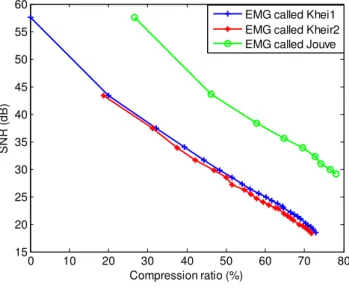
The proposed approach has been evaluated on real EMG data, baptized kheir1, kheir2 and Jouve. The signals were amplified with a total gain of 2000, and sampled at 2024 Hz using a 12 bits data acquisition. Signals were measured on the biceps muscle. The signals were collected during 40%MVC contraction, with an angle of 90° between the arm and the forearm. The biorthogonal wavelet 9/7 has been used for the wavelet transform (lifting scheme) to level 21, in order to have better results, following the cutting window size 16384 samples each. Figures 3 and 4 show variations of compression ratio as a function of SNR and PRD for EMG signals; figure 4 shows the mean PRD measured on the 3 isometric EMG signals. The quality decreases. The behavior of the compression method in terms of SNR and PRD as a function of compression ratio is quite similar for the three EMG signals considered. The SNR decreases as the compression ratio increases is explained mathematically by a rational fraction which numerator is the power of the original signal and denominator, the error between original signal and reconstructed signal. Given this result, if the SNR may be used as criteria for evaluating the method, it should be used by requiring only a minimum of around 20 dB. A minimum level of reconstruction of 20 dB corresponds to a level of error between original signal and reconstructed signal very acceptable. Similarly, when compression ratio is believed; the PRD increases. Thus, the relative error in energy is high at high compression ratio. However, a high compression ratio does not necessarily mean a good compression when the SNR and the PRD are taken into account. This compromise is sought by a good compression ratio with an acceptable signal to noise ratio. To achieve this, the subjective criteria should be used.
TABLE I
Comparison between the results obtained by the proposed algorithm and results from literature, for the compression of emg signals called kheir1 measured during isometric contractions
Method CR (%) PRD (%)
Berger et al. [4] 72,96 2,01 Berger et al. [1] 72,96 2,50 Norris et al. [17] 72,96 3,51 Filho et al. [3] 72,96 2,10 Marcus et al. [11] 72,96 4,75 Marcus et al. [19] 73,02 3,50 Proposed method 72,96 1,26
The proposed method provides a higher SNR at equal compression ratio compared to the methods using B-splines [7] and transform called “odd”, proposed by PN Eloundou et al. [5]. Compression ratios obtained are significantly higher.
Our results compared to those obtained by Berger et al. [1], [4], Marcus et al. [11], [19] showed a significant improvement in terms of PDR in compression ratio. Thus, for a compression ratio of 72.96%, we have a PRD of 1.26% with modified method JPEG 2000 called 1D.
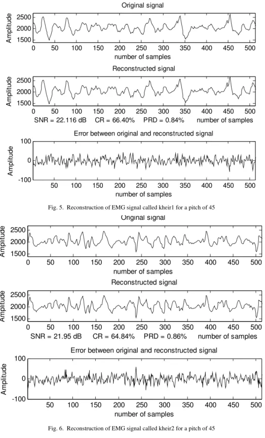
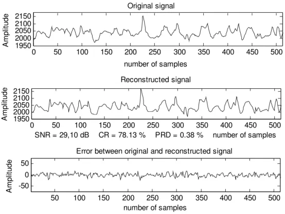
The same improvements are observed when we consider the results obtained by Filho et al. [3] at equal compression ratio. Generally, at equal compression ratio, we notice a low PRD compared to other quoted methods above, which causes minimal loss of reconstruction of EMG signals for the subjective criteria. Examples of original and reconstructed signals are shown through figures 5, 6 and 7.
0 10 20 30 40 50 60 70 80
15 20 25 30 35 40 45 50 55 60
Compression ratio (%)
SNR
(
d
B
)
EMG called Khei1 EMG called Kheir2 EMG called Jouve
Fig. 3. Variation of SNR as a function of compression ratio
0 10 20 30 40 50 60 70 80
0 0.2 0.4 0.6 0.8 1 1.2 1.4
PRD
(%
)
0 50 100 150 200 250 300 350 400 450 500 1500
2000 2500
Original signal
number of samples
A
m
pl
it
ude
0 50 100 150 200 250 300 350 400 450 500 1500
2000 2500
Reconstructed signal
SNR = 22.116 dB CR = 66.40% PRD = 0.84% number of samples
A
m
p
lit
u
d
e
50 100 150 200 250 300 350 400 450 500 -100
0 100
Error between original and reconstructed signal
number of samples
A
m
pl
it
ude
Fig. 5. Reconstruction of EMG signal called kheir1 for a pitch of 45
0 50 100 150 200 250 300 350 400 450 500 1500
2000 2500
Original signal
number of samples
A
m
p
lit
u
d
e
0 50 100 150 200 250 300 350 400 450 500 1500
2000 2500
Reconstructed signal
SNR = 21.95 dB CR = 64.84% PRD = 0.86% number of samples
Amp
lit
u
d
e
50 100 150 200 250 300 350 400 450 500 -100
0 100
Error between original and reconstructed signal
number of samples
A
m
pl
it
ude
0 50 100 150 200 250 300 350 400 450 500 1950
2000 2050 2100 2150
Original signal
number of samples
A
m
pl
it
ud
e
0 50 100 150 200 250 300 350 400 450 500
1950 2000 2050 2100 2150
Reconstructed signal
SNR = 29,10 dB CR = 78.13 % PRD = 0.38 % number of samples
Am
p
lit
u
d
e
50 100 150 200 250 300 350 400 450 500
-50 0 50
Error between original and reconstructed signal
number of samples
Am
p
lit
u
d
e
Fig. 7. Reconstruction of EMG signal called Jouve for a pitch of 24
V. CONCLUSION
In this paper, the proposed scheme was evaluated on surface EMG signals measured during isometric contraction. We had showed that, it is possible to compress EMG signals with a modified compression standard using the coding of still images JPEG 2000. The results are encouraging, if we take into account the objective and subjective criteria. The compression ratio and the PRD obtained are slightly lower in term of quality to the various methods mentioned above. It is possible to compress the EMG signals to a compression ratio of approximately 66.40% to 0.83% of a PRD and 22.15 dB for SNR. The signal can be reconstructed without noticeable degradation. The method used allows us to evaluate in the first instance the possibilities of the technique that can be used to compress EMG signals. Its extension to the EMG signals collected under dynamic conditions to better experience and generally to other electrophysiological signals could be further investigated.
REFERENCES
[1] P. A. Berger, F. A. O. Nascimento, J. C. Carmo, and A. F. Rocha, “Compression of EMG signals with wavelet transform and artificial neural networks”, Institute of physics publishing: Physiological Measurement, 27(6): 457-465, 2006 March.
[2] M. O. Diab, C. Marque, and M. Khalil, “Une approche de classification des contractions utérines basée sur la théorie des ondelettes et la statistique”, Lebanese Science Journal, 7(1): 91-103, 2006.
[3] E. B. L. Filho, E. A. B. De Silva, and M. B. De Carvalho, “On EMG Signal Compression with Recurrent Patterns”, IEEE Transactions
on biomedical engineering, vol. 55, N°12, pp. 2844-2896, January 2008.
[4] P. A. Berger, F. A. O. Nascimento, F. A. Rocha, and J. L. A. Carvalho, “A New Wavelet-Based Algorithm for Compression of Emg Signals”, Proc. of the 29th Annual International, Conf. of the IEEE EMBS, Lyon, France, pp.1554-1557, August 23-26, 2007.
[5] N. P. Eloundou, B. Marius, and P. Ele, “Compression des signaux EMG par la Transformée dite « impaire»”, JSPI, N°7, pp. 1-9, 2006. [6] S. Jalaleddine, C. G. Hutchens, R. D. Strattan, and W. A. Coberly, “ECG Data Compression Techniques - A Unified Approach”, IEEE
Trans BME, vol.37, N°4, pp.329-343, 1990.
[7] E. P. Ntsama, P. Ele, and E. Tonye, “Compression robuste du signal ElectroMyoGraphique (EMG) par la Transformée avec les B-splines”, HAMMAMET, CARI 2004, pp. 59-66, 22-25 novembre 2004.
[8] D. Tchiotsop, “Modélisations polynomiales des signaux ECG. Applications à la compression”, Thèse de Doctorat en Automatique et Traitement du Signal, Institut Polytechnique de Lorraine, 15 Novembre 2007.
[9] E. Christopoulos, J. Askelof, and M. Larsson, “Efficient encoding and Reconstruction or Regions of Interest in JPEG 2000”.
EUSIPCO-2000, Tampere, Finland, Sep. 2000.
[10] W. Fourati, and M. S. Bouhlel, “Amélioration de La norme JPEG 2000 Par un Prétraitement basé sur le filtrage”, 3rd International Conference: Sciences of Electronic, Technologies of Information and Telecommunications, March 27-31, Tunisia, 2005.
[11] V. C. C. Marcus, L. A. Joao Carvavalho, A. Pedro Berger, F. D. R. Adson, and A. O. Francisco Nascimento, “Compression of Surface Electromyographic Signals Using Two-Dimensional Techniques”, Recent Advances in Biomedical Engineering, Ganesh R Naik (Ed), InTech, ISBN: 978-953-307-004: pp.17-38, October 2009.
[14] J. C. Rolón, P. Salembier, and X. Alameda, “Image Compression with generalized Lifting and partial knowledge of the Signal PDF”, Proc. ICIP 2008, 15th IEEE Int. Conf on Image Processing, pp.129-132, 2008.
[15] W. Sweldens, “The lifting scheme: A construction of second generation wavelets”, SIAM J. Math. Anal., Vol. 29, N°2, pp. 511-546, 1997.
[16] N. Thi Hoàng Lan, “Etude de la méthode de la transformation en ondelette et l'application à la compression des images”, Rapport, Luong Hông Viêt, 15 juillet 2005.
[17] J. A. Norris, K. Englehart, and D. F. Lovely, “Steady-state and dynamic myoelectric signal compression using embedded zero-tree wavelets”, In Proc 23rd Annu. Int. Conf. IEEE Engineering in Medicine Biology Society, pp, 1879–1882, 2001.
[18] M. Fira, L. Goras, “Biomedical Signal Compression based on Basis Pursuit”, International Journal of Advanced Science and Technology, Vol. 14, pp.53-64, January, 2010.
[19] C. C. Marcus, D. B. A. Pedro, F. D. R. Adson, L. A. D. Joao, and A. D. O. N. Francisco, “Compression of Surface Electromyographic Signals Using image compression techniques”. 30th Annual International IEEE EMBS conference, Vancouver, Bristish Colimbia,