Analysis of Fibronectin-Modified
Titanium Surfaces
Yu-Chi Chang1, Wei-Fang Lee2, Sheng-Wei Feng1, Haw-Ming Huang1,3, Che-Tong Lin1,4, Nai-Chia Teng1,4, Wei Jen Chang1,5*
1School of Dentistry, College of Oral Medicine, Taipei Medical University, Taipei, Taiwan, ROC,2School of Dental Technology, College of Oral Medicine, Taipei Medical University, Taipei, Taiwan, ROC,3Graduate Institute of Biomedical Materials & Tissue Engineering, College of Oral Medicine, Taipei Medical University, Taipei, Taiwan, ROC,4Dental Department, Taipei Medical University Hospital, Taipei, Taiwan, ROC,
5Dental Department of Taipei Medical University, Shuang-Ho Hospital, Taipei, Taiwan, ROC
Abstract
Background
Glow discharge plasma (GDP) procedure is an effective method for grafting various pro-teins, including albumin, type I collagen, and fibronectin, onto a titanium surface. However, the behavior and impact of titanium (Ti) surface modification is yet to be unraveled.
Purpose
The purpose of this study is to evaluate and analyze the biological properties of fibronectin-grafted Ti surfaces treated by GDP.
Materials and Methods
Grade II Ti discs were initially cleaned and autoclaved to obtain original specimens. Subse-quently, the specimens were GDP treated and grafted with fibronectin to form Ar-GDP (Argon GDP treatment only) and GDP-fib (fibronectin coating following GDP treatment) groups. Blood coagulation test and MG-63 cell culture were performed to evaluate the bio-logical effects on the specimen.
Results
There was no significant difference between Ar-GDP and GDP-fib groups in blood compati-bility analysis. While in the MTT test, cellular proliferation was benefited from the presence of fibronectin coating. The numbers of cells on Ar-GDP and GDP-fib specimens were greater than those in the original specimens after 24 h of culturing.
Conclusions
GDP treatment combined with fibronectin grafting favored MG-63 cell adhesion, migration, and proliferation on titanium surfaces, which could be attributed to the improved surface properties. OPEN ACCESS
Citation:Chang Y-C, Lee W-F, Feng S-W, Huang H-M, Lin C-T, Teng N-C, et al. (2016)In VitroAnalysis of Fibronectin-Modified Titanium Surfaces. PLoS ONE 11(1): e0146219. doi:10.1371/journal.pone.0146219
Editor:Mohammed Yousfi, University Paul Sabatier, FRANCE
Received:April 23, 2015
Accepted:November 8, 2015
Published:January 5, 2016
Copyright:© 2016 Chang et al. This is an open access article distributed under the terms of the
Creative Commons Attribution License, which permits unrestricted use, distribution, and reproduction in any medium, provided the original author and source are credited.
Data Availability Statement:All relevant data are within the paper and its Supporting Information files.
Funding:The author(s) received no specific funding for this work.
Introduction
Osseointegration [1], which refers to the process through which the mature living bone estab-lishes a direct structural and functional connection with the implant without any intervening soft or fibrous tissue, is an important phenomenon favoring the effective fixation of implants to bone. Successful osseointegration depends on both the chemistry of the substrate and its sur-face texture. The sursur-face roughness of titanium implants greatly affects the rate of osseointegra-tion and biomechanical fixaosseointegra-tion.[2,3] In addition, cell adhesion and spreading are important parameters influencing the implant engineering.
Cellular responses in the vicinity of the implants are strongly affected by surface hydrophi-licity, roughness, texture, chemical composition, charge and morphology.[4,5] Given this fact, several studies have attempted to enhance the adhesion and activity of osteoblastic cells on the surfaces by depositing functional proteins onto the titanium surface. [6,7] Of the dif-ferent methods, glow discharge plasma (GDP) procedure is a popular method for realizing surface modification of biomaterials. Several studies have demonstrated the advantages of GDP method in sterilizing and modifying the surface of the biomaterials [8–10]. The XPS results showed that the Ti surface after plasma treatment was leaving chemistry uncharged.[11] Most importantly, this method offers the possibility of forming bio-func-tional groups and funcbio-func-tional proteins on the titanium surfaces [12,13]. Given all these advan-tages, this method can be considered as an efficient tool for improving the biocompatibility of biomaterials.
In general, contact between the implants and bone can be improved by adopting surface treatment methods, such as sand blasting, sol-gel dip coating, titanium nitride oxide (TiNOx) coating, plasma spraying, acid etching, and various combinations thereof.[14–16] Further-more, the biocompatibility of titanium surface could be improved by grafting several functional proteins, such as extracellular matrix proteins (ECM), type I collagen, and albumin using the GDP method.[12,17,18] Bioactive adhesive proteins, such as the extracellular matrix protein fibronectin, have been used to facilitate the surface properties of titanium. The titanium surface with fibronectin coating following GDP treatment were more hydrophilic and had greater sur-face roughness. Meanwhile, the FITC labeling showed that the number of fibronectin dots on the titanium surface increased positively with the concentration of fibronectin solution used. [19] The attachment of cells through fibronectin binding were measured.[20] The attachment and differentiation of osteoblast-like cells were enhanced on Ti6Al4V titanium surface through fibronectin coating.[21–24] However, the cell culture study on commercially pure titanium (cpTi) remains an important focus of investigation.
Fibronectin is known to play an important role in cell attachment, growth, migration and differentiation. In addition, it is also reported to participate in platelets adhesion and aggre-gation via the integrinαIIbβ3 (glycoprotein IIb/IIIa) receptors on platelet membrane.[25] However, with the advance of technology, the hemolysis rate and blood coagulation time could be lower and prolonged on titanium surface after fibronectin coating.[26] The blood compatibility of titanium surface with fibronectin coating after GDP treatment has not been studied yet.
Materials and Methods
Preparation of titanium surface
10 mm diameter Grade II titanium discs (BioTech One Inc., Taipei, Taiwan) were used in this study. The preparation method was clearly described previously.[18,19] The samples were cleaned in an ultrasound bath with detergent, acetone, and distilled water for 15 min separately to remove the surface contaminants. Subsequently, the cleaned titanium discs were autoclaved at 121°C for 20 min and dried at 40°C in a conventional oven. The titanium discs thus obtained are henceforth referred to as“original specimens.”
Glow discharge plasma cleaning and protein grafting
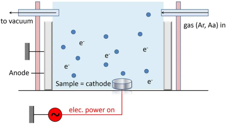
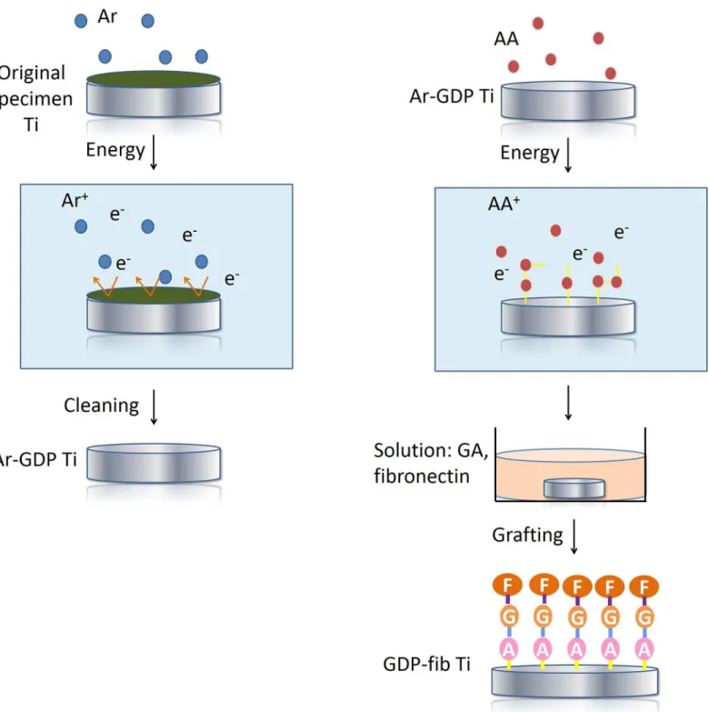
The original specimens were cleaned by GDP (PJ; AST Products Inc., North Bellericca, MA, USA) with argon. This process involves GDP treatment under argon gas at room temperature under the following conditions: power, 85 W; frequency, 13.56 MHz; pressure, 100 mtorr for 15 min (Fig 1).[19] The titanium discs thus obtained are referred to as“Ar-GDP”(Fig 2a).
Subsequently, NH groups were adhered onto the titanium surfaces in the GDP reactor fed with allylamine (AA) gas at room temperature under the same conditions as mentioned above except treatment time was increased to 30 min. Thereafter, the specimens were placed in a 3% glutaraldehyde solution (Merck, NJ, USA) for 30 min. Then, the titanium specimens were placed in a solution with 5μg/ml fibronectin (Sigma-Aldrich Co., St. Louis, MO, USA) for 24
hours to form fibronectin coating on the surface of titanium discs. A tris-phosphate buffer (pH 7.4) was used to stop the chain reaction between amine group and fibronectin (immersed for 30 min).[19] The titanium discs thus obtained are referred to as“GDP-fib”(Fig 2b).
Surface topography evaluation
The surface characteristics of Ar-GDP and GDP-fib were examined with scanning electron microscope (SEM) (Model 2400; Hitachi, Ltd) in order to evaluate the topography of Ti surface after fibronectin coating.
Blood coagulation test
Eight 5 month old New Zealand white rabbits of mean weight 3 kg were used for the analysis. All procedures were approved by the Animal Care and Ethics Committee of Taipei Medical University, Taipei, Taiwan (affidavit of approval No. LAC-2013-0265) (S1 File). The study was conducted according to the principles of the 2010 Basel Declaration. This animal research was conducted in accordance to the ARRIVE (Animal Research: Reporting ofIn VivoExperiments) guidelines developed in NC3Rs (National Centre for the Replacement, Refinement and Reduc-tion of Animal Research) (S2 File).[27]
The coagulation of the blood on the surface of blank Petri dish and Ar-GDP and GDP-fib titanium disks were evaluated by using spectrophotometry. Fresh rabbit blood was used for each experiment. During the test, 10 ml of blood from each rabbit was drawn with a syringe with 24G needle (TERUMO Co., Tokyo, Japan) from the marginal vein on the right or left ear under animal fixation with a fixation box. The rabbits were alive and kept for further study after the blood collection. Subsequently, a 200μl of fresh rabbit blood was placed on the surface
ELISA reader (Model 2020, Anthos Labtec Instruments, Eugendorf, Wals, Austria) at 540 nm. Each absorbance value represented the average of six measurements.
Blood coagulation of Ar-GDP and GDP-fib specimens were expressed as a percentage and compared to that of the blank plates (100%). The remainders of Ar-GDP and GDP-fib were used for statistical analysis. High absorbance value, which is also demonstrated as the lower absorbance value remainder, implies a low amount of blood coagulation.
Cell vitality test
In the process, MG-63 (ATCC CRL-1427) osteoblast-like cells, at a concentration of 1 × 10⁴ cells/ml, were seeded onto the surface of the original specimen, Ar-GDP and GDP-fib titanium discs in the 24-well Petri dishes (Nunclon; Nunc, Roskilde, Denmark). During all experiments, the cells were initially incubated without medium for 10 hours to ensure attachment. The cells were then cultured in Dulbecco’s modified Eagle’s medium (DMEM; HyClone, Logan, UT) supplemented with L-glutamine (4 mmol/L) with 10% fetal bovine serum (FBS), and 1% peni-cillin streptomycin. Cultures were incubated in 5% CO2atmosphere at 37°C and 100%
humid-ity. Accordingly, the time, when DMEM was added, was defined as the 0thhour (T0) for every test.[17]
At 0th(T0) and 24th(T24) hour of culture, the culture media were removed and the discs were rinsed three times with PBS. The samples were then fixed in 2.5% GA and 2% paraformal-dehyde solution for 30 min. Subsequently, for post-fixation, the samples were rinsed in 1% osmium tetroxide for 1 hour. The titanium discs were then dehydrated in 70%, 80%, 90%, 95%, and 100% series ethanol and dried in a critical point dryer (HCP-2; Hitachi Ltd, Tokyo, Japan). Fig 1. Schematic of the flow discharge plasma system.The blue circles represented the gas ions (argon or allylamine) which were used to modify the titanium surface.
Thereafter, a thin layer of palladium gold was coated on the samples with a sputter coater (IB-2; Hitachi, Ltd, Tokyo, Japan). The morphological features of the cells were examined by using a Hitachi S-2400 electron microscope (Model 2400; Hitachi, Ltd). Two samples were prepared for each group.
Fig 2. Schematic of sample preparation.(a)The surfaces of Ti discs (original specimens) were cleaned with argon-based GDP. These titanium discs were then labeled as“Ar-GDP.”(b) Amine groups were grafted onto the Ar-GDP surfaces in the GDP reactor fed with allylamine (AA) gas. These specimens were subsequently treated in glutaraldehyde (GA) and fibronectin solutions and were labeled as“GDP-fib”.
Cell vitality was determined using an MTT ((3–4,5-dimethylethiazol-2-yl)-2,5-diphenyl tet-razolium bromide) reduction assay. Tettet-razolium salt (MTT kit, Roche Applied Science, Mann-heim, Germany) was added and metabolically reduced to colored formazan by mitochondrial dehydrogenase in viable cells, as indicated in the manufacturer’s instructions. After solubiliz-ing, the formazan dye was added with 500μl of dimethyl sulfoxide for 5 min. Subsequently, the
optical density of the medium was determined by using the ELISA reader at 570 nm. Optical density results were expressed as a percentage of that of the original specimen. In this study, four samples were prepared for each group.
Statistical analysis
Biological characters of the modified titanium surfaces were evaluated by performing hemo-compatibility and MTT assay. The morphology of the MG-63 cell cultured samples was observed by using SEM. For all assays, differences between tested groups were evaluated by Stu-dent’st-test. Significance levels were set toP0.05.
Results
Surface topography evaluation
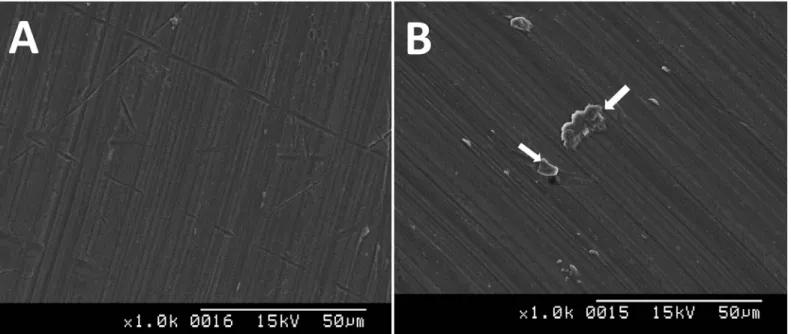
The surface of Ar-GDP discs showed planar morphology with parallel grooves (Fig 3a). How-ever, the GDP-fib samples had a homogenous layer with irregular folding structure of protein (Fig 3b).
Blood coagulation test
The results of blood coagulation test showed that the hemocompatibility of titanium discs decreased after fibronectin grafting; nevertheless, there was no significant difference between two groups (Fig 4). The hemocompatibility of Ar-GDP and GDP-fib samples were expressed as a percentage and compared to that of the blank plate (100%). The remainders of the Ar-GDP titanium discs were 22.6%, 67.5%, 80.3% and 77.3% at 10, 20, 30 and 40 min of blood coagulation, respectively. Similarly, in the case of GDP-fib discs, the remainders were 15.6%, 54.1%, 68.9% and 65.0% at 10, 20, 30 and 40 min of blood coagulation, respectively. As is seen, the remainder of the Ar-GDP titanium discs was not significantly greater than the GDP-fib discs at each set point of time.
Cell vitality test
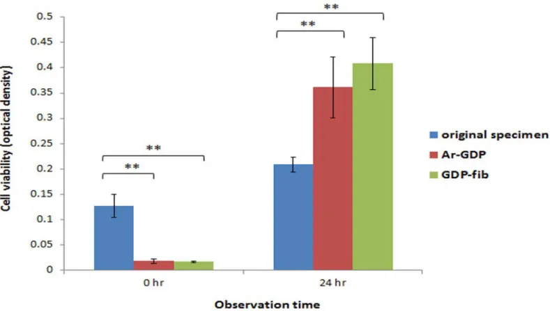
According to the MTT assay analysis, the number of MG-63 cells on Ar-GDP and GDP-fib titanium specimens after 24 hours (T24) of culture was greater than that on the original speci-mens (Fig 5). Besides, the cells on the original specimens showed significantly less cell prolifer-ation when compared to the cells on the Ar-GDP and GDP-fib titanium discs (P<0.01). There was no significant difference between the Ar-GDP and GDP-fib groups.
MG-63 cell morphology evaluation
Fig 3. Scanning electron microscopy images of the Ar-GDP (a) and GDP-fib (b) titanium discs.Irregular folding of the fibronectin was revealed on the surface of fibronectin-grafted titanium disks(white arrows).
doi:10.1371/journal.pone.0146219.g003
Fig 4. Blood coagulation of Ar-GDP and GDP-fib samples was expressed as percentage and compared to that of the blank plate (100%).There was no significant difference between two groups.
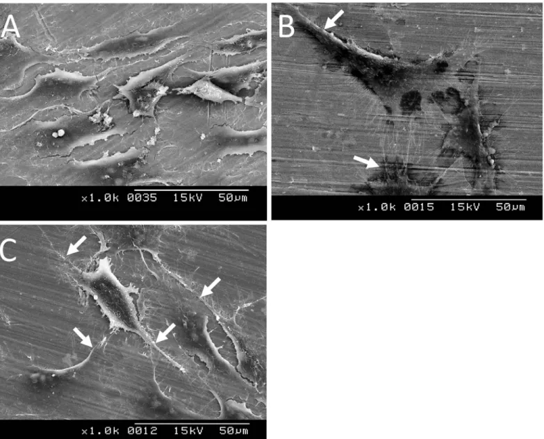
each other and with the substrate (Fig 6c), could be observed when the fibronectin-grafted tita-nium discs were used as substrates.
Discussion
The objective of this study was to gain in-depth insights of the biological effects, including hemocompatibility and cellular reaction, of the titanium surfaces after GDP and fibronectin modification. Therefore, we analyzed the surfaces prepared using different methods. The sur-face roughness and wettability of the modified titanium sursur-faces have already been reported in previous studies. In the study, fibronectin-grafted specimens had higher hydrophilicity and greater surface roughness than the GDP-treated specimens.[19] The FITC labeling ensured the formation of fibronectin coating. Grafted fibronectin on titanium surface was spotty distribu-tion instead of monolayer formadistribu-tion. In addidistribu-tion, the number of fibronectin dots on the tita-nium surface increased positively with the concentration of fibronectin solution used.[19]
The normal wound healing process is divided into four stages, namely, hemostasis, inflam-matory, proliferative and remodeling phases. Coagulation (thrombogenesis), the process by which blood forms clots, is an important part of hemostasis.[28] The GDP treatment could enhance the adhesion of blood cells onto the surface of the biomaterial, thereby favoring aggre-gation and blood coagulation on polymer-based biomaterials.[29] Because of the effect, the wound healing and osseointegration will be promoted. In order to improve hemocompatibility, various proteins, such as collagen, were adsorbed onto the titanium surfaces.[12,30,31] Accord-ing to this study, the blood coagulation decreased on the surface of fibronectin-grafted titanium Fig 5. The MTT assay of MG-63 cells.Compared to the original specimen, both on the surface of GDP-treated and fibronectin-modified titanium, the MG-63 cells viability was significantly enhanced after 24 hours of culture (**P<0.01).
discs. In other words, there might be more blood cells in contact with the surface of the fibro-nectin-grafted discs and not trapped into the clot. This might have accelerated the differentia-tion. In addition, there is no significant difference between Ar-GDP and GDP-fib groups. Overall, the adhesion and aggregation of platelets were not significantly minimized by the pres-ence of the thin fibronectin layer, but cellular proliferation and differentiation might have been benefited.
In general, MG-63 cells, a human osteoblast-like cell line, are being used to evaluate the bio-logical effects of different implant surface designs. Despite proliferating more rapidly than the other human bone-derived cells, MG-63 cells perform many osteoblastic traits that are charac-teristic of bone-forming cells. MG-63 cells show enhanced alkaline phosphatase activity and actin-ring formation following 1α, 25-dihydeoxyvitamin D3administration. This is again a
typical characteristic of bone-derived cells.[31,32] Thus far, severalin vitrostudies with Fig 6. Scanning electron microscopy images of MG-63 cells cultured on original specimen (a), Ar-GDP (b), and GDP-fib (c) titanium discs after 24 h.Cells showed morphological alternation from spindle to more-stellar shapes, and extensive process (white arrows).
different cell culture analysis have shown that the osteoblast proliferation, differentiation, and matrix synthesis is greatly influenced by the nature of implant surface.[33,34] In general, cells cultured on rough surfaces tended to demonstrate more differentiated osteoblasts than those cultured on smoother surfaces. [35,36]
The results performed in the present study supported the abovementioned reports. The sur-faces of GDP-fib titanium discs (0.400μm; 5.4 ± 0.8°) were significantly rougher and more
hydrophilic than Ar-GDP specimens (0.170μm; 36.8 ± 8.5°).[19] The MTT assay (Fig 5) and
SEM images (Fig 6) demonstrated that the number of cells on Ar-GDP and GDP-fib specimens were greater than that on the original specimens after 24 hours of culture. The results of MTT assay showed that the cell number was greater on original specimen at T0; nevertheless, the number of MG-63 cells on Ar-GDP and GDP-fib titanium specimens was greater than that on the original specimens at T24. The result might imply that, during the first 10 hours of attach-ment without DMEM, MG-63 cells tended to proliferate on original specimens while tended to differentiate on other two groups due to the different contact surface. The hypothesis was sup-ported by the results of SEM morphology evaluation. The result of T24 culture also suggested that the GDP treatment and fibronectin grafting could enhance the proliferation rate of MG-63 cells. Besides, the cell viability of GDP-fib group was more than that of Ar-GDP group. On the basis of these results, it is reasonable to conclude that the fibronectin-grafted titanium spec-imens that are treated by GDP method could reduce the osseointegration period and improve osseointegration in the early stages of dental implant fixation.
In conclusion, the results obtained in this study indicate that the blood coagulation was not significantly different with fibronectin coating after GDP pretreatment. However, the cell adhe-sion, migration, and proliferation were benefited from the presence of fibronectin grafting. This approach allows to find the biological modified implant surface and to examine the effects that could possibly occur during the material-cell interaction. Morein vitroandin vivostudies are certainly necessary for the identification of future endosseous implant design.
Supporting Information
S1 File. LAC-2013-0265.The affidavit of approval from the Animal Care and Ethics
Commit-tee of Taipei Medical University, Taipei, Taiwan. (PDF)
S2 File. The ARRIVE checklist.
(PDF)
Acknowledgments
The authors would like to thank Enago (www.enago.tw) for the English language review.
Author Contributions
Conceived and designed the experiments: WJC. Performed the experiments: YCC SWF. Ana-lyzed the data: YCC WFL. Contributed reagents/materials/analysis tools: HMH CTL NCT. Wrote the paper: YCC WJC. Obtained permission for use of cell line: HMH.
References
2. Wennerberg A.; Hallgren C.; Johansson C.; Danelli S. A histomorphometric evaluation of screw-shaped implants each prepared with two surface roughnesses.Clin Oral Implants Res1998, 9, 11–19. PMID: 9590940
3. Lazzara R.J.; Testori T.; Trisi P.; Porter S.S.; Weinstein R.L. A human histologic analysis of osseotite and machined surfaces using implants with 2 opposing surfaces.Int J Periodontics Restorative Dent 1999, 19, 117–129. PMID:10635177
4. Oshida Y.; Hashem A.; Nishihara T.; Yapchulay M.V. Fractal dimension analysis of mandibular bones: Toward a morphological compatibility of implants.Biomed Mater Eng1994, 4, 397–407. PMID: 8000293
5. Lampin M.; Warocquier C.; Legris C.; Degrange M.; Sigot-Luizard M.F. Correlation between substratum roughness and wettability, cell adhesion, and cell migration.J Biomed Mater Res1997, 36, 99–108. PMID:9212394
6. Schwarz F.; Wieland M.; Schwartz Z.; Zhao G.; Rupp F.; Geis-Gerstorfer J., et al. Potential of chemi-cally modified hydrophilic surface characteristics to support tissue integration of titanium dental implants.J Biomed Mater Res B Appl Biomater2009, 88, 544–557. doi:10.1002/jbm.b.31233PMID: 18837448
7. Gronowicz G.; McCarthy M.B. Response of human osteoblasts to implant materials: Integrin-mediated adhesion.J Orthop Res1996, 14, 878–887. PMID:8982129
8. Aronsson B.O.; Lausmaa J.; Kasemo B. Glow discharge plasma treatment for surface cleaning and modification of metallic biomaterials.J Biomed Mater Res1997, 35, 49–73. PMID:9104698
9. Czarnowska E.; Wierzchon T.; Maranda-Niedbala A.; Karczmarewicz E. Improvement of titanium alloy for biomedical applications by nitriding and carbonitriding processes under glow discharge conditions. J Mater Sci Mater Med2000, 11, 73–81. PMID:15348050
10. Duske K.; Wegner K.; Donnert M.; Kunert U.; Podbielski A.; Kreikemeyer B. et al. Comparative in vitro study of different atmospheric pressure plasma jets concerning their antimicrobial potential and cellular reaction.Plasma Processes and Polymers2015.
11. Cools P.; Vanderleyden E.; Dubruel P.; Morent R. Surface analysis of titanium cleaning and activation processes: Non-thermal plasma versus other technique.Plasma Chemistry Plasma Processing2014, 34, 917–932.
12. Yamamoto H.; Shibata Y.; Miyazaki T. Anode glow discharge plasma treatment of titanium plates facili-tates adsorption of extracellular matrix proteins to the plates.J Dent Res2005, 84, 668–671. PMID: 15972599
13. Alves C.M.; Yang Y.; Carnes D.L.; Ong J.L.; Sylvia V.L.; Dean D.D., et al. Modulating bone cells response onto starch-based biomaterials by surface plasma treatment and protein adsorption. Bioma-terials2007, 28, 307–315. PMID:17011619
14. Singh R.G. Evaluation of the bioactivity of titanium after varied surface treatments using human osteo-sarcoma osteoblast cells: An in vitro study.Int J Oral Maxillofac Implants2011, 26, 998–1003. PMID: 22010082
15. Durual S.; Pernet F.; Rieder P.; Mekki M.; Cattani-Lorente M.; Wiskott H.W. Titanium nitride oxide coat-ing on rough titanium stimulates the proliferation of human primary osteoblasts.Clin Oral Implants Res 2011, 22, 552–559. doi:10.1111/j.1600-0501.2010.02033.xPMID:21087318
16. da Silva J.S.; Amico S.C.; Rodrigues A.O.; Barboza C.A.; Alves C. Jr.; Croci A.T. Osteoblastlike cell adhesion on titanium surfaces modified by plasma nitriding.Int J Oral Maxillofac Implants2011, 26, 237–244. PMID:21483875
17. Huang H.M.; Hsieh S.C.; Teng N.C.; Feng S.W.; Ou K.L.; Chang W.J. Biological surface modification of titanium surfaces using glow discharge plasma.Med Biol Eng Comput2011, 49, 701–706. doi:10. 1007/s11517-011-0742-2PMID:21286829
18. Chang W.J.; Ou K.L.; Lee S.Y.; Chen J.Y.; Abiko Y.; Lin C.T., et al. Type i collagen grafting on titanium surfaces using low-temperature glow discharge.Dent Mater J2008, 27, 340–346. PMID:18717160
19. Chang Y.C.; Feng S.W.; Huang H.M.; Teng N.C.; Lin C.T.; Lin H.K., et al. Surface analysis of titanium biological modification with glow discharge.Clin Implant Dent Relat Res2013.
20. Rapuano B.E.; Lee J.J.; MacDonald D.E. Titanium alloy surface oxide modulates the conformation of adsorbed fibronectin to enhance its binding to alpha(5) beta(1) integrins in osteoblasts.Eur J Oral Sci 2012, 120, 185–194. doi:10.1111/j.1600-0722.2012.954.xPMID:22607334
22. Rapuano B.E.; Hackshaw K.; Macdonald D.E. Heat or radiofrequency plasma glow discharge treat-ment of a titanium alloy stimulates osteoblast gene expression in the mc3t3 osteoprogenitor cell line. Journal of periodontal & implant science2012, 42, 95–104.
23. Rapuano B.E.; Singh H.; Boskey A.L.; Doty S.B.; MacDonald D.E. Heat and radiofrequency plasma glow discharge pretreatment of a titanium alloy: Evidence [corrected] for enhanced osteoinductive properties.J Cell Biochem2013, 114, 1917–1927. doi:10.1002/jcb.24536PMID:23494951
24. Shibata Y.; Hosaka M.; Kawai H.; Miyazaki T. Glow discharge plasma treatment of titanium plates enhances adhesion of osteoblast-like cells to the plates through the integrin-mediated mechanism.Int J Oral Maxillofac Implants2002, 17, 771–777. PMID:12507235
25. Cho J.; Mosher D.F. Enhancement of thrombogenesis by plasma fibronectin cross-linked to fibrin and assembled in platelet thrombi.Blood2006, 107, 3555–3563. PMID:16391013
26. Li G.; Yang P.; Qin W.; Maitz M.F.; Zhou S.; Huang N. The effect of coimmobilizing heparin and fibro-nectin on titanium on hemocompatibility and endothelialization.Biomaterials2011, 32, 4691–4703. doi:10.1016/j.biomaterials.2011.03.025PMID:21463893
27. Kilkenny C.; Browne W.J.; Cuthill I.C.; Emerson M.; Altman D.G. Improving bioscience research report-ing: The arrive guidelines for reporting animal research.PLoS Biol2010, 8, e1000412. doi:10.1371/ journal.pbio.1000412PMID:20613859
28. Stadelmann W.K.; Digenis A.G.; Tobin G.R. Physiology and healing dynamics of chronic cutaneous wounds.Am J Surg1998, 176, 26S–38S. PMID:9777970
29. Sharma C.P.; Hari P.R. Adhesion and stability of blood cells onto polymer substrates: Effect of glow dis-charge.J Biomater Appl1991, 6, 72–79. PMID:1920070
30. Kottke-Marchant K.; Anderson J.M.; Umemura Y.; Marchant R.E. Effect of albumin coating on the in vitro blood compatibility of dacron arterial prostheses.Biomaterials1989, 10, 147–155. PMID: 2524222
31. Kawai H.; Shibata Y.; Miyazaki T. Glow discharge plasma pretreatment enhances osteoclast differenti-ation and survival on titanium plates.Biomaterials2004, 25, 1805–1811. PMID:14738844
32. Clover J.; Gowen M. Are mg-63 and hos te85 human osteosarcoma cell lines representative models of the osteoblastic phenotype?Bone1994, 15, 585–591. PMID:7873286
33. Feng B.; Weng J.; Yang B.C.; Qu S.X.; Zhang X.D. Characterization of titanium surfaces with calcium and phosphate and osteoblast adhesion.Biomaterials2004, 25, 3421–3428. PMID:15020115
34. Kurachi T.; Nagao H.; Nagura H.; Enomoto S. Effect of a titanium surface on bone marrow-derived oste-oblastic cells in vitro.Arch Oral Biol1997, 42, 465–468. PMID:9382711
35. Owen T.A.; Aronow M.; Shalhoub V.; Barone L.M.; Wilming L.; Tassinari M.S., et al. Progressive devel-opment of the rat osteoblast phenotype in vitro: Reciprocal relationships in expression of genes associ-ated with osteoblast proliferation and differentiation during formation of the bone extracellular matrix.J Cell Physiol1990, 143, 420–430. PMID:1694181