Comparison between Pre-Therapeutic Gene Expression
Profiles and
In Vitro
Chemosensitivity Assay
Christian F. Singer1*, Florian Klinglmu¨ller2, Rembert Stratmann3, Christine Staudigl1, Anneliese Fink-Retter1, Daphne Gschwantler1, Samir Helmy1, Georg Pfeiler1, Anne Catharina Dressler1,
Christian Sartori3, Martin Bilban4
1Department of Gynecology and Obstetrics and Comprehensive Cancer Center, Medical University of Vienna, Vienna, Austria,2Institute for Medical Statistics and Informatics, Medical University of Vienna, Vienna, Austria,3DCS Innovative Diagnostik-Systeme GmbH, Hamburg, Germany,4Department of Laboratory Medicine, Medical University of Vienna, Vienna, Austria
Abstract
Although the use of (neo-)adjuvant chemotherapy in breast cancer patients has resulted in improved outcome, not all patients benefit equally. We have evaluated the utility of anin vitro chemosensitivity assay in predicting response to
neoadjuvant chemotherapy. Pre-therapeutic biopsies were obtained from 30 breast cancer patients assigned to neoadjuvant epirubicin 75 mg/m2 and docetaxel 75 mg/m2 (Epi/Doc) in a prospectively randomized clinical trial. Biopsies were subjected to a standardized ATP-based Epi/Doc chemosensitivity assay, and to gene expression profiling. Patients then received 3 cycles of chemotherapy, and response was evaluated by changes in tumor diameter and Ki67 expression. The efficacy of Epi/Docin vitrowas correlated with differential changes in tumor cell proliferation in response to Epi/Docin vivo
(p = 0.0011; r = 0.73670, Spearmans rho), but did not predict for changes in tumor size. While a pre-therapeutic gene expression signature identified tumors with a clinical response to Epi/Doc, no such signature could be found for tumors that responded to Epi/Doc in vitro, or tumors in which Epi/Doc exerted an antiproliferative effect in vivo. This is the first
prospective clinical trial to demonstrate the utility of a standardized in vitro chemosensitivity assay in predicting the
individual biological response to chemotherapy in breast cancer.
Citation:Singer CF, Klinglmu¨ller F, Stratmann R, Staudigl C, Fink-Retter A, et al. (2013) Response Prediction to Neoadjuvant Chemotherapy: Comparison between Pre-Therapeutic Gene Expression Profiles andIn VitroChemosensitivity Assay. PLoS ONE 8(6): e66573. doi:10.1371/journal.pone.0066573
Editor:Abdelilah Aboussekhra, King Faisal Specialist Hospital & Research center, Saudi Arabia
ReceivedMarch 9, 2013;AcceptedMay 9, 2013;PublishedJune 24, 2013
Copyright:ß2013 Singer et al. This is an open-access article distributed under the terms of the Creative Commons Attribution License, which permits unrestricted use, distribution, and reproduction in any medium, provided the original author and source are credited.
Funding:This work was supported by the Privatstiftung fu¨r Brustgesundheit, by the Jubila¨umsfonds des Bu¨rgermeisters der Stadt Wien, and by a research grant of the Austrian Society of Senology. The funders had no role in study design, data collection and analysis, decision to publish, or preparation of the manuscript.
Competing Interests:Rembert Stratman and Christian Sartori are employed by DCS Diagnostics, whose company provided the ATP-TCA used within this study. There are no further patents, products in development or marketed products to declare. This does not alter the authors’ adherence to all the PLOS ONE policies on sharing data and materials.
* E-mail: [email protected]
Introduction
Neo-adjuvant chemotherapy has traditionally been used to treat locally advanced and initially inoperable breast cancer, but is now increasingly also used in the treatment of smaller tumors [1]. One of the main reasons for applying a systemic therapy prior to rather than after curative surgery is the potential reduction in the size of a malignant tumor which is thought to permit less invasive curative surgery. In addition, women in whom a clinical, and more so, a pathological remission can be achieved prior to surgery, enjoy an improved outcome [2]. Unfortumately, while in many cases a clinically meaningful remission can be achieved, not all patients benefit equally. Some tumors even increase in size despite ongoing chemotherapy, thereby suggesting a priori resistance [3]. Never-theless, neoadjuvant and adjuvant chemotherapy is still applied empirically, since until today, no clinical tests exists that would allow to reliably predict response to and benefit from a particular chemotherapy.
The difficulty to adequatly measure tumor response has hampered resarch in the field of neoadjuvant systemic therapy
considerably. Clinical evaluation of tumor response to neoadju-vant treatment is usually performed by caliper, ultrasound, or mammography [4]. While these techniques are readily available and can easily be applied, they are subject to considerable methodological inaccuracy, and are not sensitive enough to detect early biological effects such as proliferative changes and tumor cell apoptosis – both cellular responses that are thought to be underlie tumor shrinkage [5,6].
Ki-67 is a nuclear antigen which is expressed during G1, S and
G2phase and which has been established as a proliferation marker
in many solid tumors, since it can be detected immunohistochem-ically in a semiquantitative manner. A high level of pre-therapeutic proliferation activity in breast cancer has been associated with poor prognosis, but is also predictive for response to preoperative chemotherapy [7]. Furthermore, changes in the relative propor-tion of Ki-67 positive cells can be observed during preoperative chemotherapy and correlate well with clinical and pathological response in early breast cancer [8,9].
relapse-free survival, and both pre- and post therapy values have been demonstrated to be significant independent predictors for overall survival on multivariate analysis [10–13]. It is mainly because of these reasons that tumor response in many neo-adjuvant endocrine and chemotherapy trials is now mainly evaluated by Ki67 immunohistochemistry.
While the individual response of a specific chemotherapy in a particular patient can be assessedin vivoby measuring biological parameters or by following clinical endpoints, we are still far from being able to predict an individual response to a given regimen, or to single out the one particular antineoplastic drug that will be the most effective out of a number of other substances. This dilemma has lead to the utilization of gene expression arrays, in which specific gene expression signatures have been proposed to predict response to selective chemotherapies [14,15].
Another strategy is the development ofin vitro drug assays, in which short-term cultures of breast cancer-derived cell suspensions are subjected to a number of cytotoxic drugsin vitro. Among these, the microplate adenosine triphosphate (ATP)-based tumor che-mosensitivity assay (ATP-TCA) has gained particular interest forex vivo chemosensitivity testing of native nonhematological tumors [16]. It has been hypothesized that the most effective drugin vitro
would also result in a superior responsein vivo.Indeed, in primary ovarian cancers, the ATP-TCA has been reported to accurately predict both clinical response and survival: In two prospectively designed clinical trials in patients with heavily pretreated ovarian cancer, chemotherapy regimen that were individually selected by the ATP-TCA have been found to triple the response rates and nearly double the survival compared to empirically chosen regimens [17,18]. However, this has never been confirmed in breast cancer.
We have used the ATP-TCA in order to measure the effect of Epi/Doc on the survival of tumor cell suspensions in vitro, and
compared the results with thein vivoeffect of the same regimen on Ki67 reduction.
Materials and Methods
Patients and Treatment Intervention
This prospective clinical investigation followed Good Clinical Practice guidelines, and all patients provided written informed consent prior to enrolment. The study was approved by the IRB of the Medical University of Vienna and was conducted in Austria. The clinical investigation has been conducted according to the principles expressed in the Declaration of Helsinki. Eligible patients had to have histologically proven invasive breast cancer stage I-III, and the presence of distant metastasis as evaluated by chest x-ray, liver sonography and bone scan had to be ruled out prior to study entry. All patients received three cycles of epirubicin 75 mg/m2 IV followed by docetaxel 75 mg/m2 (1-hour IV infusion) on day 1 and granulocyte colony-stimulating factor (G-CSF) 5mg/kg/d subcutaneously on days 3 to 10 (filgrastim, Neupogen; Amgen, Thousand Oaks, CA).
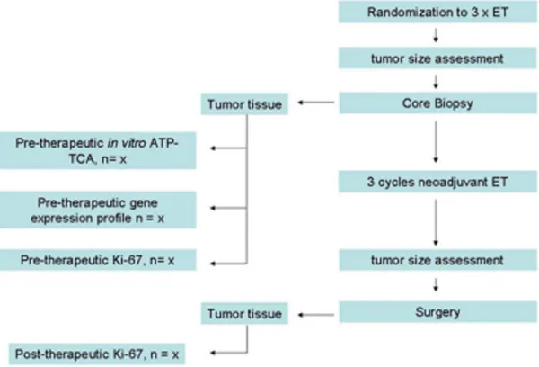
Tumor diameter were measured by ultrasound before systemic therapy and immediately prior to curative surgery, and clinical responses were evaluated according to RECIST criteria [19]. A reduction in the longest tumor diameter of.30% was required for classifying a tumor as responsive. Pre-treatment tumor samples were obtained by three to six 14-gauge core needle biopsies, and were equally split in three portions: one portion was used for histological confirmation of invasive breast cancer and for evaluation of Ki67 protein expression by IHC. The second portion was subjected to ATP-TCA, while the third portion was immediately snap-frozen in dry ice for gene expression profiling. Post-treatment tumor samples for Ki67 determination were obtained during curative surgery, which was performed within 2 to 4 weeks after the last scheduled chemotherapy cycle. Tumors
Figure 1. Study flow chart.
which had a decrease in Ki67 expression of.30% in response to Epi/Doc were considered sensitive. The procedural flow is shown in figure 1.
ATP-TCA Chemosensitivity Testing
Primary human breast cancer cells were isolated from pre-treatment tumor samples, and ATP-TCA was performed as previously described [20]. In brief, 10 000–20 000 tumor cells per well were seeded in tumor cell-supporting growth medium in 96-well microtiter plates. Epirubicin and Docetaxel were used in combination at test drug concentrations (TDCs) of 200, 100, 50, 25, 12.5 and 6.25% (with 100% TDC corresponding to peak plasma concentrations). All tests were performed in triplicates. Tumor cells were lysed after 7 days of incubation and the amount of cellular ATP was determined with a luciferin/luciferase reaction. Cell preparation and ATP-TCA were performed using commercially available reagents (TCA-100, DCS, Hamburg, Germany). Luminescence was measured with either a LB953 luminometer (Berthold Technologies, Bad Wildbad, Germany) or a MPL2 Microplate Luminometer (Berthold Detection Systems, Pforzheim, Germany). Chemosensitivity across the tested concen-tration range was evaluated by using the Sensitivity Index (‘‘SI’’) [21]. The SI is obtained by measuring the area under the inhibition curve for multiple dilutions of the effective chemother-apy dose. A low SI indicates good chemosensitivity to a particular antineoplastic compoundin vitro, while a high SI value describes poor chemosensitivity.
RNA Extraction, Amplification and GeneChip Hybridization
RNA extraction, amplification and GeneChip hybridization was performed according to the manufacturers protocol (Affyme-trix Inc., Santa Clara, CA). In brief, double-stranded cDNA was synthesized from one patient by a chimeric oligonucleotide with an oligo-dT and a T7 RNA polymerase promoter at a concentration of 100 pmol/ml. Reverse transcription was performed as recom-mended by Affymetrix by using commercially available reagents (Invitrogen). The reaction products were cleaned by phenol-chloroform extraction before biotin-labelling and an approximate-ly 250-fold amplification were performed. 10mg of each of the 6 labelled cRNA samples was then hybridized onto a separate Affymetrix U133 GeneChipH Set. The set consists of two GeneChipH arrays and contains almost 45,000 probe sets representing more than 39,000 transcripts derived from approx-imately 33,000 well-substantiated human genes. The set design uses sequences selected from GenBankH, dbEST, and RefSeq. Prehybridization, hybridization, washing and staining with streptavidin-phycoerythrin were carried out according to the manufacturers protocol. Antibody amplification was accomplished using a biotin-linked antistreptavidin antibody (Vector Laborato-ries, Buringame, CA). The goat immunoglobulin G-blocking (Sigma, St Louis, MO) antibody was used for blocking unspecific binding. The arrays were then stained and washed as recommen-det by Affymetrix, before being scanned on an Affymetrix GeneChip Scanner (Agilent, Palo Alto, CA). The gene expression
Table 1.Patient characteristics.
Characteristics n = 26 (100%)
Median age in years (range) 47 (33–77)
Menopausal stage
Premenopausal 16 (62%) Postmenopausal 10 (38%)
Tumor stage, n (%)
T1 10 (38%)
T2 9 (35%)
T3 5 (19%)
T4 2 (8%)
Nodal stage, n (%)
Negative 13 (50%)
Positive 12 (46%) Not evaluated 1 (4%)
Estrogen receptor status
Positive 16 (62%) Negative 10 (38%)
Progesteron receptor status
Positive 13 (50%) Negative 13 (50%)
HER-2 overexpression
Positive 6 (23%) Negative 20 (77%)
Grading
G1-2 10 (38%)
G3 16 (62%)
doi:10.1371/journal.pone.0066573.t001
Figure 2. Sensitivity index (SI) describing in vitro chemosensi-tivity to Epi/Doc over a concentration range of a panel of 65 breast cancer samples.Tumor samples are arranged from left to right according to increasing SI.
doi:10.1371/journal.pone.0066573.g002
Table 2.262 table showing tumor response to 3 cycles of neoadjuvant Epi/Doc as defined in Materials and Methods and chemosensitivity to the dichotomized Ki67 change (,30% vs .30%).
Ki67 changein vivo ATP-TCA
no yes sum
No 6 2 8
Yes 1 7 8
Sum 7 9 16
data presented in our manuscript have been deposited in a publicly available data base (Gene Expression Omnibus) and can be accessed under the following weblink: http://www.ncbi.nlm. nih.gov/geo/query/acc.cgi?acc = GSE29561.
Statistical Analysis
Microarray data were preprocessed using quantile normaliza-tion and RMA [22]. In order to test the null hyothesis of no differential expression between tumors that showed considerable size reduction after treatment and those that did not, for each gene Welch‘s two sample t-test was computed. Marginal p-values were adjusted using methods previously described in order to provide control over the local false discovery rate (locFDR). A cut-off of 0.15 was considered to achieve similar control as conventional.05
a-levels when using the false discovery rate. For the sake of
exploration, genes with a locFDR of up to.25 were considered. All computations were implemented using R (http://cran.r-project. org) and Bioconductor (http://www.bioconductor.org) [23,24].
Results
Patient Characteristics
Of the 30 patients who were randomized, 26 received three cycles of Epi/Doc according to protocol. Their
clinico-patholog-ical characteristics are shown in table 1. Three patients (12%) experienced a pCR (as defined by the absence of invasive tumor components), while invasive tumor components were detected in the surgical specimen in the remaining 23 cases. In all of these cases, both pre- and post-therapeutic tumor measurements and Ki67 values were available. The ATP-TCA could be peformed in 18 cases. However, due to the fact that some patients experienced a pCR post-therapeutic Ki67 was not available in all cases. Results for both, differential pre- vs post-treatment Ki67 values, and ATP-TCA, were evaluated in 16 cases. Pre-treatment gene expression profiles were available in 22 of the 26 patients who had received Epi/Doc according to protocol.
In vitro Chemosensitivity of Epi/Doc in the ATP-TCA
18 freshly isolated pre-therapeutic tumor samples from patients subsequently undergoing neoadjuvant Epi/Doc were subjected to Epi/Doc in the ATP-TCA as described in Materials and Methods. Chemosensitivity along a dilution series of the cytotoxic drug combination was evaluated for each sample by using the sensitivity index (SI), as previously described by Kurbacheret al.ATP-TCA data from 47 additional tumor cell isolates from breast cancer patients not enroled into the trial were also available and were included for figure 2. The sensitivity curve depicts individual SI values ordered by increasing in vitro sensitivity to Epi/Doc and demonstrates a highly heterogenous sensitivity to Epi/Doc.
Effect of 3 Cycles of Neoadjuvant Epi/Doc on Tumor Size and Intratumoral Ki67 Expression
Three cycles of Epi/Doc lead to a reduction in tumor size of
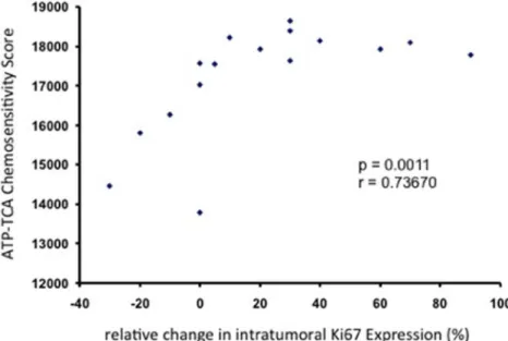
.30% in the longest tumor diameter in 11 out of 23 patients (48%). In the 23 tumors in which intratumoral Ki67 protein expression was measured before and after neoadjuvant treatment with 3 cycles of Epi/Doc, we found a reduction in Ki67 of.30% in 10 cases (43%). Interestingly, although both measurements yielded comparable rates in terms of antitumoral effects (response vs non-response) in vivo, we did not find a significant association when the relative reductions in tumor size (mm) were correlated Figure 3. Correlation between the relative changes (%) in intratumoral Ki67 expression in response to 3 cycles of neoadjuvant Epi/ Doc, and in vitro chemosensitivity to Epi/Doc as measured by ATP-TCA.
doi:10.1371/journal.pone.0066573.g003
Table 3.262 table showing tumor response to 3 cycles of neoadjuvant Epi/Doc as defined in Materials and Methods and chemosensitivity to Epi/Docin vitro(ATP-TCA).
Change in tumor size ATP-TCA
no yes sum
no 3 5 8
yes 5 5 10
Sum 8 10 18
with the relative reduction in Ki67 expression (%) (r = 0.1797, p = 0.3176; Kendall tau, data not shown).
Effect of Epi/Doc on Tumor Cell Proliferation in vitro (ATP-TCA)
We then investigated the response to Epi/Doc in short term primary cultures which had been obtained from malignant breast tumors of individual patients who had subsequently undergone neoadjuvant Epi/Doc treatment. ATP-TCA results were available in 16 cases, and 9 (56%) tumor tisues were found to be responsive according to the pre-defined cut-off values described in Materials and Methods. Tumor cells which responded to Epi/Doc in the ATP-TCA in vitroalso responded to 3 cycles of Epi/Doc in vivo
(p = 0.041, Fishers Exact Test, table 2). In addition, the antiproliferative effect of Epi/Doc in the ATP-TCA assay was significantly correlated to the decrease in Ki67 protein expression in the same patients (p = 0.001, r = 0.737, Kendalls Tau, figure 3). When we investigated whether tumor sensitivity to Epi/Doc determined by ATP-TCA was able to predict a clinically meaninful remission in response to neoadjuvant Epi/Doc if tumor response was measured by breast ultrasound, we did not observe a comparable pattern (p = 0.999, Fishers Exact Test, table 3).
Gene Expression Profile and Response to Epi/Doc in vitro and in vivo
We then investigated whether we could identify gene expression profiles in the initial core biopsy that would permit to a) predict a clinically meaningful response to Epi/Doc in terms of tumor size reduction of .30%, b) predict a profound reduction in
intratumoral Ki67 protein expression, and c) predict an in vitro
response to Epi/Doc in the ATP-TCA. When the analyses were carried out as described in Materials and Methods, we were unable to identify a gene expression signature that was associated with a reduction in Ki67in vivo, or chemosensitivityin vitroin our training set. We did, however, identify a set of 5 genes that were significantly up-regulated (locFDR,15%) in chemosensitive tumors. Table 4 provides annotated results for genes which show expression differences with a locFDR level of 25% and less. A Heatmap of the corresponding expression profiles is shown in figure 4.
Discussion
Pathological complete response (pCR) following neoadjuvant chemotherapy is seen in 3–26% of patients, and is a good predictor of survival [25]. Although the majority of neoadjuvant regimen do not achieve pCR, tumors can still respond in a clinically meaningful way. Response to neoadjuvant chemotherapy has traditionally been evaluated by ultrasound, mammography, or caliper measurements. The value of these techniques in predicting response, however, is now being increasingly challenged because they do not provide adequate information about the biological activity of a particular tumor [4]. It is not uncommon to experienced surgeons to discover the absence of viable invasive tumor cells in the specimen of a breast cancer that had not appreciably changed its size under systemic therapy. Other diagnostic techniques such as FDG-PET, which measures the uptake of labeled glucose into a tumor, are thus better suited to evaluate biological activity at a given point in time and are
Table 4.Genes expression of genes with a local false discovery rate (FDR),0.25 for the discrimination between tumors with a clinically meaningful reduction in tumor size under neoadjuvant Epi/Doc from those which do not benefit from therapy.
Gene.Name Gene.Sbol locFDR logFC
zinc finger protein 238 ZNF238 0.18 21.74
homeobox C10 HOXC10 0.18 21.26
B-cell CLL/lymphoma 2 BCL2 0.24 20.99
tumor protein D52 TPD52 0.25 20.96
lectin, galactoside-binding, soluble, 8 LGALS8 0.19 20.93
homeobox C6 HOXC6 0.21 20.92
tetratricopeptide repeat domain 3 TTC3 0.15 20.88
transmembrane protein 106B TMEM106B 0.22 20.86
chromosome 20 open reading frame 103 C20orf103 0.12 20.76
lysophospholipase I LYPLA1 0.20 20.72
solute carrier family 7 (cationic amino acid transporter, y+system), member 8 SLC7A8 0.14 20.69
F-box protein 28 FBXO28 0.24 20.68
chromosome 1 open reading frame 27 C1orf27 0.12 20.66
solute carrier family 7 (cationic amino acid transporter, y+system), member 8 SLC7A8 0.19 20.62
KIAA0040 KIAA0040 0.24 20.47
RAS-like, family 11, member B RASL11B 0.19 20.46
vacuolar protein sorting 37 homolog C (S. cerevisiae) VPS37C 0.15 20.38
tripartite motif-containing 45 TRIM45 0.23 20.33
LYR motif containing 4 LYRM4 0.24 20.33
TBC1 domain family, member 19 TBC1D19 0.18 20.30
acetyl-Coenzyme A carboxylase alpha ACACA 0.17 20.28
phenylalanyl-tRNA synthetase 2, mitochondrial FARS2 0.21 20.26
currently evaluated [26,27]. An alternative to evaluate the presence of viable tumor cells is by measuring the readily available immunohistochemical analysis of the nuclear antigen Ki67 [28]. There is now increasing interest in the nuclear proliferation antigen Ki67, since allows to distinguish the relatively chemo-resistant luminal A from the more sensitive luminal B breast cancer subtype [29]. It is now well established that higher levels of post-treatment Ki67 values are associated with poorer survival in breast cancer, while higher pre-therapeutic intratumoral Ki67 levels are associated with improved response [30,31].
The value of Ki67 in assessing neoadjuvant response has recently been demonstrated by Dowsettet al., who reported that a 2 week pre-operative treatment with the aromatase inhibitor anastrozole was more effective in reducing intratumoral Ki67 levels than tamoxifen, or a combination of anastrozole and tamoxifen, in postmenopausal receptor-positive breast cancer patients (‘‘IMPACT’’ trial). Their findings exactly matched the results of the much larger adjuvant ATAC trial, in which disease-free survival in response to the same three regimen was chosen as primary endpoint. Interestingly, the group also used ultrasound to measure tumor size alterations in response to the three treatment strategies, and – similar to our results – also failed to detect differential changes across the three arms, thus suggesting that ultrasound might simply not be sensitive enough to measure biological response [32]. This hypothesis is, however, somewhat challenged by the fact that, similar to Chang et al., we also identified a particular gene expression signature that is
character-istic of chemosensitive tumors when measured by ultrasound, while no such pattern was seen when the tumor response was evaluated by Ki67 expression [15].
Tumor shrinkage is a robust, albeit relatively insensitive parameter which was readily available also in tumors with pCR. In these tumors, however the relatively sensitive Ki67 values were no longer available and a differential Ki67 expression could thus not be determined. This circumstance reduced the number of evaluable tumors and might thus have impacted on the statistical significance. It is well possible that with a larger sample size, a particular expression profile might have emerged. Alternatively, individual pre- and posttreatment Ki67 measurements can be difficult to assess, and the measurement of a differential Ki67 expression might result in even greater imprecision, which could also have contributed to our inability to identify a Ki67 response signature.
We were also unable to identify a ATP-TCA chemosensitivity-associated expression profile by using the human genome Affymetrix U133 GeneChipH set which comprises more than 33.000 well characterized genes. This is in sharp contrast to two recent publications in non small-cell lung cancer (NSCLC) and in platinum-pretreated ovarian cancer, in which the RT-PCR based expression signature derived from a panel of only 23 resistance-associated genes was resistance-associated with ATP-TCA-measured che-mosensitivity [33,34]. In our case, the genetic heterogeneity of malignant breast tumors, the fact that the tissue preparation that precedes the ATP-TCA alters the cellular composition of the Figure 4. Heatmap of Genes with local FDR less than 0.25.Red corresponds to high, blue to low signals. Genes are annotated using gene symbols. Color code at the top indicates treatment success. Corresponding fold changes are depicted in the right slide panel.
initial tumor, and the relatively small sample size may have all contributed to the absence of a clear chemosensitivity signature.
Our work has several limitations: The difficulty in obtaining sufficient pre-therapeutic tissue by core biopsy has allowed us to perform both ATP-TCA and gene expression profiling in only 16 out of 30 patients, thus resulting in a comparatively small number of samples which makes this a pilot trial. Also, while tumor response was evaluated according to RECIST criteria, the 30% cut-off level of for response to Ki67 was chosen arbitrarily to allow for equal group sizes. In addition, only one chemotherapy regimen (Epi/Doc) was evaluated in vivo. While we have also generated
in vitro data for Epirubicin, Docetaxel, and for several other chemotoxic drugs individually (data not shown), we do not know whether their efficacy in the assay also translates into a comparable reduction in Ki67 in vivo. Furthermore, while it is tempting to speculate that the most effective drug in the ATP-TCA is also the most potent drug in vivo, this requires further prospective evaluation.
Finally, in contrast to analytical tools such as gene expression profiling or proteomic analysis, the ATP-TCA offers little mechanistic insight into why a particular drug is effective in one
tumor but fails to do so in another for it reflects the net sum of cellular responses to chemotherapy. Nevertheless, the results of our pilot trial suggest, for the first time, the utility of a standardized
in vitrochemosensitivity assay in predicting the individual biolog-ical response to chemotherapy in breast cancer patients in a prospective setting.
Acknowledgments
The authors wish to acknowledge the technical expertise of Ms Daniela Muhr. This work was supported by the Privatstiftung fu¨r Brustgesundheit, by the Jubila¨umsfonds des Bu¨rgermeisters der Stadt Wien, and by a research grant of the Austrian Society of Senology.
Author Contributions
Conceived and designed the experiments: CFS FK RS C. Sartori MB. Performed the experiments: CFS FK RS C. Staudigl AF DG SH GP AD C. Sartori MB. Analyzed the data: FK CFS MB C. Sartori CD. Contributed reagents/materials/analysis tools: RS C. Sartori AF DG SH GP AD. Wrote the paper: CFS FK RS AF SH AD C. Staudigl MB. Obtained permission for use of cell line: C. Staudigl DG. Statistical Analysis: FK AF CFS.
References
1. Gralow JR, Burstein HJ, Wood W, Hortobagyi GN, Gianni L, et al. (2008) Preoperative therapy in invasive breast cancer: pathologic assessment and systemic therapy issues in operable disease. J Clin Oncol 26(5): 814–9. 2. Liedtke C, Mazouni C, Hess KR, Andre´ F, Tordai A, et al. (2008) Response to
neoadjuvant therapy and long-term survival in patients with triple-negative breast cancer. J Clin Oncol 26(8): 1275–81.
3. Bear HD (2010) Neoadjuvant chemotherapy for operable breast cancer: individualizing locoregional and systemic therapy. Surg Oncol Clin N Am. 19(3): 607–26.
4. Keune JD, Jeffe DB, Schootman M, Hoffman A, Gillanders WE, et al. (2010) Accuracy of ultrasonography and mammography in predicting pathologic response after neoadjuvant chemotherapy for breast cancer. Am J Surg 199(4): 477–84.
5. Dowsett M, Dunbier AK (2008) Emerging biomarkers and new understanding of traditional markers in personalized therapy for breast cancer. Clin Cancer Res 14(24): 8019–26.
6. Miller WR (2010) Clinical, pathological, proliferative and molecular responses associated with neoadjuvant aromatase inhibitor treatment in breast cancer. J Steroid Biochem Mol Biol 118(4–5): 273–6.
7. Dowsett M, A’Hern R, Salter J, Zabaglo L, Smith IE (2009) Who would have thought a single Ki67 measurement would predict long-term outcome? Breast Cancer Res. 11 Suppl 3: S15.
8. Bottini A, Berruti A, Bersiga A, Brizzi MP, Bruzzi P, et al. (2001) Relationship between tumour shrinkage and reduction in Ki67 expression after primary chemotherapy in human breast cancer. Br J Cancer 19;85(8): 1106–12. 9. Assersohn L, Salter J, Powles TJ, A’hern R, Makris A, et al. (2003) Studies of the
potential utility of Ki67 as a predictive molecular marker of clinical response in primary breast cancer. Breast Cancer Res Treat 82(2): 113–23.
10. Takada M, Kataoka A, Toi M, Bando H, Toyama K,et al. (2004) A close association between alteration in growth kinetics by neoadjuvant chemotherapy and survival outcome in primary breast cancer. Int J Oncol 25(2): 397–405. 11. Billgren AM, Rutqvist LE, Tani E, Wilking N, Fornander T, et al. (1999)
Proliferating fraction during neoadjuvant chemotherapy of primary breast cancer in relation to objective local response and relapse-free survival. Acta Oncol 38(5): 597–601.
12. Jones RL, Salter J, A’Hern R, Nerurkar A, Parton M, et al. (2009) The prognostic significance of Ki67 before and after neoadjuvant chemotherapy in breast cancer. Breast Cancer Res Treat 116(1): 53–68.
13. Lee J, Im YH, Lee SH, Cho EY, Choi YL, et al. (2008) Evaluation of ER and Ki-67 proliferation index as prognostic factors for survival following neoadjuvant chemotherapy with doxorubicin/docetaxel for locally advanced breast cancer. Cancer Chemother Pharmacol 61(4): 569–77.
14. Chang JC, Wooten EC, Tsimelzon A, Hilsenbeck SG, Gutierrez MC (2005) Patterns of resistance and incomplete response to docetaxel by gene expression profiling in breast cancer patients.,et al. J Clin Oncol 20;23(6): 1169–77. 15. Cleator S, Tsimelzon A, Ashworth A, Dowsett M, Dexter T, et al. (2006) Gene
expression patterns for doxorubicin (Adriamycin) and cyclophosphamide (cytoxan) (AC) response and resistance. Breast Cancer Res Treat 95(3): 229–33. 16. Kurbacher CM, Cree IA (2005) Chemosensitivity testing using microplate adenosine triphosphate-based luminescence measurements. Methods Mol Med. 110: 101–20.
17. Solomayer E, Meisner C, Zwirner M, Wallwiener D, Fehm T et al. (2008) Predicting resistance to platinum-containing chemotherapy with the ATP tumor
chemosensitivity assay in primary ovarian cancer. Anticancer Res 28(2A): 949– 55.
18. Sharma S, Neale MH, Di Nicolantonio F, Knight LA, Whitehouse PA, et al. (2003) Outcome of ATP-based tumor chemosensitivity assay directed chemo-therapy in heavily pre-treated recurrent ovarian carcinoma. BMC Cancer 3;3: 19.
19. Jaffe N (2006) Measures of Response: RECIST, WHO, and New Alternatives Journal of Clinical Oncology, 24 (20): 3245–3251.
20. Ugurel S, Schadendorf D, Pfo¨hler C, Neuber K, Thoelke A, et al. (2006) In vitro drug sensitivity predicts response and survival after individualized sensitivity-directed chemotherapy in metastatic melanoma: a multicenter phase II trial of the Dermatologic Cooperative Oncology Group. Clin Cancer Res 15;12(18): 5454–63.
21. Kurbacher CM, Bruckner HW, Cree IA, Kurbacher JA, Wilhelm L, et al. (1997) Mitoxantrone Combined with Paclitaxel as Salvage Therapy for Platinum-refractory Ovarian Cancer: Laboratory Study and Clinical Pilot Trial, Clinical Cancer Research 3: 1527–1533.
22. Irizarry RA, Hobbs B, Collin F, Beazer-Barclay YD, Antonellis KJ, et al. (2003) Exploration, normalization, and summaries of high density oligonucleotide array probe level data. Biostatistics 4: 249–64.
23. Efron B (2007) Size, power, and false discovery rates. Ann Statist 35: 1351–1377. 24. Benjamini Y, Hochberg Y (1995) Controlling the false discovery rate: a practical and powerful approach to multiple testing. Journal of the Royal Statistical Society B 57 (1): 289–300.
25. Montagna E, Bagnardi V, Rotmensz N, Viale G, Pruneri G, et al. (2010) Pathological complete response after preoperative systemic therapy and outcome: relevance of clinical and biologic baseline features. Breast Cancer Res Treat 124(3): 689–9.
26. Ueda S, Tsuda H, Saeki T, Osaki A, Shigekawa T, et al. (2010) Early reduction in standardized uptake value after one cycle of neoadjuvant chemotherapy measured by sequential FDG PET/CT is an independent predictor of pathological response of primary breast cancer. Breast J 16(6): 660–2. 27. Groheux D, Giacchetti S, Moretti JL, Porcher R, Espie´ M, et al. (2011)
Correlation of high (18)F-FDG uptake to clinical, pathological and biological prognostic factors in breast cancer. Eur J Nucl Med Mol Imaging 38(3): 426–35. 28. Ueda S, Tsuda H, Saeki T, Omata J, Osaki A, et al. (2010) Early metabolic response to neoadjuvant letrozole, measured by FDG PET/CT, is correlated with a decrease in the Ki67 labeling index in patients with hormone receptor-positive primary breast cancer: a pilot study. Breast Cancer 18(4): 299–308. 29. Cheang MC, Chia SK, Voduc D, Gao D, Leung S, et al. (2009) Ki67 index,
HER2 status, and prognosis of patients with luminal B breast cancer. J Natl Cancer Inst 101(10): 736–50.
30. Azambuja E, Cardoso F, de Castro G Jr, Colozza M, Mano MS, et al. (2997) Ki-67 as prognostic marker in early breast cancer: a meta-analysis of published studies involving 12,155 patients. Br J Cancer 96(10): 1504–13.
31. Urruticoechea A, Smith IE, Dowsett M (2005) Proliferation marker Ki-67 in early breast cancer. J Clin Oncol 23(28): 7212–20.
33. Glaysher S, Yiannakis D, Gabriel FG, Johnson P, Polak ME (2009) Resistance gene expression determines the in vitro chemosensitivity of non-small cell lung cancer (NSCLC). BMC Cancer 27;9: 300.