Arabidopsis thaliana
Young-Hee Cho, Sang-Dong Yoo*Department of Biological Science, Sungkyunkwan University, Suwon, Korea
Abstract
Sugars are evolutionarily conserved signaling molecules that regulate the growth and development of both unicellular and multicellular organisms. As sugar-producing photosynthetic organisms, plants utilize glucose as one of their major signaling molecules. However, the details of other sugar signaling molecules and their regulatory factors have remained elusive, due to the complexity of the metabolite and hormone interactions that control physiological and developmental programs in plants. We combined information from a gain-of-function cell-based screen and a loss-of-function reverse-genetic analysis to demonstrate that fructose acts as a signaling molecule inArabidopsis thaliana. Fructose signaling induced seedling developmental arrest and interacted with plant stress hormone signaling in a manner similar to that of glucose. For fructose signaling responses, the plant glucose sensor HEXOKINASE1 (HXK1) was dispensable, while FRUCTOSE INSENSITIVE1 (FINS1), a putative FRUCTOSE-1,6-BISPHOSPHATASE, played a crucial role. Interestingly, FINS1 function in fructose signaling appeared to be independent of its catalytic activity in sugar metabolism. Genetic analysis further indicated that FINS1– dependent fructose signaling may act downstream of the abscisic acid pathway, in spite of the fact that HXK1–dependent glucose signaling works upstream of hormone synthesis. Our findings revealed that multiple layers of controls by fructose, glucose, and abscisic acid finely tune the plant autotrophic transition and modulate early seedling establishment after seed germination.
Citation:Cho Y-H, Yoo S-D (2011) Signaling Role of Fructose Mediated by FINS1/FBP in Arabidopsis thaliana. PLoS Genet 7(1): e1001263. doi:10.1371/ journal.pgen.1001263
Editor:Gregory P. Copenhaver, The University of North Carolina at Chapel Hill, United States of America
ReceivedApril 30, 2010;AcceptedNovember 30, 2010;PublishedJanuary 6, 2011
Copyright:ß2011 Cho, Yoo. This is an open-access article distributed under the terms of the Creative Commons Attribution License, which permits unrestricted use, distribution, and reproduction in any medium, provided the original author and source are credited.
Funding:This work was supported by Korean National Research Foundation to S-DY (2010-0015456, 2010-0016509) and Y-HC (2010-0007068). The funder had no rule in study design, data collection and analysis, decision to publish, or preparation of the manuscript.
Competing Interests:The authors have declared that no competing interests exist.
* E-mail: sangdong@skku.edu
Introduction
Myriad metabolic pathways enable cells to sustain life with basic carbon and nitrogenous compounds. Thus, the integration of metabolite status, which reflects external and internal living conditions, into cellular activities (e.g., gene expression) is a pivotal process that equips organisms with the ability to survive and proliferate. For example, cellular metabolites often serve regulatory roles in modulating organism growth and development, from unicellular bacteria and yeasts to multicellular animals and plants [1–6]. To sense and transduce such metabolite signals, organisms have developed sophisticated biochemical and cellular mechanisms. Glucose is an evolutionarily conserved regulatory sugar molecule in many different organisms [1–6]. It has multiple roles as an energy source, building block, and osmotic regulator, and also acts as a potent signaling molecule that regulates gene expression and controls organism growth and development. For example, in yeast, glucose is sensed by at least four different types of sensors, Hxk2, Snf3, Rgt2 and Gpr1, and regulates gene expression and cell growth [4]. In mammalian pancreatic isletb
cells, glucose signaling may be a function of the total amount of ATP generated via catabolism [6].
In plants, glucose [7–9], sucrose [10–12], trehalose-6-phosphate [13], and low energy/high AMP concentrations [14,15] function as cellular signaling molecules in specific regulatory pathways that modulate plant growth and development. Of these signaling metabolites, glucose has been studied the most comprehensively in
plants. Glucose signaling modulates the gene expression of enzymes in the glyoxylate cycle [16] and the photosynthesis pathway [17], and is also involved in the developmental decision of whether to progress to normal seedling establishment after seed germination [18].
Glucose-mediated developmental repression is largely depen-dent on HEXOKINASE1 (HXK1) [7–9]. HXK1’s function in glucose-mediated developmental repression is mostly independent of its catalytic activity and integrates glucose signaling with other plant hormone such as auxin and cytokinin. HXK1-independent glucose signaling has also been reported in plants. For instance, expression of the genes encoding chalcone synthase, phenylalanine ammonia-lyase, and asparagine synthase responds to glucose signaling, but their regulation is independent of HXK1 activity [3,19]. A recent study further demonstrated that a refined low-glucose condition can uncouple HXK1-dependent and -indepen-dent glucose signaling responses during earlyA. thalianaseedling establishment [9,20].
In both animals and plants, the developmental roles and regulatory functions of hexoses other than glucose have remained largely unknown. However, within the last few years, dietary fructose was implicated in mammalian cell signaling perturbation and metabolic syndromes such as insulin resistance, obesity, type 2 diabetes, and high blood pressure [21,22].
carried out by fructose-1,6-bisphosphatase (FBP), UDP-glucose pyrophosphorylase, sucrose phosphate synthase, and sucrose phosphatase [2]. Sucrose is then stored in vacuoles or cleaved into glucose and fructose by invertases or UDP-glucose and fructose by sucrose synthases [23]. Thus, following sucrose hydrolysis, fructose becomes one of the prevalent hexoses in plants and has long been proposed as a possible signaling molecule [24]. Nevertheless, fructose signaling in plants has remained largely unexplored. Recently, Kato-Naguchi et al. [25] showed that the fructose analog psicose induced root growth inhibition in lettuce. Fructokinase (FRK), which performs the same catalytic function as HXK, but with fructose as the substrate rather than glucose, was the first fructose enzyme to be studied for a putative regulatory role in fructose signaling [24,26,27]. Although FRK is involved in modulation of plant growth, a regulatory role in fructose signaling was ruled out [28]; hence, little is known about fructose signaling and its regulatory pathways.
In this study, we used a cell-based functional screen and a reverse genetics assay to investigate the signaling role of fructose in A. thaliana. We identified FRUCTOSE INSENSITIVE1 as an indispensable regulatory factor in the signaling pathway. Here, we report the molecular and genetic characterization offins1in a fructose signaling context, and its close interactions with ABA signaling during early seedling development.
Results/Discussion
Fructose signaling modulates early seedling development and interacts with ABA and ethylene
To evaluate the regulatory role of fructose signaling in plant
developmental modulation, we examined A. thaliana seedling
growth on 6% (w/v) fructose agar medium with full-strength
Murashige and Skoog (MS) salts. Wild-type (WT; Ler and Col
accession) seedlings grown on high-fructose medium exhibited a typical early developmental arrest, which was manifested by inhibition of hypocotyl and root growth and repression of
cotyledon expansion and chlorophyll accumulation (Figure 1A and 1B). Although the seedling development repression pattern caused by high fructose was similar to that caused by high glucose (6%) [7,20], fructose caused slightly more root growth inhibition than glucose (Figure S1). Mannitol, an osmotic control, did not induce the same seedling repression, suggesting that the observed phenotype was a developmental response to fructose signaling.
Recently, we refined glucose assay conditions for growing A. thalianaseedlings and showed that the high glucose requirement is due to the high nitrogen content in MS media [9,20]. When MS salts were omitted, 2% glucose induced equivalent seedling growth repression to 6% glucose media including MS. However, decreasing the concentration of fructose in the absence of MS salts had little effect on seedling growth (Figure S2); this suggested that nitrogen had a different effect on fructose and glucose signaling.
Indeed, further experiments indicated that fructose and glucose signaling does rely on distinct sensors. The glucose-insensitive
HXK1-null mutant gin2-1 (gin2) exhibited normal fructose
sensitivity, as did transgenic gin2-expressing WT HXK1 and its
catalytically inactive mutants HXK1S177A or HXK1G104D
(Figure 1A). These data confirmed that the glucose sensor HXK1 was dispensable in fructose signaling. Although HXK1 carries out metabolic activities for both glucose and fructose, it does not appear to be involved in fructose signaling. This may reflect the fact that HXK1 has an approximately 100-fold higher Author Summary
Among the many plant sugar metabolites, glucose signaling has received the most attention. Although fructose is also an abundant hexose, its signaling role in plant growth and development has not been addressed clearly and systematically to date. We found that fructose functions as a regulatory sugar metabolite and interacts with signaling by the plant hormones abscisic acid (ABA) and ethylene in A. thaliana. The fructose-dependent growth response is mediated by FRUCTOSE INSENSITIVE1 (FINS1), which encodes an ancient metabolic enzyme, putative fructose-1,6-bisphosphatase. Interestingly, the catalytic function of FINS1 in sucrose biosynthesis is dispensable for its regulatory role in fructose signaling. FINS1 appears to act downstream of GLUCOSE-INSENSI-TIVE1, which is involved in ABA synthesis. Overall, it is evident that although fructose and glucose have unique regulatory pathways, they also share some signaling interactions with plant stress and defense hormones and coordinate early seedling establishment of A. thaliana. Fructose affects cell signaling in mammals and causes various metabolic syndromes. However, a direct relation-ship between fructose and physiological diseases has not been established yet. Because FINS1 is evolutionarily conserved, our genetic evaluation of its signaling function may provide useful information about fructose signaling in animals as well as plants.
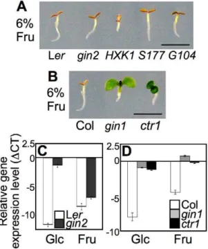
Figure 1. Differential seedling response to fructose signaling. (A) WT (Ler),gin2, andHXK1WT,S177A, andG104D-complementedgin2
showed seedling developmental arrest phenotypes on MS agar media containing 6% fructose. The seedlings were grown for 5 d under constant light. Scale bar, 5mm. (B) Unlike WT (Col), gin1 and ctr1
affinity for glucose compared to fructose [28–30]. In a previous study, root growth inhibition in lettuce was reported in the presence of either the fructose analog psicose or the glucose analog mannose [25]. However, the HXK inhibitor mannoheptulose restored root growth in the presence of mannose, but not psicose. These results are further evidence that psicose/fructose signaling is independent of HXK function.
Plant sugar signaling, mainly glucose and sucrose, interacts with stress and defense hormone signaling pathways and coordinates seedling growth and development [1–3,23,31]. For glucose signaling,gin1,gin5, andgin6were respectively identified as alleles of aba-deficient2(aba2),aba3, andaba-insensitive4(abi4) in the ABA pathway, andgin4was found to be a new allele ofconstitutive triple response1 (ctr1) in the ethylene pathway [31–36]. These mutants have been selected repeatedly from various independent screens for sugar responses, further confirming that sugar signaling interacts with ABA and ethylene response pathways during early seedling development [37–40].
To test whether fructose signaling interacts with plant stress/ defense hormones, we observed the early developmental response of ABA and ethylene mutants on a 6% fructose agar medium with MS salts. Unlike WT andgin2, bothgin1-3(gin1) andctr1-1(ctr1) seedlings were not only insensitive to high glucose, but also overcame fructose repression and developed green cotyledons (Figure 1B). GIN1/ABA2 encodes a short-chain dehydrogenase/ reductase in ABA synthesis, and CTR1/GIN4 encodes a putative mitogen-activated protein kinase kinase kinase that functions as a negative regulator of ethylene signaling [31,33]. Therefore, fructose signaling appears to interact positively with ABA signaling via hormone biosynthesis, whereas it is likely antagonized by ethylene signaling. Interestingly, in ABA-deficient gin1 mutants, cotyledon repression was de-repressed by fructose, but root repression was not (Figure 1B); however, glucose relieved both cotyledon and root growth repression ingin1mutants (Figure S1) [20]. This indicated that fructose repression of root growth was independent of ABA biosynthesis, unlike cotyledon greening. This observation revealed differential seedling responses to fructose and glucose in an organ-specific manner.
We further monitored marker gene expression using real-time PCR with cDNA templates generated from mRNA of five-d-old seedlings grown on MS agar medium containing 6% glucose, fructose, or mannitol. Expression of the photosynthesis-related CHLOROPHYLL A/B BINDING PROTEIN2(CAB2/AT1G29920) gene was markedly repressed in WT by both glucose and fructose (Figure 1C and 1D). Gene expression was similarly repressed in
gin2 seedlings by fructose, but not by glucose (Figure 1C).
However, CAB2 expression was de-repressed in both gin1 and
ctr1seedlings (Figure 1D). TheCAB2gene expression patterns in the mutants reflected the fructose resistance revealed by their phenotypes (Figure 1B). Taken together, these data indicated that fructose signaling was mediated through a unique/unknown sensor, but shared a downstream pathway with glucose signaling, which interacted with the plant stress and defense hormones ABA and ethylene to modulate early seedling development inA. thaliana. Although the application of high sugar to A. thaliana growth media has been criticized because it is not a normal physiological condition, it is unclear how much sugar is actually taken up by roots, how fast it is metabolized or fluxed, and in which suborganelles the sugar is partitioned. These factors could affect developmental responses to high sugar levels. Glucose and sucrose nanosensors, which detect cytoplasmic levels of sugar content, have demonstrated that plant roots take up sugars supplied in growth media rather efficiently [41]. To comprehensively understand sugar uptake and allocation in plants, apoplasmic
sugar levels, sugar distribution in subcellular organelles, and fluxes for specific sugars need to be monitored more closely. Further development of sugar nanosensors will hopefully lead to a better understanding of sugar sensing and signaling [42].
FINS1/FBP mediates fructose signaling
To learn more about the specific regulatory components involved in fructose signaling, we took advantage of a cell-based functional screen using transient expression of the A. thaliana mesophyll protoplast system [43]. Because fructose caused deficient chlorophyll accumulation inA. thaliana (Figure 1A and 1B), we reasoned that fructose signaling may affect photosynthetic gene expression in a manner similar to that of glucose signaling [7,17]. To monitor the fructose signaling response in leaf mesophyll protoplasts, we generated a reporter construct with an approximately 0.5 kbCAB2promoter fused to the firefly luciferase
gene (CAB2-fLUC). In leaf mesophyll protoplasts, CAB2-fLUC
activity was downregulated by fructose, but not by the osmo-tic control mannitol (Figure S3). We then screened several enzymes involved in fructose metabolism, including putative cytoplasmic FBP (AT1G43670), FRK1 (AT5G51830), and PFK1 (AT4G29220) for their potential roles in fructose signaling (Figure 2A). Of these enzymes, putative FBP (we tested two
independent constructs, FBP_3 and FBP_4) had the greatest
suppressive effect on CAB2-fLUC activity (Figure 2B). CAB2
promoter activity seemed to be suppressed even without high-fructose treatment, possibly because plant cells became
hypersen-sitized to endogenous fructose when putative FBP was
overex-pressed. To test if putative FBP enzyme activity is required for CAB2gene repression, we generated a catalytically inactive form, FBPS126AS127A(SSM), based on domain conservation in plant and animal FBPs (Figure S4). The dual mutation of S126A and S127A in FBP caused a loss of FBP enzymatic activity in protoplasts (Figure S5). This mutation probably distorted the local structure and prevented FBP121Dfrom associating with a divalent ion that is necessary for the enzyme activity [45]. Interestingly, the catalytically inactive form FBPS126AS127Asuppressed CAB2-fLUC activity in the same manner as the wild-type FBP (Figure 2B). This result indicates that the regulatory function of putative FBP in fructose signaling may be independent of its catalytic activity in sugar metabolism, similar to how HXK1 functions in glucose signaling [9,19].
Surprisingly, we observed putative FBP in both the cytoplasm and nucleus (Figure 2C). We were not able to determine whether the nuclear localization of putative FBP depends on cellular fructose signaling (Figure S6), since it is almost impossible to generate zero-fructose conditions in plant cells. However, the nuclear localization of putative FBP certainly suggests that it could be directly involved in fructose-dependent gene regulation. Based on the initial functional screen and localization test in plant cells, we hypothesized that putative FBP was a regulatory factor in fructose signaling.
Infins1protoplasts, FINS1expression (using the two
indepen-dent constructs FBP/FINS1_3 and FBP/FINS1_4) clearly
sup-pressed CAB2-fLUC activity (Figure 3C), which was similar to the effect of HXK1 [7]. To investigate FINS1 function in fructose-mediated gene regulation, we examined marker gene expression in WT andfins1seedlings grown on 6% fructose agar media with MS salts. Consistent with their growth phenotypes (Figure 3B),CAB2 expression was markedly repressed by fructose in WT, but not in fins1 seedlings (Figure 3D). A key transcription factor in ABA
signaling, ABI4 (also known as GIN6/AT2G40220) [44] was
induced by fructose in WT but not infins1seedlings. In contrast, ETHYLENE RESPONSE FACTOR1 (ERF1/AT3G23240), an ethylene response transcription factor [46], was repressed by fructose in WT, but de-repressed infins1seedlings. However, the change inERF1expression levels was relatively weak compared to other marker gene responses. These data showed that FINS1 had a central role in fructose-inducible gene regulation.
To verify that the fructose insensitivity exhibited byfins1 was due to the loss ofFINS1, we complemented thefins1mutant with FINS1cDNA using anAgrobacteriumsystem. Transgenic lines with FINS1 expression levels similar to that of WT were selected by
gene transcript and protein levels using reverse transcriptase– dependent PCR and protein blot analysis, respectively (Figure 3E). The selected complementation lines had restored sensitivity to fructose and exhibited seedling developmental arrest similar to that of WT Col seedling (Figure 3F); this confirmed that loss of FINS1 function in fins1 seedling was responsible for fructose insensitivity. Furthermore, afins1mutant expressing catalytically inactiveFBPS126AS127Aalso restored fructose sensitivity WT levels (Figure 3G and 3H and Figure S8A). The seedling response was specific to fructose, and did not occur in the presence of mannitol (Figure S8B). This response verified that the function of FINS1/ FBP in fructose signaling was independent of its catalytic activity in sugar metabolism, as shown by the results of the cellular assay (Figure 2B).
As stated previously, unlike in the glucose assay, in which the high nitrogen levels of MS salts necessitated a high concentration of glucose, 2% fructose without MS salts did not cause the same phenotypic effect as 6% fructose with MS salts (Figure S2). Consequently, it was not clear whether fructose signaling was related to nitrogen signaling. To address this, we tested the effect of different concentrations of fructose on fructose-mediated seedling developmental responses without osmotic pressure, as well as the sugar-antagonistic effect of nitrate (Figure S9). At 3% fructose, fins1, FINS1-complemented fins1, gin2, HXK1-complemented gin2, and WT seedlings did not exhibit any obvious developmental phenotype (Figure S9), as was the case for 2% fructose (Figure S2). However, all of these seedlings exhibited severe growth repression at 5% fructose. Strikingly, at 4% fructose, fins1 showed a clear insensitivity, and FINS1-complemented fins1 restored seedling developmental arrest to a WT-like phenotype (Figure S9). The glucose-insensitive gin2 seedlings displayed consistent fructose-mediated developmental arrest phenotypes. Some of the extreme sensitivity ofgin2could have been due to its accession, because Ler was hypersensitive compared to Col at the same fructose concentration. These results confirmed that nitrogen affects fructose and glucose signaling in different ways [20]. Together with the initial cell-based functional screen, the reverse genetics analysis revealed the regulatory role of FINS1 in fructose signaling during earlyA. thalianaseedling establishment.
Regulatory role of FINS1/FBP in fructose signaling is independent of its sucrose metabolic activity
FBP isozymes have multiple roles in plant sugar metabolic pathways at different subcellular locales [47]. Chloroplast-localized FBP (AT3G54050) has 50% sequence homology to cytoplasmic FBP in A. thalianaand is mainly involved in starch biosynthesis [48]. Cytoplasmic FBP is involved in sucrose metabolism and is inactivated under dark conditions, mainly due to the increase in fructose-2,6-bisphosphate in some species [47,49]. Consistent with these previous findings, etiolated WT, fins1, andFINS1-complemented fins1seedlings did not show any striking phenotypic differences when they were grown on MS agar medium containing 6% glucose, fructose, or mannitol in completely dark conditions (Figure 4A–4C). This result suggested that FINS1 mainly mediated fructose signaling under light conditions (Figure 3B, 3F, and 3H).
The genetic repression of FINS1 results in shifting sugar
metabolism in favor of starch over sucrose synthesis, but does not affectA. thalianagrowth [47]. To physiologically compensate for the decrease in sucrose content during the day, starch breakdown and sugar export are enhanced at night inA. thaliana [47] and tobacco [50], but not in rice [49]. Because FBP/FINS1 plays a central role in sucrose synthesis, we tested whether low sucrose in fins1 was a direct cause of its fructose insensitivity Figure 2. Function and localization of FBP in response to
fructose. (A) A simplified schematic diagram of the sucrose biosynthesis pathway. The fructose metabolic pathway includes fructokinase1 (FRK1), phosphofructokinase1 (PFK1), and putative fructose-1,6-bisphopatase (FBP). (B) FBP suppressed CAB2-fLUC activity. Leaf mesophyll protoplasts were cotransfected withCAB2-fLUCtogether withPFK1,FRK1,FBP(FBP_3orFBP_4), orSSM(FBPS126AS127A). UBQ10-rLUCwas cotransfected as a transfection control. An empty vector was used as a control for effectors. Protein expression was analyzed by protein blotting using an anti-HA antibody. (C) FBP was localized to both the nucleus and cytoplasm. Leaf mesophyll protoplasts were transfected withFBP-GFPand then incubated for 5 h or 18 h.GFP-only and EIN3-GFP constructs were transfected and examined as control proteins localized to the cytosol and nucleus, respectively. GFP was observed under a fluorescence microscope (2006magnification). Cell images were also taken under white light as a control.
[47,50,51]. When we observed seedling growth phenotypes on MS agar media containing 6%, 10%, or 12% sucrose in the presence of light, fins1seedlings were resistant to developmental arrest at high concentrations of sucrose. However,gin2 was resistant only up to 10% sucrose (Figure 4D–4F), indicating that sucrose levels were irrelevant to the fructose insensitivity offins1seedlings.
Sucrose is converted to fructose and glucose or UDP-glucose and fructose in plant cells and then is likely integrated into FINS1-dependent or HXK1-FINS1-dependent signaling. Thus, the strong sucrose resistance of fins1 seedlings (Figure 4F) indicated that fructose became a predominant hexose after sucrose hydrolysis [2,23,24]. This finding was supported by a previous observation
using a fluorescence resonance energy transfer–based nanosensor, which showed that a measurable cytoplasmic glucose level was induced within 10–20 s of sucrose application toA. thaliana roots [41]. To obtain further molecular insights into the interconnected nature of sugar signaling, we have currently performing a comprehensive analysis of transcriptome changes.
In summary, the fructose insensitivity offins1seedlings was most likely not caused by the loss of FBP catalytic activity or by lower sucrose in the mutant [47], because (1) the fructose-responsive CAB2promoter activity was modulated by FINS1/FBP, but not by FRK1, which is also involved in the sucrose synthesis (Figure 2B); (2) the fructose signaling response was modulated similarly by Figure 3. FINS1/FBP in fructose signaling.(A) Molecular analysis offins1. T-DNA insertion sites and primer (LP, RP, and LB) locations are indicated. Expression ofFINS1was shown by reverse transcription-PCR with a gene-specific primer set (Table S1).UBQ10served as an internal control. (B) Thefins1
andgin2mutants showed different sensitivities to 6% glucose (Glc) and fructose (Fru). The seedlings were grown for 5 d under a 16 h photoperiod. Scale bar, 5 mm. (C) FINS1 and HXK1 expression suppressed CAB2-fLUC activity. Protoplasts isolated fromfins1seedlings were cotransfected withCAB2-fLUC
andHXK1,FINS1/FBP_3, orFINS1/FBP_4.UBQ10-rLUCwas used as a transfection control. An empty vector served as a treatment control. (D) Marker gene expression was compromised infins1. The gene expression was measured using 5-d-old seedlings grown on MS agar media containing 6% fructose or mannitol. (E) Expression analysis ofFINS1transcripts (RT-PCR and DNA-PCR) and proteins (protein blot) in a selectedFINS1-complementedfins1(cFINS1). (F) cFINS1seedlings were fructose sensitive similar to WT (7 d). Scale bar, 5mm. (G) Expression analysis of catalytically inactiveFINS1_ssmin a selected
catalytically active or inactive forms of FBP (Figure 2B and Figure 3H); and (3) high sucrose did not induce fins1 seedling developmental arrest (Figure 4F).
FINS1–dependent fructose signaling acts downstream of ABA signaling
Upon fructose treatment, we noted a slightly more inhibition of root growth (Figure 3B, Figure S1) and a marked ABA-dependent gene response (Figure 3D). These results led us to examine the interaction between fructose and ABA signaling. To do so, we generated transgenic gin1 seedlings that overexpress FINS1. We then analyzed the epistatic relationship between FINS1 in fructose
signaling and GIN1 in the ABA pathway.FINS1-overexpressing
gin1seedlings exhibited a seedling developmental arrest phenotype like that observed in WT seedlings on 6% fructose agar medium with MS salts (Figure 5A). The fructose-dependent seedling response was not due to high osmotic effects, because seedlings grew similarly on 6% mannitol agar medium with MS salts (Figure S10). Thus, fructose signaling appears to be integrated into FINS1 downstream of GIN1, which is involved in ABA synthesis. Interestingly, gin1 seedlings that overexpress the plant glucose
sensor AtHXK1 display glucose insensitivity, suggesting that
glucose sensing by AtHXK1 occurs upstream of ABA synthesis [32]. Taken together, these findings indicate that although both fructose and glucose signaling crosstalk with ABA signaling during early seedling establishment, FINS1 and HXK1 function down-stream and updown-stream of the ABA pathway, respectively.
To further test whether FINS1 has a critical role in the ABA pathway, WT,gin1, andFINS1-overexpressinggin1seedlings were grown on MS agar media containing different concentrations of ABA (Figure 5B–5D). All of the seedlings displayed characteristic developmental arrest phenotypes at a saturated level of 1mM ABA (Figure 5B). Notably,FINS1-overexpressinggin1seedlings, but not WT orgin1seedlings, displayed similar growth inhibition at a sub-potent level of 0.5mM ABA (Figure 5C). This result supports the notion that FINS1-dependent fructose signaling worked down-stream of ABA synthesis (Figure 5A). Because these transgenic
lines did not show any growth inhibition in the absence ABA (Figure 5D), it is unlikely that the growth response of theFINS1
-overexpressing gin1 was caused by accelerated ABA synthesis
rather than increased sensitivity to ABA.
Based on the results shown in Figure 5, we decided to investigate the definitive role of FINS1 in ABA signaling. When seedling growth was observed on MS agar media containing 1mM ABA, fins1 and the constitutive ethylene signaling mutant ctr1
exhibited ABA insensitivity compared to WT, gin1, and gin2
(Figure 6A). Nevertheless, thefins1phenotype clearly differed from that ofctr1seedlings, suggesting that the ABA insensitivity offins1 may not be directly related to an alteration in ethylene sensitivity. To elucidate the function of FINS1 in ABA-mediated gene
regulation, we monitored the gene expression ofABI1 (an ABA
negative regulator) and ABI3, ABI4, and ABI5 (ABA positive
regulators) in fructose-insensitivefins1, fructose/glucose-insensitive gin1andctr1, and glucose-insensitivegin2seedlings, and as well as in WT seedlings.ABI1expression was higher infins1,ctr1, andgin2
compared to its expression in WT and gin1 (Figure 6B). In
contrast, expression of the ABA positive regulators (ABI3, ABI4, and ABI5) was suppressed in fins1, and suppressed to an even greater extent in ctr1 (Figure 6C–6E). The higher level of gene suppression in ctr1 correlated with its stronger ABA-insensitive response (Figure 6A). The ABA-dependent seedling phenotypes and gene expression patterns offins1further supported the idea that fructose signaling closely interacted with ABA signaling through FINS1. Unlike HXK1 in glucose signaling, FINS1 may not acts as a fructose sensor, because FINS1 binds more readily to fructose-1,6-bisphosphate than to fructose for its catalytic activity (Figure S5). However, it remains to be determined if fructose directly binds to putative FBP and acts as an allosteric regulator of the protein. Further elucidation of the biochemical and cellular processes underlying the interactions between GIN1 and FINS1 will provide a better mechanistic understanding of how fructose signaling controls early seedling establishment.
We have identified fructose as a novel hexose signal that modulates early establishment ofA. thalianaseedlings via a pathway Figure 4. FINS1 in fructose signaling is independent of its sucrose metabolic activity.(A–C) WT,fins1, and cFINS1seedlings (3.5 d) showed a similar growth phenotype on MS agar plates with glucose or fructose in the dark. Mannitol served as an osmotic control. Scale bar, 5 mm. (D–F)
that is distinct from glucose signaling (Figure 1). Genetic analyses revealed that fructose signaling interacted positively with ABA and negatively with ethylene, similar to high glucose signaling. Using a cell-based functional screen and reverse genetic analysis, we uncovered a regulatory role for FINS1/FBP in fructose signaling that is independent of its catalytic activity (Figure 2 and Figure 3). fins1 seedlings also showed sucrose insensitivity, indicating that alteration of sucrose content by loss ofFINS1is irrelevant to the fructose insensitivity offins1(Figure 4).
The growth response of transgenic gin1 seedlings expressing FINS1to fructose and ABA indicated that fructose signaling was acting downstream of ABA synthesis (Figure 5). The ABA response was consistently compromised infins1seedlings (Figure 6). Further explorations of the biochemical connections among GIN1/ABA2, GIN2/HXK1, and FINS1/FBP within a sugar-signaling context will provide a better mechanistic understanding of hexose signaling processes during early seedling establishment (Figure 7). However, it is apparent that multiple layers of
interactions/cross-talk among glucose, fructose, and ABA signaling pathways tightly modulate plant growth promotion and inhibition, and provide developmental plasticity during the plant autotrophic transition following seed germination.
Materials and Methods
Plasmid constructs
Approximately 0.5 kb of theCAB2promoter was amplified by
PCR and fused to LUC to create the CAB2-fLUC reporter
construct [38]. All of the effector constructs were generated by
inserting the cDNA between the 35SC4PPDKpromoter and the
NOS terminator in a plant expression vector for protoplast
transient assays and then verifying by DNA sequencing.
A. thalianamesophyll protoplast transient expression assay
Plants were grown in soil at 23uC for 20–22 d under 60mmol/ m2/s with a 13 h photoperiod. Protoplast isolation and transient
expression assays were carried out as described previously [38]. All of the protoplasts transient assays were performed with UBQ10-renillaLUC (UBQ10-rLUC) as an internal control. The reporter activities were calculated based on the fLUC/rLUC ratio and normalized to the values obtained without treatment or effector expression.
Figure 5. FINS1 in fructose signaling acts downstream of ABA signaling. (A) FINS1-overexpressing gin1 (ovFINS1 gin1) seedlings showed fructose sensitivity. Seedlings were grown for 4 d under a 16 h photoperiod. HA-tagged FINS1 expression was shown by protein blot analysis. (B, C) ovFINS1 gin1seedlings showed hypersensitive responses to subsaturated (0.5mM) but not to saturated (1mM) levels of ABA. The
seedlings were grown for 9 d under a 16 h photoperiod. (D) All seedlings exhibited normal growth without ABA (5 d). Scale bar, 5 mm. doi:10.1371/journal.pgen.1001263.g005
Figure 6. Function of FINS1 in ABA signaling.(A)fins1seedlings showed ABA insensitivity during seedling establishment similar toctr1. The seedlings were grown for 5 d under a 16 h photoperiod. Scale bar, 5 mm. (B–E) Marker gene expression for ABA signaling (1mM) was
altered infins1. The experiments were repeated three times with similar results.
Transgenic plants
Plasmid constructs for transgenic plants were generated by
inserting the cDNA of FINS1 between the35SC4PPDKpromoter
and theNOSterminator in a mini-binary vector pCB302[8] and expressing it in fins1orgin1 mutant plants. The transgenic lines expressing transgenes at levels similar to those of WT were selected and used for further analyses. We analyzed the phenotypes of transgenic plants/seedlings from at least two independent lines at the T2 or T3 generation, except for catalytically inactive FINS1_ssm-complemented fins1 (cSSM), which was used at the T1 generation. FINS1/FBP protein expression was analyzed using a cytoplasmic fructose-1,6-bisphosphatase–specific antibody (Agri-sera,#AS04043) or HA antibody (Roche).
Sugar and ABA response assays
For sugar repression assays, seedlings were grown on MS (Caisson Laboratories) agar medium containing 6% glucose (Sigma), fructose (Sigma), or mannitol (Sigma) for 5 d under constant light (60mmol/m2/s). A germination test was performed to determine the ABA sensitivity of each genotype grown on half-strength MS agar medium containing 1% sucrose and a designated amount of ABA under a photoperiod of 16 h light/ 8 h dark. For the sucrose assay, seedlings were grown on 6, 10, or 12% sucrose MS agar medium with a photoperiod of 16 h light/ 8 h dark until they showed a clear phenotype. For each experiment, seeds were stratified at 4uC for 4 d before plating. The results were confirmed through several replications.
RNA isolation and transcript measurement
For gene expression analysis, total RNA was isolated by the Trizol method (Invitrogen) and 1mg of total RNA was used for cDNA synthesis [15]. We investigated glucose- and fructose-mediated gene regulation and their interactions with ABA and ethylene signaling by monitoring marker gene expression in WT and hormone mutants. Gene expression was quantitatively measured using real-time PCR with cDNA templates generated from the RNA of 5-d-old seedlings grown on MS media containing 6% glucose, fructose, or mannitol. Gene expression values in seedlings grown on mannitol served as osmotic controls.
Real-time PCR was carried out with iQ SYBR Green dye-added PCR mix (Bio-Rad).Tubulin4(AT1G04820) orelongation initiation factor4a(ELF4a, AT3G13920) transcript was used as a real-time PCR control with gene-specific primers. Detailed primer sequenc-es are listed in Table S1. Each primer set was pretsequenc-ested by PCR for a single gene product. Experiments were repeated three times with consistent results.
Supporting Information
Figure S1 Quantitative analysis of root growth repression in response to fructose. The seedlings were grown on MS agar medium containing 6% glucose, fructose, or mannitol for 5 d under constant light. Each value represents the mean of the primary root length of 20 samples with an error bar indicating standard deviation.
Found at: doi:doi:10.1371/journal.pgen.1001263.s001 (0.10 MB TIF)
Figure S2 Seedling establishment was not suppressed in 2% fructose (without MS) assay. Unlikegin2, which showed glucose insensitivity, HXK1 WT, HXKS177A, and HXK1G104A-expressing gin2, gin1, ctr1 seedlings and their corresponding WTs were sensitive to 2% glucose (Glc). However, in the presence of 2% fructose (Fru) or mannitol (Man), all seedlings displayed similar growth phenotypes. The seedlings were grown for 5 d under constant light. Scale bar, 5 mm.
Found at: doi:doi:10.1371/journal.pgen.1001263.s002 (1.71 MB TIF)
Figure S3 Fructose suppresses CAB2-fLUC activity. Protoplasts
were transfected with CAB2-fLUC and then treated with
designated concentrations of fructose (Fru) for 6 h.UBQ10-rLUC was cotransfected as a transfection control. An equal concentration of mannitol (Man) was used as an osmotic control.
Found at: doi:doi:10.1371/journal.pgen.1001263.s003 (0.13 MB TIF)
Figure S4 Evolutionarily conserved domains of FBPs in plants and animals. Amino acid sequences of plant and animal
FBPs: poplar (Populus), XP_002306693.1; grape (Vitis),
XP_002269230.1; Arabidopsis, At1G43670; human (Homo), AAA35817.1; rabbit (Oryctolagus), P00637. Alignments were made with ClustalX. Conserved amino acids are denoted with an asterisk. The highly conserved S126 and S127 are marked with a red box. D121 is a key amino acid for divalent ion association that is important for FBP enzyme activity.
Found at: doi:doi:10.1371/journal.pgen.1001263.s004 (2.81 MB TIF)
Figure S5 FINS/FBP1 expression increased enzyme activity in leaf mesophyll protoplasts despite its high background. The activity of putative fructose-1,6-bisphosphatase (FBPase) was measured using total enzymes extracted from protoplasts express-ing FINS1, FINS1ssm, PFK1, or FRK1, accordexpress-ing to the method described previously [49]. The experiments were repeated twice with consistent results.
Found at: doi:doi:10.1371/journal.pgen.1001263.s005 (0.20 MB TIF)
Figure S6 FINS1/FBP localized in the nucleus as well as the cytoplasm. Protoplasts were transfected withFINS1-GFPand then incubated with or without 2% fructose (Fru) for 8 h. GFP localization was observed under a fluorescence microscope (2006
magnification). Cell images were also taken under white light as a control.
Found at: doi:doi:10.1371/journal.pgen.1001263.s006 (0.51 MB TIF)
Figure S7 fins1 and gin2 mutants showed similar growth responses to 6% mannitol (Man). Seedlings were grown for 5 d under a 16 h photoperiod. Scale bar, 5 mm.
Found at: doi:doi:10.1371/journal.pgen.1001263.s007 (0.39 MB TIF)
Figure S8 Genetic background of transgenicfins1seedlings and growth response to 6% mannitol. (A) Genetic background of transgenic fins1 seedlings expressing the catalytically inactive FBPS126AS127A (cSSM) was examined using gene specific primer sets (Table S1). (B) WT, fins1, and cSSM showed similar growth responses to 6% mannitol (Man). Seedlings were grown for 5 d under a 16 h photoperiod. Scale bar, 5 mm.
Found at: doi:doi:10.1371/journal.pgen.1001263.s008 (0.40 MB TIF)
Figure S9 Fructose signaling interacts with nitrogen signaling. Seedlings offins1,FINS1-complementedfins1(cFINS1),gin2,HXK1
-complemented gin2 (cHXK1), and WT (5 d) showed different
seedling responses to 3%, 4%, and 5% fructose signaling without MS salts under constant light. Scale bar, 1 mm.
Found at: doi:doi:10.1371/journal.pgen.1001263.s009 (1.78 MB TIF)
Figure S10 Five-day-old WT, gin1, and FINS1-overexpressing gin1(ovFINS1 gin1) seedlings showed similar growth responses on 6% mannitol (Man) media with MS salts. Scale bar, 5 mm. Found at: doi:doi:10.1371/journal.pgen.1001263.s010 (0.49 MB TIF)
Table S1 Oligonucleotides used in this study.
Found at: doi:doi:10.1371/journal.pgen.1001263.s011 (0.03 MB DOC)
Acknowledgments
We thank Jong-Sung Jeon for generously sharing SALK_064456 seeds, Jun-Seop Um and Jong-Sung Jeon for support with the FBP activity assay, Woo-Sung Lee forUBQ10-rLUCplasmid, Wan-Hsing Cheng for sharing information about thegin2response on 6% fructose, and Jen Sheen for suggestions to improve the manuscript.
Author Contributions
Conceived and designed the experiments: YHC SDY. Performed the experiments: YHC SDY. Analyzed the data: YHC SDY. Contributed reagents/materials/analysis tools: YHC SDY. Wrote the paper: YHC SDY.
References
1. Smeekens S (2000) Sugar-induced signal transduction in plants. Annu Rev Plant Physiol Plant Mol Biol 51: 49–81.
2. Rolland F, Baena-Gonzalez E, Sheen J (2006) Sugar sensing and signaling in plants: Conserved and novel mechanisms. Annu Rev Plant Biol 57: 675–709. 3. Ramon M, Rolland F, Sheen J (2008) Sugar sensing and signaling. The
Arabidopsisbook (TAB), ISSN: 1543–8120, 1–22.
4. Santangelo GM (2006) Glucose signaling inSaccharomyces cerevisiae. Microbiology and Mol Biol Reviews 70: 253–282.
5. Herman MA, Kahn BB (2006) Glucose transport and sensing in the maintenance of glucose homeostasis and metabolic harmony. J Clin Invest 116: 1767–1775.
6. Schuit FC, Huypens P, Heimberg H, Pipeleers DG (2001) Glucose sensing in pancreaticb-cells: A model for the study of other glucose regulated cells in gut, pancrease, hypothalamus. Diabetes 50: 1–11.
7. Moore B, Zhou L, Rolland F, Hall Q, Cheng WH, et al. (2003) Role of the Arabidopsisglucose sensor HXK1 in nutrient, light, and hormonal signaling. Science 300: 332–336.
8. Cho YH, Yoo SD, Sheen J (2006) Regulatory functions of neclear hexokinase1 complex in glucose signaling. Cell 127: 579–589.
9. Cho YH, Yoo SD, Sheen J (2007) Glucose signaling through nuclear hexokinase1 complex inArabidopsis. Plant Signaling and Behavior 2: 123–124. 10. Chiou TJ, Bush DR (1998) Sucrose is a signal molecule in assimilate
partitioning. Proc Natl Acad Sci USA 95: 4784–4788.
11. Rook F, Gerrits N, Kortstee A, vanKampen M, Borrias M, et al. (1998) Sucrose-specific signaling represses translation of theArabidopsis ATB2bZIP transcription factor gene. Plant J 15: 253–263.
12. Vaughn MW, Harrington GN, Bush DR (2002) Sucrose-mediated transcrip-tional regulation of sucrose symporter activity in the phloem. Proc Natl Acad Sci USA 99: 10876–10880.
13. Paul MJ, Primavesi LF, Jhurreea D, Zhang Y (2008) Trehalose metabolism and signaling. Annu Rev Plant Biol 59: 417–441.
14. Baena-Gonza´lez E, Sheen J (2008) Convergent energy and stress signaling. Trends in Plant Sci 13: 474–482.
15. Halford NG, Hey SJ (2009) Snf1-related protein kinases (SnRKs) act within an intricate network that links metabolic and stress signalling in plants. Biochem J 419: 247–259.
16. Graham IA, Denby KJ, Leaver CJ (1994) Carbon catabolite repression regulates glyoxylate cycle gene expression in cucumber. Plant Cell 6: 761–772. 17. Jang JC, Sheen J (1994) Sugar sensing in higher plants. Plant Cell 6: 1665–1679. 18. Jang JC, Leon P, Zhou L, Sheen J (1997) Hexokinase as a sugar sensor in higher
plants. Plant Cell 9: 5–19.
19. Baena-Gonza´lez E, Rolland F, Thevelein JM, Sheen J (2007) A central integrator of transcription networks in plant stress and energy signalling. Nature 448: 938–943.
20. Cho YH, Sheen J, Yoo SD (2010) Low glucose uncouples hexokinase1-dependent sugar signaling from stress and defense hormone abscisic acid and C2H4responses inArabidopsis. Plant Physiol 152: 1180–1182.
21. Rutledge AC, Adeli K (2007) Fructose and the metabolic syndrome: Pathophysiology and molecular mechanisms. Nutrition Reviews 65: S13–S23.
22. Wei Y, Wang D, Topczewski F, Pagliassotti MJ (2007) Fructose-mediated stress signaling in the liver: implications for hepatic insulin resistance. J Nutr Biochem 18: 1–9.
23. Koch K (2004) Sucrose metabolism: regulatory mechanisms and pivotal roles in sugar sensing and plant development. Curr Opin Plant Biol 7: 235–246. 24. Pego JV, Smeekens S (2000) Plant fructokinases: A sweet family get together.
Trends in Plant Sci 5: 531–536.
25. Kato-Noguchi H, Takaoka T, Izumori K (2005) Psicose inhibits lettuce root growth via a hexokinase-independent pathway. Physiologia Plantarum 125: 293–298.
26. Odanaka S, Bennett AB, Kanayama Y (2002) Distinct physiological roles of fructokinase isozymes revealed by gene-specific suppression of Frk1 and Frk2 expression in tomato. Plant Physiol 129: 1119–1126.
27. German MA, Dai N, Matsevitz T, Hanael R, Petreikov M, et al. (2003) Suppression of fructokinase encoded by LeFRK2 in tomato stem inhibits growth and causes wilting of young leaves. Plant J 34: 837–846.
28. Dai N, Kandel-Kfir M, Petreikov M, Hanael R, Levin I, et al. (2002) The tomato hexokinase LeHXK1 cloning, mapping, expression pattern and phylogenetic relationships. Plant Sci 163: 581–590.
29. Gonzali S, Alpi A, Blando F, De Bellis L (2002)Arabidopsis thaliana(HXK1 and HXK2) and yeast (HXK2) hexokinases overexpressed in transgenic lines are characterized by different catalytic properties. Plant Sci 163: 943–954. 30. Granot D (2007) Role of tomato hexose kinases. Functional Plant Biol 34:
564–570.
31. Leon P, Sheen J (2003) Sugar and hormone connections. Trends in Plant Sci 8: 110–116.
32. Zhou L, Jang JC, Jones T, Sheen J (1998) Glucose and ethylene signal transduction cross-talk revealed by anArabidopsis glucose-insensitive mutant. Proc Natl Acad Sci USA 95: 10294–10299.
33. Cheng WH, Endo A, Zhou L, Penney J, Chen H, et al. (2002) A unique short-chain dehydrogenase/reductase inArabidopsisglucose signaling and abscisic acid biosynthesis and functions. Plant Cell 14: 2723–2743.
34. Lin PC, Hwang SG, Endo A, Okamoto M, Koshiba T, et al. (2007) Ectopic expression of ABSCISIC ACID 2/GLUCOSE INSENSITIVE 1 in Arabidopsispromotes seed dormancy and stress tolerance. Plant Physiol 143: 745–758.
35. Arenas-Huertero F, Arroyo-Becerra A, Zhou L, Sheen J, Leon P (2000) Analysis ofArabidopsisglucose insensitive mutants, gin5 and gin6, reveals a central role of the plant hormone ABA in the regulation of plant vegetative development by sugar. Genes and Dev 14: 2085–2096.
36. Arroyo A, Bossi F, Finkelstein RR, Leon P (2003) Three genes that affect sugar sensing abscisic acid insensitive 4, abscisic acid insensitive 5, and constitutive triple response 1 are differentially regulated by glucose inArabidopsis. Plant Physiol 133: 231–242.
38. Laby RJ, Kincaid MS, Kim D, Gibson SI (2000) TheArabidopsissugar-insensitive mutants sis4 and sis5 are defective in abscisic acid synthesis and response. Plant J 23: 587–596.
39. Gibson SI, Laby RJ, Kim D (2001) The sugar-insensitive1 (sis1) mutant of Arabidopsisis allelic to ctr1. Biochem Biophys Res Commun 280: 196–203. 40. Rook F, Corke F, Card R, Munz G, Smith C, et al. (2001) Impaired
sucrose-induction mutants reveal the modulation of sugar-induced starch biosynthetic gene expression by abscisic acid signalling. Plant J 26: 421–433.
41. Chaudhuri B, Ho¨rmann F, Lalonde S, Brady SM, Orlando DA, et al. (2008) Protonophore- and pH-insensitive glucose and sucrose accumulation detected by FRET nanosensors inArabidopsisroot tips. Plant J 56: 948–962.
42. Lalonde S, Ehrhardt DW, Frommer WB (2005) Shining light on signaling and metabolic networks by genetically encoded biosensors. Curr Opin Plant Biol 8: 574–581.
43. Yoo SD, Cho YH, Sheen J (2007)Arabidopsismesophyll protoplasts: A versatile cell system for transient gene expression analysis. Nature Protocols 2: 1565–1572.
44. Finkelstein RR, Wang ML, Lynch TJ, Rao S, Goodman HM (1998) The Arabidopsisabscisic acid response locus ABI4 encodes an APETALA 2 domain protein. Plant Cell 10: 1043–1054.
45. Weeks CM, Roszak AW, Erman M, Kaiser R, Jornvall H, et al. (1999) Structure of rabbit liver fructose1,6-bisphosphatase at 2.3A resolution. Acta Cryst D55: 93–102.
46. Solano R, Stepanova A, Chao Q, Ecker JR (1998) Nuclear events in ethylene signaling: a transcriptional cascade mediated by ETHYLENE-INSENSITIVE3 and ETHYLENE-RESPONSE-FACTOR1. Genes Dev 12: 3703–3714. 47. Strand A, Zrenner R, Trevanion S, Stitt M, Gustafsson P, et al. (2000)
Decreased expression of two key enzymes in the sucrose biosynthesis pathway, cytoplasmic fructose-1-6-bisphosphatase and sucrose phosphate synthase, has remarkably different consequences for photosynthetic carbon metabolism in transgenicArabidopsis thaliana. Plant J 23: 759–770.
48. Sahrawy M, Avila C, Chueca A, Ca´novas FM, Lo´pez-Gorge´ J (2004) Increased sucrose level and altered nitrogen metabolism inArabidopsis thalianatransgenic plants expressing antisense chloroplastic fructose-1,6-bisphosphatase. J Exp Bot 55: 2495–2503.
49. Lee SK, Jeon JS, Bo¨rnke F, Voll L, Cho JI, et al. (2008) Loss of cytosolic fructose-1,6-bisphosphatase limits photosynthetic sucrose synthesis and causes severe growth retardations in rice (Oryza sativa). Plant Cell Environ 31: 1851–1863.
50. Ha¨usler RE, Schlieben NH, Schulz B, Flu¨gge UI (1998) Compensation of decreased triose phosphate/phosphate translocator activity by accelerated starch turnover and glucose transport in transgenic tobacco. Planta 204: 366–376. 51. Lundmark M, Cavaco AM, Trevanion S, Hurry V (2006) Carbon partitioning