Direct Detection and Differentiation of Pathogenic
Leptospira
Species Using a Multi-Gene Targeted Real
Time PCR Approach
Ana Sofia Ferreira1,2*, Pedro Costa1,3, Teresa Rocha1, Ana Amaro1, Maria Luı´sa Vieira3, Ahmed Ahmed5, Gertrude Thompson2,4, Rudy A. Hartskeerl5, Joa˜o Ina´cio1,6*
1Instituto Nacional de Investigac¸a˜o Agra´ria e Veterina´ria, I.P. (INIAV, I.P.), Unidade Estrate´gica de Investigac¸a˜o e Servic¸os em Produc¸a˜o e Sau´de Animal, Lisboa, Portugal, 2Instituto de Cieˆncias Biome´dicas de Abel Salazar, Universidade do Porto, Porto, Portugal,3Unidade de Microbiologia Me´dica, Instituto de Higiene e Medicina Tropical, Universidade Nova de Lisboa, Lisboa, Portugal,4Research Center in Biodiversity and Genetic Resources (CIBIO-ICETA), Universidade do Porto, Porto, Portugal,5WHO/ FAO/OIE and National Collaborating Centre for Reference and Research on Leptospirosis, KIT Biomedical Research, Amsterdam, The Netherlands,6School of Pharmacy and Biomolecular Sciences, University of Brighton, Brighton, United Kingdom
Abstract
Leptospirosis is a growing public and veterinary health concern caused by pathogenic species ofLeptospira. Rapid and reliable laboratory tests for the direct detection of leptospiral infections in animals are in high demand not only to improve diagnosis but also for understanding the epidemiology of the disease. In this work we describe a novel and simpleTaqMan -based multi-gene targeted real-time PCR approach able to detect and differentiateLeptospira interrogans,L. kirschneri,L. borgpetereseniiandL. noguchii, which constitute the veterinary most relevant pathogenic species ofLeptospira. The method uses sets of species-specific probes, and respective flanking primers, designed fromompL1and secYgene sequences. To monitor the presence of inhibitors, a duplex amplification assay targeting both the mammalb-actinand the leptospiral lipL32genes was implemented. The analytical sensitivity of all primer and probe sets was estimated to be,10 genome equivalents (GE) in the reaction mixture. Application of the amplification reactions on genomic DNA from a variety of pathogenic and non-pathogenicLeptospirastrains and other non-related bacteria revealed a 100% analytical specificity. Additionally, pathogenic leptospires were successfully detected in five out of 29 tissue samples from animals (Musspp., Rattus spp., Dolichotis patagonum and Sus domesticus). Two samples were infected with L. borgpetersenii, two with L. interrogansand one withL. kirschneri. The possibility to detect and identify these pathogenic agents to the species level in domestic and wildlife animals reinforces the diagnostic information and will enhance our understanding of the epidemiology of leptopirosis.
Citation:Ferreira AS, Costa P, Rocha T, Amaro A, Vieira ML, et al. (2014) Direct Detection and Differentiation of PathogenicLeptospiraSpecies Using a Multi-Gene Targeted Real Time PCR Approach. PLoS ONE 9(11): e112312. doi:10.1371/journal.pone.0112312
Editor:Brian Stevenson, University of Kentucky College of Medicine, United States of America
ReceivedJuly 21, 2014;AcceptedOctober 3, 2014;PublishedNovember 14, 2014
Copyright:ß2014 Ferreira et al. This is an open-access article distributed under the terms of the Creative Commons Attribution License, which permits unrestricted use, distribution, and reproduction in any medium, provided the original author and source are credited.
Data Availability:The authors confirm that all data underlying the findings are fully available without restriction. All relevant data, including accession numbers, are within the paper and its Supporting Information files.
Funding:Ana Sofia Ferreira and Pedro Costa are recipients of PhD grants from FCT (www.fct.pt) (SFRH/BD/64136/2009 and SFRH/BD/62317/2009, respectively). The funders had no role in study design, data collection and analysis, decision to publish, or preparation of the manuscript.
Competing Interests:The authors confirm that the corresponding author (Joa˜o Ina´cio) and Rudy Hartskeerl are listed in the PLOS ONE online databases as Academic Editors. This does not alter the authors’ adherence to PLOS ONE Editorial policies and criteria.
* Email: [email protected] (ASF); [email protected] (JI)
Introduction
Leptospirosis is a growing and underestimated public health and veterinary concern, caused by pathogenic spirochetes belonging to the familyLeptospiracea, genusLeptospira[1,2]. The disease is an important cause of abortion, stillbirths, infertility, poor milk production and death amongst livestock, harboring a significant economic impact [3–5]. Its transmission requires circulation of the agents among domestic and wild animal reservoirs, with rodents recognized as the most important sources that establish persistent renal carriage and urinary shedding of Leptospira. Humans are incidental hosts acquiring a systemic infection upon direct or indirect exposure to the urine, blood or tissue of an infected animal. Farmers, veterinarians, sewer workers, pet keepers, rodent catchers and those persons participating in aquatic leisure activities are more prone to acquire the disease.
Conventional classification ofLeptospirais based on serological criteria, using the serovar as the basic taxon. To date over 250 pathogenic serovars separated into 25 serogroups are known [6]. The serological classification system is complemented by a genotypic one, in which 21 genetic species are currently recognized, including pathogenic, intermediate and non-patho-genic (or saprophytic) species [7–10]. Genetic species boundaries hardly correlate with the serological classification [8].
Serological approaches are used commonly for diagnosis of leptospirosis in animals. The reference method is the Microscopic Agglutination Test (MAT), which has the advantage of being specific for serogroups [3] but has several drawbacks of being laborious and requiring a panel of viable Leptospira cultures. Isolation of leptospires, from suspect clinical specimens, constitutes the definitive diagnosis but is also technically demanding, time consuming and subject to contamination and high rates of failure
[4]. Isolates are traditionally classified to the serovar level by the Cross Agglutinin Absorption Test (CAAT) [8] which is cumber-some for routine use and is only performed in a few reference laboratories worldwide.
Rapid and reliable laboratory tests for the direct detection of leptospiral infections in animals are in high demand, particularly to support suitable control measures. Serology does not corrob-orate well with the presence of pathogenic viable leptospires in the kidneys or urine and detection of the agents is necessary to identify healthy animal carriers. Molecular-based assays have been previously described for detecting leptospires in clinical samples. Most approaches are PCR-based and target specific genes or polymorphisms in the genome of pathogenic leptospires. Several real time PCR assays have been described predominantly for use with human samples such as whole-blood, serum or urine [11–17] but only few have been plentifully validated [18,19]. A few assays were evaluated or used for detectingLeptospirain kidney tissue, blood, urine and other clinical specimens from animals such as sheep [20], dogs [21,22], pigs [5], deer [23], flying foxes [24] and rodents [25,26]. Most assays rely on SYBR green detection chemistry and only differentiate between pathogenic and non-pathogenic leptospires, lacking the ability to distinguish between different species. Nevertheless, speciation of infecting Leptospira from clinical material may be important for determining the clinical significance, the probable source of infection, to distinguish sporadic cases from possible outbreaks and to better access the epidemiology of the disease.
In the present work we have developed a novel and simple TaqMan-based multi-gene targeted real-time PCR approach yielding high sensitivity and specificity for the direct detection and differentiation of the most relevant pathogenic Leptospira species in animal samples, suitable for introduction into the routine diagnostics of veterinary laboratories.
Materials and Methods
Bacterial strains
Eighty five reference strains and clinical and environmental isolates ofLeptospira spp. belonging to pathogenic, intermediate and non-pathogenic phylogenetic clades were used in this study (Table 1). Strains were obtained from the collection maintained by the Instituto Nacional de Investigac¸a˜o Agra´ria e Veterina´ria (INIAV), Portugal, which is the Portuguese reference laboratory for animal diseases, from the Leptospirosis Laboratory at the Instituto de Higiene e Medicina Tropical(IHMT/UNL), Portugal, and from the WHO/FAO/OIE and National Leptospirosis Reference Centre in Amsterdam, The Netherlands. Strains were grown in liquid Ellinghausen-McCullough-Johnson-Harris (EMJH) medium for up to 7 days.
Culturing Leptospira from tissue samples was performed as described by the OIE Manual of Diagnostic Tests and Vaccines for Terrestrial Animals [27]. Other bacterial strains were provided by INIAV for assessing the analytical specificity of the amplifica-tion reacamplifica-tions, representing the species: Acinetobacter baumannii (LNIV 1628/12), Bacillus licheniformis (VLA 1831), Klebsiella pneumoniae (VLA 1643), Salmonella Dublin (VLA 1272), Streptococcus agalactiae (VLA 33), Proteus mirabilis (LNIV 2269/II), Yersinia enterocolitica (VLA 1884), Staphylococcus aureus (VLA 1032), Pseudomonas aeruginosa (VLA 67), Arcanobacterium pyogenes(VLA 1321) andListeria monocytogenes (VLA 1774).
Spiked tissue samples
A sample of kidney tissue from a bovine was used for testing as spiked sample. The kidney was acquired from a local official slaughterhouse (Raporal, Portugal), obtained from a bovine intended for normal human consumption, with no signs of leptospirosis. The bovine was not killed specifically for the purpose of this study. Approximately 200 mg portions of kidney tissue were excised with a sterile scalpel and homogenized with 5 ml of PBS buffer in a sterile plastic bag (Whirl-Pak bags) using a stomacher lab-blender. Kidney samples were individually spiked with the following strains, in order to determine the analytical detection sensitivity: Leptospira interrogans (serovar Autumnalis, strain Akiyami), L. kirschneri (serovar Mozdok, strain Portugal 1990) [28], L. noguchii (serovar Panama, strain CZ 214K) and L. borgpetersenii (serovar Tarassovi, strain Mitis Johnson). All the strains were grown at 29uC and the concentrations of leptospires were determined using a Petroff-Hausser counting chamber and adjusted to 108cells/ml with PBS buffer. For each strain, tenfold serial dilutions from 107 to 100 cells/ml were prepared in PBS buffer and 0.1 ml aliquots were used to spike 0.9 ml of tissue homogenates. Tissue homogenate spiked with 0.1 ml PBS buffer was used as negative control. DNA extraction was performed as described in the paragraph ‘‘Genomic DNA extraction’’ below.
Tissue samples
INIAV IP is the Portuguese Reference Laboratory for animal diseases and provides diagnostic services to national veterinary authorities and private clients. Twenty seven dead wild rodents (25 Musspp. and 2Rattus spp.) were sent to the INIAV laboratory during the year 2011 for analysis and further used in this study (Table 2). The rodents were captured in the Lisbon Zoo under routine operations for rodent population control, by the local veterinary authorities. No animals were sacrificed for the only purposes of research. Additionally, a Patagonian mara (Dolichotis patagonum), also from the zoo, and a swine (Sus domesticus) stillbirth fetus, from a private client, both suspect of dying with leptospirosis, were submitted for analysis to our reference laboratory and later included in this study (Table 2). On arrival to the laboratory, animals were given a reference number and sent to the pathology where kidney, liver and/or lung tissue samples were collected. Specimens were then analysed using culture-based methods according to the OIE standard procedures for leptospi-rosis [27]. Briefly, specimens were aseptically collected at necropsy, immediately emulsified in sterile buffered saline solution in a 10% tissue suspension, two to three drops were inoculated in a first tube of medium and two more tubes were similarly inoculated with increasing 10-fold dilutions of the tissue suspension. For the tissue culture, a semisolidLeptospiraEMJH medium was used by adding 0.1% agar to commercial EMJH (Difco), to which rabbit serum (0.4%) and 5-Fluorouracil (100mg/ml) were further added [27].
DNA was extracted directly from tissues homogenates as described below.
Genomic DNA extraction
Genomic DNA was extracted from both bacterial liquid cultures and tissue homogenates using the QIAamp DNA extraction kit according to the manufacturer’s instructions (Qiagen, Hilden, Germany), with a final elution volume of 200ml. The DNA concentration from the pure cultures was estimated spectropho-tometrically using a Nanodrop 1000 spectrophotometer (Nano-drop Technologies, Wilmington, DE) and standardized to a concentration of 104 genome equivalents (GE)/ml for use in the reactions. The number of GE was estimated using an average
PLOS ONE | www.plosone.org 2 November 2014 | Volume 9 | Issue 11 | e112312
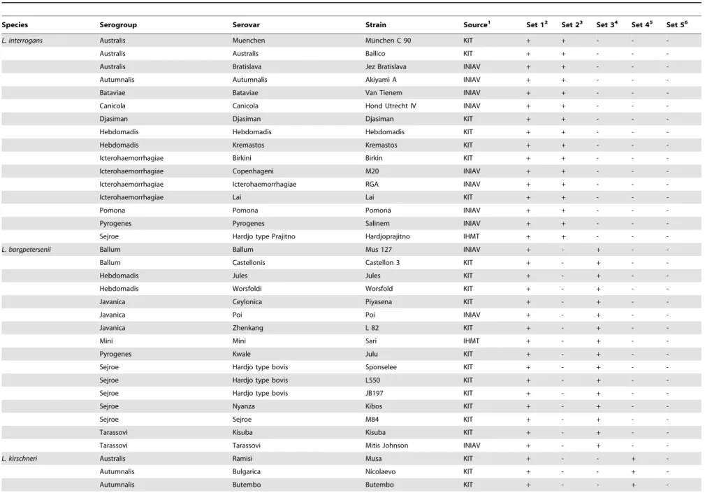
Table 1.Leptospirastrains used in the present study and results of the real time PCR assays using the species-specific probes and flanking primers.
Species Serogroup Serovar Strain Source1 Set 12 Set 23 Set 34 Set 45 Set 56
L. interrogans Australis Muenchen Mu¨nchen C 90 KIT + + - -
-Australis Australis Ballico KIT + + - -
-Australis Bratislava Jez Bratislava INIAV + + - -
-Autumnalis Autumnalis Akiyami A INIAV + + - -
-Bataviae Bataviae Van Tienem INIAV + + - -
-Canicola Canicola Hond Utrecht IV INIAV + + - -
-Djasiman Djasiman Djasiman KIT + + - -
-Hebdomadis Hebdomadis Hebdomadis KIT + + - -
-Hebdomadis Kremastos Kremastos KIT + + - -
-Icterohaemorrhagiae Birkini Birkin KIT + + - -
-Icterohaemorrhagiae Copenhageni M20 INIAV + + - -
-Icterohaemorrhagiae Icterohaemorrhagiae RGA INIAV + + - -
-Icterohaemorrhagiae Lai Lai KIT + + - -
-Pomona Pomona Pomona INIAV + + - -
-Pyrogenes Pyrogenes Salinem INIAV + + - -
-Sejroe Hardjo type Prajitno Hardjoprajitno IHMT + + - -
-L. borgpetersenii Ballum Ballum Mus 127 INIAV + - + -
-Ballum Castellonis Castellon 3 KIT + - + -
-Hebdomadis Jules Jules KIT + - + -
-Hebdomadis Worsfoldi Worsfold KIT + - + -
-Javanica Ceylonica Piyasena KIT + - + -
-Javanica Poi Poi INIAV + - + -
-Javanica Zhenkang L 82 KIT + - + -
-Mini Mini Sari IHMT + - + -
-Pyrogenes Kwale Julu KIT + - + -
-Sejroe Hardjo type bovis Sponselee KIT + - + -
-Sejroe Hardjo type bovis L550 KIT + - + -
-Sejroe Hardjo type bovis JB197 KIT + - + -
-Sejroe Nyanza Kibos KIT + - + -
-Sejroe Sejroe M84 KIT + - + -
-Tarassovi Kisuba Kisuba KIT + - + -
-Tarassovi Tarassovi Mitis Johnson INIAV + - + -
-L. kirschneri Australis Ramisi Musa KIT + - - +
-Autumnalis Bulgarica Nicolaevo KIT + - - +
-Autumnalis Butembo Butembo KIT + - - +
-Detect
ion
and
Differentiati
on
of
Pathogenic
Leptospires
ONE
|
www.ploson
e.org
3
November
2014
|
Volume
9
|
Issue
11
|
Table 1.Cont.
Species Serogroup Serovar Strain Source1 Set 12 Set 23 Set 34 Set 45 Set 56
Cynopteri Cynopteri 3522C IHMT + - - +
-Grippotyphosa Grippotyphosa type Moskva Moskva V IHMT + - - +
-Grippotyphosa Ratnapura Wumalasena KIT + - - +
-Grippotyphosa Vanderhoedeni Kipod 179 KIT + - - +
-Icterohaemorrhagiae Bogvere LT 60-69 KIT + - - +
-Pomona Mozdok 5621 KIT + - - +
-Pomona Mozdok Portugal 1990 INIAV + - - +
-Pomona Tsaratsovo B 81/7 KIT + - - +
-L. noguchii Australis Nicaragua 1011 KIT + - - - +
Autumnalis Fortbragg Fort Bragg KIT + - - - +
Bataviae Argentiniensis Peludo KIT + - - - +
Djasiman Huallaga M 7 KIT + - - - +
Louisiana Louisiana LSU 1945 KIT + - - - +
Panama Panama CZ 214 INIAV + - - - +
Pomona Proechimys 1161 U KIT + - - - +
Pyrogenes Myocastoris LSU 1551 KIT + - - - +
Shermani Carimagua 9160 KIT + - - - +
L. santarosai Ballum Peru MW 10 KIT + - - -
-Bataviae Balboa 735 U KIT + - - -
-Bataviae Kobbe CZ 320 KIT + - - -
-Grippotyphosa Canalzonae CZ 188 KIT + - - -
-Hebdomadis Borincana HS 622 KIT + - - -
-Hebdomadis Maru CZ 285 KIT + - - -
-Javanica Fluminense Aa 3 KIT + - - -
-Mini Beye 1537 U KIT + - - -
-Sarmin Rio Rr 5 KIT + - - -
-Sejroe Guaricura Bov.G. KIT + - - -
-Shermani Babudieri CI 40 KIT + - - -
-Shermani Shermani 1342 K KIT + - - -
-Tarassovi Atchafalaya LSU 1013 KIT + - - -
-L. weilii Celledoni Celledoni Celledoni INIAV + - - -
-Celledoni Mengding M 6906 KIT + - - -
-Javanica Coxi Cox KIT + - - -
-Javanica Mengma S 590 KIT + - - -
-Javanica Mengrun A 102 KIT + - - -
-Detect
ion
and
Differentiati
on
of
Pathogenic
Leptospires
PLOS
ONE
|
www.ploson
e.org
4
November
2014
|
Volume
9
|
Issue
11
|
Table 1.Cont.
Species Serogroup Serovar Strain Source1 Set 12 Set 23 Set 34 Set 45 Set 56
Mini Hekou H 27 KIT + - - -
-Pyrogenes Menglian S 621 KIT + - - -
-Sarmin Sarmin Sarmin KIT + - - -
-Tarassovi Topaz 94-79970/3 KIT + - - -
-Tarassovi Vughia LT 89-68 KIT + - - -
-L. alexanderi Hebdomadis Manzhuang A 23 KIT nd - - -
-Javanica Mengla A 85 KIT nd - - -
-Manhao Manhao 3 L 60 KIT nd - - -
-Mini Yunnan A 10 KIT nd - - -
-L. meyeri Ranarum Ranarum ICF KIT nd - - -
-Semaranga Semaranga Veldrat Semaranga 173 KIT nd - - -
-L. inadai Manhao Lincang L 14 KIT nd - - -
-L.fainei Hurstbridge Hurstbridge BUT 6T KIT nd - - -
-L.biflexa Andaman Andamana CH 11 KIT - - - -
-Semaranga Patoc Patoc I KIT - - - -
-1INIAV - Instituto Nacional de Investigac¸a˜o Agra´ria e Veterina´ria, Lisbon, Portugal. IHMT - Instituto de Higiene e Medicina Tropical, Lisbon, Portugal. KIT - Royal Tropical Institute, Amsterdam, The Netherlands; 2Set 1 targets the
lipL32gene of pathogenicLeptospiraspp.;
3Set 2 targets thesecYgene ofL. interrogans; 4Set 3 targets theompL1gene ofL. borgpetersenii; 5Set 4 targets the
secYgene ofL. kirschneri;
6Set 5 targets thesecYgene ofL. noguchii; nd - not done; Amplification (
+) or no amplification (2). doi:10.1371/journal.pone.0112312.t001
Detect
ion
and
Differentiati
on
of
Pathogenic
Leptospires
ONE
|
www.ploson
e.org
5
November
2014
|
Volume
9
|
Issue
11
|
genome size of 4.6 Mb [29]. Genomic DNA suspensions were stored at220uC until further use.
Design ofTaqManprobes and flanking primers
DNA sequences of representative strains and species of Leptospirawere retrieved from NCBI-GenBank and aligned using the ClustalW algorithm implemented in the program MegAlign (vers. 5.03) (DNAStar, USA). Primers and dual labeled hydrolysis probes (TaqManprobes) were designed to target selected species-specific genetic polymorphisms of the following pathogenic Leptospira spp.: L. interrogans, L. borgpetersenii, L. kirschneri and L. noguchii (Table 3). Probes and primers specificities were assessedin silicousing the BLAST tools from NCBI-GenBank. All probes and primers were synthesized by MWG Biotech (Ger-many).
Real-time PCR assays
We have implemented the following assay format for testing DNA templates extracted from biological samples: (i) a first duplex amplification step aiming the detection of pathogenicLeptospira spp. (by targeting the leptospiral lipL32 gene; Table 3) and including an internal control to monitor the presence of potential amplification inhibitors (by targeting the mammal b-actingene; Table 3); (ii) if pathogenic leptospires are detected in the first reaction, these may be further discriminated by testing each of the L. interrogans, L. borgpetersenii, L. kirschneri and L. noguchii targeted probes/primers (Table 3). The CFX96 real-time PCR detection system (Bio-Rad, USA) was used for all assays. The amplification reactions were optimized individually for all the probes and associated primers using the SsoFast Probes Supermix (Bio-Rad, USA), according to the manufacturer’s instructions. Each reaction was conducted in a total volume of 20ml consisting of 16SsoFast Probes Supermix, 400 nM of each primer, 150 nM
ofTaqManprobe, DNase free water (GIBCO) and 5ml of DNA template solution (extracted from pure cultures or tissues samples). Non-template negative controls (with PCR grade water) were included in each run to rule out the possibility of cross-contamination. The assay thermal conditions were as follows: 95uC for 2 min, followed by 45 cycles of 5 s at 95uC and 15 s at the optimized annealing temperature for each probe (Table 3). The thermal cycling conditions for the duplex amplification targetingb-actinandlipL32were 95uC for 2 min, followed by 45 cycles of 5 s at 95uC and 35 s at 60uC. Reproducibility of the assays was assessed by repeating the assays at least twice. Data analyses were performed by the detection system of the real-time PCR equipment, according to the manufacturer’s instructions.
Analytical specificity and sensitivity
In order to determine if each set of probe and associated primers was specific for the respectiveLeptospiratarget species, the amplification assays were tested on DNA templates extracted from different strains belonging to pathogenic, intermediate and non-pathogenic Leptospira species (Table 1), and from other non-related bacteria previously mentioned in ‘‘bacterial strains’’ section. The analytical sensitivity of the amplification assays (limits of detection – LODs) were determined using 10-fold serial dilutions of genomic DNA extracted from pure cultures of L. interrogans (serovar Autumnalis, strain Akiyami), L. kirschneri (serovar Mozdok, strain Portugal 1990), L. noguchii (serovar Panama, strain CZ 214K) and L. borgpetersenii (serovar Tarassovi, strain Mitis Johnson). LODs on tissue samples were assessed using DNA extracted from the serially diluted spiked macerates. Each template was tested in triplicate.
Sequencing
Leptospiraisolates obtained from tissue samples were identified by comparative sequence analysis of a 245 bp region of the secY gene, as described by Victoria et al.[30]. Briefly, the region of interest was amplified using primers SecYII (59-GAA TTT CTC TTT TGA TCT TCG-39) and SecYIV (59-GAG TTA GAG CTC AAA TCT AAG-39), which amplifysecYsequences from all pathogenic strains of Leptospira. PCR amplifications were performed on a C1000 thermocycler (Bio-Rad) using the following program: an initial step of denaturation for 5 min at 95uC, followed by 34 cycles consisting of annealing, 45 sec at 54uC, extension, 2 min at 72uC, and denaturation, 30 sec at 94uC. Nucleotide sequences were determined, using the same primers, by commercially available sequencing services. Nucleotide se-quence analysis and comparison with other relevant reference sequences were performed using the BLAST suite at NCBI-GenBank and aligned using Clustal X or MEGA software (version 5.0).
Results
Design of probes and primers
Species-specific sets of primers and probes targeting L. interrogans, L. borgpetersenii, L. kirschneri and L. noguchiiare listed in Table 3. As shown in Figures S1, S2, S3 and S4 in File S1, these sets of probes and primers contained sufficient polymor-phisms to warrant ‘in silico’ species specific amplification.
Analytical specificity and sensitivity
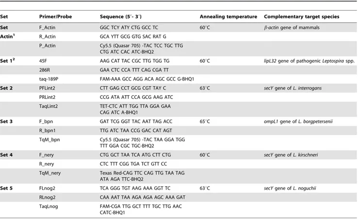
Execution of the PCRs on DNA extracted from various bacteria, revealed a highly specific amplification from any of the pathogenic strains belonging to the respective targetLeptospira spp., i.e. L. interrogans, L. kirschneri, L borgpetersenii and L. noguchii. None of the other strains yielded a positive amplification reaction (Table 1; Fig. 1A). The analytical sensitivity (LOD) of the amplification assays were found to be between 1 and 10 genome copies in the PCR mixture for each probe and primer set.
Spiked tissue samples
The LOD of the PCRs on spiked tissue samples was similar for all probe/primers sets targeting the respective target species, and estimated to be 103 leptospires/ml of tissue homogenate (<per
20 mg of tissue) (Fig. 1B). Furthermore, the same LOD was estimated for the lipL32-targeted probe/primers when used in duplex amplification reactions with the mammal b-actin probe (not shown).
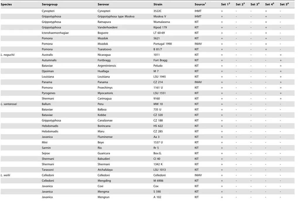
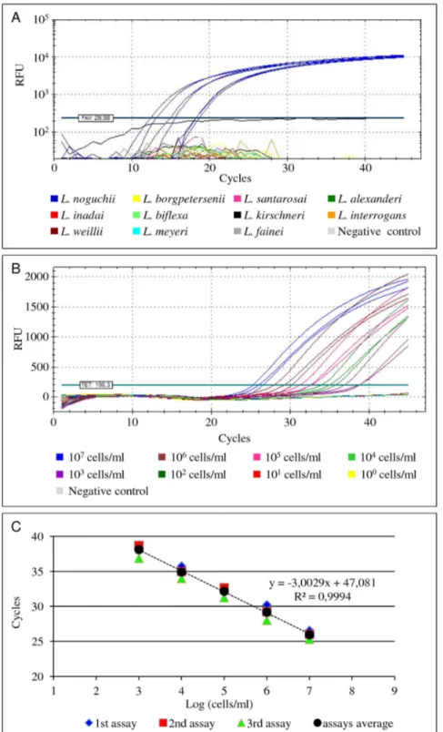
Clinical tissue samples
DNA extracted from 27 kidney samples of wild rodents were analysed with thelipL32 and mammalb-actin targeted duplex assay (Table 2; Fig. 2A). Leptospiral DNA was detected in three samples, as demonstrated by a positive amplification of thelipL32 gene region (Table 2; Fig. 2A). Furthermore, the partialb-actin gene was amplified from all samples, showing that the PCR reactions were not significantly inhibited by potential contami-nants. When tested with each of the L. interrogans, L. borgpetersenii, L. kirschneri and L. noguchii targeted probes/ primers, only these three samples showed amplification (Table 2; Fig. 2B). Two of these DNA samples were identified as L. borgpeterseniiand one sample asL. interrogans. Testing a pooled sample of kidney and liver tissues from a Patagonian mara, and a lung sample from an aborted swine fetus with the duplex PCR revealed a positive amplification for both samples (Table 2). Subsequent testing with the species-specific sets of probes and Detection and Differentiation of PathogenicLeptospires
Table 2.Results of the bacteriological culture and of the real time amplification assays for the tissue samples analyzed in the present study.
Sample Origin Set Actin1 Set 12 Set 23 Set 34 Set 45 Set 56 Bacteriological analysis7
12-17433-Z1 Mussp. + + - + - - L. borgpetersenii
12-18078-Z6 Mussp. + + - + - - L. borgpetersenii
12-18458-Z13 Mussp. + - - - Negative
12-18458-Z14 Mussp. + - - - Negative
12-19472-Z15 Mussp. + - - - Negative
12-20553-Z16 Mussp. + - - - Negative
12-22955-Z17 Mussp. + - - - Negative
12-22955-Z18 Mussp. + - - - Negative
12-22955-Z19 Mussp. + - - - Negative
12-22955-Z20 Mussp. + - - - Negative
12-22955-Z22 Mussp. + - - - Negative
12-22955-Z23 Mussp. + - - - Negative
12-22955-Z24 Mussp. + - - - Negative
12-22955-Z25 Mussp. + - - - Negative
12-22955-Z26 Mussp. + - - - Negative
12-22955-Z27 Mussp. + - - - Negative
12-22955-Z28 Mussp. + - - - Negative
12-22955-Z29 Mussp. + - - - Negative
12-22955-Z30 Mussp. + - - - Negative
12-22955-Z31 Mussp. + - - - Negative
12-22955-Z32 Mussp. + - - - Negative
12-22955-Z33 Mussp. + - - - Negative
12-22955-Z34 Mussp. + - - - Negative
12-22955-Z36 Mussp. + - - - Negative
12-22955-Z37 Rattussp. + + + - - - L. interrogans
12-22955-Z38 Mussp. + - - - Negative
12-22955-Z39 Rattussp. + - - - Negative
11-36840 Dolichotis patagonum + + + - - - L. interrogans
12-494 Sus domesticus(fetus) + + - - + - L. kirschneri
1Set Actin targets theb-actingene of mammals, 2Set 1 targets thelipL32gene of pathogenicLeptospira; 3Set 2 targets the
secYgene ofL. interrogans;
4Set 3 targets the
ompL1gene ofL. borgpetersenii;
5Set 4 targets thesecYgene ofL. kirschneri; 6Set 5 targets thesecYgene ofL. noguchii; 7The analysis of the partial sequences of the
secYgene of each isolate allowed to identify theLeptospiraspecies; Amplification (+) or no amplification (2). doi:10.1371/journal.pone.0112312.t002
Detect
ion
and
Differentiati
on
of
Pathogenic
Leptospires
ONE
|
www.ploson
e.org
7
November
2014
|
Volume
9
|
Issue
11
|
primers showed that the Patagonian mara was infected with L. interrogansand the swine fetus withL. kirschneri.
Leptospiraisolates were only cultured from the samples that also yielded PCR-positive results, thus confirming the presence of viable leptospires (Table 2).
Molecular speciation through analysis of the partial sequences of thesecYgene was in concordance with the results obtained by the species-specific PCRs. Two isolates were identified as L. borgpetersenii (from wild rodents; GenBank accession numbers KM066006 and KM066007), one asL. kirschneri(from the swine fetus; accession number KM066009) and two as L. interrogans (from a wild rodent and the Patagonian mara; accession numbers KM066008 and KM066010, respectively).
Discussion
In this work we present a two step real-time PCR strategy to infer the presence of pathogenic leptospires in clinical and veterinary samples. In the first step, we assess if an animal tissue sample is infected with a pathogenic leptospire by targeting its lipL32 gene. The lipL32 gene encodes an outer membrane lipoprotein that is confined to pathogenicLeptospiraspecies [16]. The second step identifies the four most common and veterinary relevant pathogenic Leptospira species, L. interrogans, L. borgpetersenii,L. kirschneriand L. noguchiiusing dedicated sets of probes and primers.
Probes and flanking primers were developed byin silicoanalysis and further tested for their practical utility on DNA extracted from cultured bacteria, spiked tissues and clinical specimens. The amplification assays have proved to be specific to the respective
targeted species, with no cross-reactions when non-pathogenic leptospires or other pathogens were tested. The amplification of theb-actingene was included in the initiallipL32-based PCR to assess the presence of amplification inhibitors in tissue samples [31]. However, the abundant presence ofb-actingene copies in DNA samples extracted from tissues may ensure some amplifica-tion even when low levels of potential inhibitors are present (but amplification curves are usually weaker and anomalous). The analytical sensitivity deduced for the amplification assays, i.e. 1 to 10 GE on DNA extracted from cultured leptospires and 103 leptospires/ml tissue homogenate, were similar to the ones of other previous studies concerning the molecular detection of leptospires [15–17,19,22].
The panel of species-specific probes and flanking primers may be extended with the design of novel oligonucleotides, e.g. for use in regions where the occurrence of additional species of pathogenic leptospires is common. As far as we know, this is the first report describing a strategy capable of clearly identify four most frequently found pathogenicLeptospiraspecies based on the use ofTaqManprobes.
From 27 kidney samples of wild rodents, and samples from a Patagonian mara and a porcine fetus suspected of leptospirosis, three rodent samples and the samples from the Patagonian mara and fetus all yielded a positive PCR test for the presence of pathogenic leptospires. In concordance, these samples were also positive by culture. Culture provides proof of infection and thus is an ideal reference standard. Consequently, these results are consistent with a 100% clinical sensitivity and specificity of the PCR. Subsequent prospective analysis of a larger sample set would allow substantiating this conclusion.
Table 3.Primers and probes used in this study targeting selected genes of pathogenic species ofLeptospira.
Set Primer/Probe Sequence (59- 39) Annealing temperature Complementary target species
Set F_Actin GGC TCY ATY CTG GCC TC 60uC b-actingene of mammals
Actin1 R_Actin GCA YTT GCG GTG SAC RAT G
P_Actin Cy5.5 (Quasar 705) -TAC TCC TGC TTG CTG ATC CAC ATC-BHQ2
Set 12 45F AAG CAT TAC CGC TTG TGG TG 60uC lipL32gene of pathogenicLeptospiraspp. 286R GAA CTC CCA TTT CAG CGA TT
taq-189P FAM-AAA GCC AGG ACA AGC GCC G-BHQ1
Set 2 PFLint2 CTT GAG CCT GCG CGT TAY C 63uC secYgene ofL. interrogans PRLint2 CCG ATA ATT CCA GCG AAG ATC
TaqLint2 TET-CTC ATT TGG TTA GGA GAA CAG ATC A-BHQ1
Set 3 F_bpn GAT TCG GGT TAC AAT TAG ACC 65uC ompL1gene ofL. borgpetersenii R_bpn1 TTG ATC TAA CCG GAC CAT AGT
TqM_bpn Cy5.5 (Quasar 705) -TAC TAA GGA TGG TTT GGA CGC TGC-BHQ2
Set 4 F_nery CTG GCT TAA TCA ATG CTT CTG 60uC secYgene ofL. kirschneri R_nery CTC TTT CGG TGA TCT GTT CC
TqM_nery Texas Red-CAG TTC CAG TTG TAA TAG ATA AGA TTC-BHQ2
Set 5 FLnog2 TCA GGG TGT AAG AAA GGT TC 63uC secYgene ofL. noguchii
RLnog2 CAA AAT TAA AGA AGA AGC AAA GAT
TaqLnog FAM-CGA TTG GCT TTT TGC TTG AAC CATC-BHQ1
1Retrieved from Costa et al. [31]; 2Retrieved from Stoddard et al. [16]. doi:10.1371/journal.pone.0112312.t003
Detection and Differentiation of PathogenicLeptospires
Figure 1. Illustration of the real-time PCR amplification curves obtained during the optimization of the assays.(A) Specificity tests of theL. noguchiitargeted amplification assay using the TaqLnog probe combined with the flanking primers FLnog2 and RLnog2. Blue amplification curves representL. noguchiistrains. All other non-target strains yielded no amplification results. (B) Estimation of the limit of detection of the amplification assay targetingL. interrogans(serovar Autumnalis, strain Akiyami) using DNA extracted directly from spiked bovine kidney samples as template as a typical example of allLeptospiraprobe and primer sets. The amplification curves obtained from different ten-fold serial dilutions of the targetLeptospiraare represented by different colours. Unspiked tissue homogenate (grey line) was used as negative control. (C) Standard curve obtained from the analysis of the amplification curves mentioned in the previous panel B. RFU - Relative Fluorescence Units.
doi:10.1371/journal.pone.0112312.g001
Phylogenetic identification of the cultures also allowed support-ing the findsupport-ings obtained with the species-specific PCRs. Indeed, speciation by phylogeny was in all cases in concordance with the results obtained via the PCR method.
Initially, we anticipated that more samples would be positive by the real time PCR assay than by culture [5,32–34]. Recently, Fornazariet al.[20] reported that quantitative PCR presented the highest sensitivity among several techniques to detect leptospires in tissues samples, the bacteriological culture being the least sensitive. Apparently, our procedure of culturing, using macerated fresh tissue has been highly effective. Alternatively, it cannot be excluded that the bacterial load of the tissues might have been very high. Nevertheless, the low rate of positive animals (11%) is not too discrepant from the prevalence values found in other studies where leptospiral DNA was detected in rodents tissues by PCR-based assays, which ranged from 13% to 20% [25,35,36]. Furthermore, as far as we know, the region of Lisbon, where the rodents were captured, is not usually regarded as having major leptospirosis problems [2], which may also reflect a lower prevalence of the agent in reservoirs such as wild rodents. We anticipate that our assays may be useful in studies inferring the prevalence of pathogenic leptospires in wild rodents and other animals, with the advantage of differentiating the infecting Leptospiraspecies.
The amplification assays described were able to detect pathogenic leptospires in samples of animal tissues, such as kidney or lung. Although the analysis of this kind of samples is not essential for an early diagnosis of leptospirosis, it has a great value in situations such as epidemiological and post-mortem investigations. The last situation is very well illustrated in this work with the detection of pathogenic leptospires in tissues of a Patagonian mara and a swine fetus. Both animals were suspect of having leptospirosis, which was confirmed by this study. The porcine fetus was infected with a strain belonging to L. kirschneri. Pigs may be infected by several Leptospira species (and serovars) that may cause infertility, fetal death and abortion.Leptospira kirschnerihas been reported but seems to be less frequently found in pigs in Portugal than other species [37]. The Patagonian mara, a relatively large rodent that lived in the local zoo, was found to be infected withL. interrogans. To our knowledge, this is the first report describing the molecular detection or the isolation of a pathogenic leptospire from that rodent, which proved to have died of leptospirosis. Zoos are often infested with rats that are notorious reservoirs ofL. interrogans. We hypothesise that this Patagonian mara has been infected by rats as the primary infection reservoir, which would support the potential hazard of rodents in zoos for both (exotics) animals and public.
The amplification assay described in this work is able to indentify the four most relevant pathogenic species ofLeptospira infecting farm and wild animals. While the approach can be extended to otherLeptospiraspecies, it is important to continually evaluate the specificity of previously designed probes and primers and, if necessary, modify and improve the sequences, in order to ensure an effective and specific detection and identification of the circulatingLeptospiraspecies.
Conclusions
The molecular assays presented in this work allow the detection and identification of four relevant pathogenic species ofLeptospira, directly from animal tissues. The assays proved to be specific and sensitive, and much faster than the bacteriological culture, reducing the time for confirmatory leptospirosis diagnosis. The assays are amenable to future automation possibilities and will reinforce the diagnostic information and enhance our knowledge about the epidemiology of leptopirosis.
Supporting Information
File S1 Sequence alignments showing the
complemen-tary targets of the species-specific Leptospira interro-gans, L. kirschneri, L. noguchii and L. borgpetersenii
probes and respective flanking primers.
(PDF)
Acknowledgments
Madalena Monteiro (INIAV, I.P.) is acknowledged for excellent technical assistance in performing pathological analysis. Lurdes Clemente and Ana
Botelho are acknowledged for providing non-Leptospirabacterial strains.
Narciso Lapa˜o, Nuno Gaspar and Anto´nio Crespo are acknowledged for their help in providing samples and respective information.
Author Contributions
Conceived and designed the experiments: ASF JI. Performed the experiments: ASF TR. Analyzed the data: ASF TR A. Amaro JI. Contributed reagents/materials/analysis tools: TR PC MLV GT RAH. Wrote the paper: ASF JI. Critical discussion during data analysis and during preparation of the paper: ASF TR PC A. Amaro MLV A. Ahmed GT RAH JI. Revised the manuscript: TR A. Amaro A. Ahmed MLV GT RAH.
Figure 2. Illustration of the real-time PCR amplification curves obtained during the testing of naturally-infected tissue samples. (A) Results of the b-actin and lipL32 targeted duplex amplification assay when testing representative samples from the wild rodents. The partialb-actingene was amplified from all tissue samples (dark pink lines). Leptospiral DNA was detected in three samples by a positive amplification of thelipL32gene (blue lines). A spiked positive control withL. interrogans(serovar Autumnalis, strain Akiyami) is shown (green line). (B) From the previous leptospiral positive amplification results, two samples were assessed as infected withL. borgpetersenii using the respective targeted amplification assay with probe TqM_bpn and flanking primers F_bpn and R_bpn1 (blue lines). The positive and negative controls are illustrated by the orange and red lines, respectively.
doi:10.1371/journal.pone.0112312.g002
Detection and Differentiation of PathogenicLeptospires
References
1. Dupouey J, Faucher B, Edouard S, Richet H, Kodjo A, et al. (2014) Human leptospirosis: An emerging risk in Europe?. Comp. Immunol. Microbiol. Infects Dis. 37: 77–83.
2. Vieira ML, Gama-Simo˜es MJ, Collares-Pereira M (2006) Human leptospirosis in Portugal: A retrospective study of eighteen years. Int J Infect Dis. 10: 378– 386.
3. Adler B, Moctezuma AP (2010) Leptospira and leptospirosis. Vet. Microbiol. 140: 287–296.
4. Bomfim MR, Barbosa-Stancioli EF, Koury MC (2008) Detection of pathogenic leptospires in urine from naturally infected cattle by nested PCR. Vet J. 178: 251–256.
5. Fearnley C, Wakeley PR, Gallego-Beltran J, Dalley C, Williamson S, et al. (2008) The development of a real-time PCR to detect pathogenicLeptospira species in kidney tissue. Res Vet Sci. 85: 8–16.
6. Picardeau M (2013) Diagnosis and epidemiology of leptospirosis. Med Mal Infect. 43: 1–9.
7. Brenner DJ, Kaufmann AF, Sulzer KR, Steigerwalt AG, Rogers FC, et al. (1999) Further determination of DNA relatedness between serogroups and serovars in the family Leptospiraceae with a proposal forLeptospira alexanderi sp. nov. and four newLeptospiragenomospecies. Int J Syst Bacteriol. 49: 839– 858.
8. Cerqueira GM, Picardeau M (2009) A century ofLeptospirastrain typing. Infect Genet Evol. 9: 760–768.
9. Morey RE, Galloway RL, Bragg SL, Steigerwalt AG, Mayer LW, et al. (2006) Species-specific identification of Leptospiraceae by 16S rRNA gene sequencing. J Clin Microbiol. 44: 3510–3516.
10. Saito M, Villanueva SY, Kawamura Y, Iida K, Tomida J, et al. (2013) Leptospira idoniisp. nov., isolated from environmental water. Int J Syst Evol Microbiol. 63: 2457–2462.
11. Agampodi SB, Matthias MA, Moreno AC, Vinetz JM (2012) Utility of quantitative polymerase chain reaction in leptospirosis diagnosis: association of level of leptospiremia and clinical manifestations in Sri Lanka. Clin Infect Dis. 54: 1249–1255.
12. Bourhy P, Bremont S, Zinini F, Giry C, Picardeau M (2011) Comparison of real-time PCR assays for the detection of pathogenicLeptospiraspp. in blood and identification of variations in target sequences. J Clin Microbiol. 49: 2154–2160. 13. Levett PN, Morey RE, Galloway RL, Turner DE, Steigerwalt AG, et al. (2005) Detection of pathogenic leptospires by real-time quantitative PCR. J Med Microbiol. 54: 45–49.
14. Merien F, Portnoi D, Bourhy P, Charavay F, Berlioz-Arthaud A, et al. (2005) A rapid and quantitative method for the detection ofLeptospiraspecies in human leptospirosis. FEMS Microbiol Lett. 249: 139–147.
15. Smythe LD, Smith IL, Smith GA, Dohnt MF, Symonds ML, et al. (2002) A quantitative PCR (TaqMan) assay for pathogenicLeptospiraspp. BMC Infect Dis. 2: 13.
16. Stoddard RA, Gee JE, Wilkins PP, McCaustland K, Hoffmaster AR (2009) Detection of pathogenicLeptospira spp. through TaqMan polymerase chain reaction targeting thelipL32gene. Diagn Microbiol Infect Dis. 64: 247–255. 17. Villumsen S, Pedersen R, Borre MB, Ahrens P, Jensen JS, et al. (2012) Novel
TaqMan PCR for detection ofLeptospiraspecies in urine and blood: Pit-falls of in silico validation. J Microbiol Methods. 91: 184–90.
18. Ahmed A, Engelberts MF, Boer KR, Ahmed N, Hartskeerl RA (2009) Development and validation of a real-time PCR for detection of pathogenic Leptospiraspecies in clinical materials. PLoS One. 4: e7093.
19. Slack A, Symonds M, Dohnt M, Harris C, Brookes D, et al. (2007) Evaluation of a modified Taqman assay detecting pathogenicLeptospiraspp. against culture
andLeptospira-specific IgM enzyme-linked immunosorbent assay in a clinical environment. Diagn Microbiol Infect Dis. 57: 361–366.
20. Fornazari F, da Silva RC, Richini-Pereira VB, Beserra HE, Luvizotto MC, et al. (2012) Comparison of conventional PCR, quantitative PCR, bacteriological culture and the Warthin Starry technique to detectLeptospiraspp. in kidney and liver samples from naturally infected sheep from Brazil. J Microbiol Methods. 90: 321–326.
21. Ahmed A, Klaasen HLBM, van der Veen M, van der Linden H, Goris MGA, et al. (2012) Evaluation of real-time PCR and culturing for the detection of leptospires in canine samples. Adv. Microbiol. 2: 162–170.
22. Rojas P, Monahan AM, Schuller S, Miller IS, Markey BK, et al. (2010) Detection and quantification of leptospires in urine of dogs: a maintenance host for the zoonotic disease leptospirosis. Eur J Clin Microbiol Infect Dis. 29: 1305– 1309.
23. Subharat S, Wilson PR, Heuer C, Collins-Emerson JM (2011) Evaluation of a SYTO9 real-time polymerase chain reaction assay to detect and identify pathogenicLeptospiraspecies in kidney tissue and urine of New Zealand farmed deer. J Vet Diagn Invest. 23: 743–752.
24. Cox TE, Smythe LD, Leung LKP (2005) Flying foxes as carriers of pathogenic Leptospiraspecies. J. Wildlife Dis. 41: 753–757.
25. Levieuge A, Aboubaker MH, Terrier O, Drancourt M, Davoust B (2010) Real-time PCR detection ofLeptospirasp. in rodents from Toulon harbour (France). Revue Me´d. Ve´t. 161: 264–266.
26. Palaniappan RU, Chang YF, Chang CF, Pan MJ, Yang CW, et al. (2005) Evaluation of lig-based conventional and real time PCR for the detection of pathogenic leptospires. Mol Cell Probes. 19: 111–117.
27. OIE 2008. Manual of Diagnostic Tests and Vaccines for Terrestrial Animals (Chapter 2.1.9), World Organization for Animal Health.
28. Rocha T (1990) Isolation ofLeptospira interrogansserovarmozdokfrom aborted swine fetuses in Portugal. Vet Record. 126: 602.
29. Nascimento AL, Verjovski-Almeida S, Van Sluys MA, Monteiro-Vitorello CB, Camargo LE, et al. (2004) Genome features ofLeptospira interrogansserovar Copenhageni. Braz J Med Biol Res. 37: 459–477.
30. Victoria B, Ahmed A, Zuerner RL, Ahmed N, Bulach DM, et al. (2008) Conservation of the S10-spc-alpha locus within otherwise highly plastic genomes provides phylogenetic insight into the genusLeptospira. PLoS One. 3: e2752. 31. Costa P, Ferreira AS, Amaro A, Albuquerque T, Botelho A, et al. (2013)
Enhanced detection of tuberculous mycobacteria in animal tissues using a semi-nested probe-based real-time PCR. PLoS ONE 8: e81337.
32. Boqvist S, Montgomery JM, Hurst M, Thu HT, Engvall EO, et al. (2003) Leptospirain slaughtered fattening pigs in southern Vietnam: presence of the bacteria in the kidneys and association with morphological findings. Vet Microbiol. 93: 361–368.
33. Grooms DL, Bolin CA (2005) Diagnosis of fetal loss caused by bovine viral diarrhea virus andLeptospiraspp. Vet Clin North Am Food Anim Pract. 21: 463–472.
34. Lilenbaum W, Varges R, Ristow P, Cortez A, Souza SO, et al. (2009) Identification ofLeptospiraspp. carriers among seroreactive goats and sheep by polymerase chain reaction. Res Vet Sci. 87: 16–19.
35. Foronda P, Martin-Alonso A, Del Castillo-Figueruelo B, Feliu C, Gil H, et al. (2011) PathogenicLeptospiraspp. in wild rodents, Canary Islands, Spain. Emerg Infect Dis. 17: 1781–1782.
36. Latifah I, Rahmat MS, Hayarti KB, Paramasvaran S, Azizah MR, et al. (2012) Prevalence of leptospiral DNA among wild rodents from a selected area in Beguk Dam Labis, Segamat, Johor, Malaysia. Malays J Pathol. 34: 157–159. 37. Rocha T (1998) A review of leptospirosis in farm animals in Portugal. Rev Sci
Tech. 17: 699–712.