Quinoline Compound KM11073 Enhances
BMP-2-Dependent Osteogenic Differentiation
of C2C12 Cells via Activation of p38
Signaling and Exhibits
In Vivo
Bone Forming
Activity
Seung-hwa Baek1,2☯, Sik-Won Choi1☯, Sang-Joon Park3, Sang-Han Lee2, Hang-Suk Chun4, Seong Hwan Kim1*
1Laboratory of Translational Therapeutics, Pharmacology Research Center, Korea Research Institute of Chemical Technology, Daejeon, 305-600, Republic of Korea,2Department of Food Science &
Biotechnology, Kyungpook National University, Daegu, 702-701, Republic of Korea,3Department of Histology, College of Veterinary Medicine, Kyungpook National University, Daegu, 702-701, Republic of Korea,4Alternative Toxicological Methods Research Center, Department of Predictive Toxicology, Korea Institute of Toxicology, Daejeon, 305-600, Republic of Korea
☯These authors contributed equally to this work.
Abstract
Recombinant human bone morphogenetic protein (rhBMP)-2 has been approved by the FDA for clinical application, but its use is limited due to high cost and a supra-physiological dose for therapeutic efficacy. Therefore, recent studies have focused on the generation of new therapeutic small molecules to induce bone formation or potentiate the osteogenic ac-tivity of BMP-2. Here, we show that [4-(7-chloroquinolin-4-yl) piperazino][1-phenyl-5-(tri-fluoromethyl)-1H-pyrazol-4-yl]methanone (KM11073) strongly enhances the BMP-2-stimulated induction of alkaline phosphatase (ALP), an early phase biomarker of osteoblast differentiation, in bi-potential mesenchymal progenitor C2C12 cells. The KM11073-mediat-ed ALP induction was inhibitKM11073-mediat-ed by the BMP antagonist noggin, suggesting that its osteogen-ic activity occurs via BMP signaling. In addition, a pharmacologosteogen-ical inhibition study
suggested the involvement of p38 activation in the osteogenic action of KM11073 accompa-nied by enhanced expression of BMP-2, -6, and -7 mRNA. Furthermore, thein vivo osteo-genic activity of KM11073 was confirmed in zebrafish and mouse calvarial bone formation models, suggesting the possibility of its single use for bone formation. In conclusion, the combination of rhBMP-2 with osteogenic small molecules could reduce the use of expen-sive rhBMP-2, mitigating the undesirable side effects of its supra-physiological dose for therapeutic efficacy. Moreover, due to their inherent physical properties, small molecules could represent the next generation of regenerative medicine.
OPEN ACCESS
Citation:Baek S-h, Choi S-W, Park S-J, Lee S-H, Chun H-S, Kim SH (2015) Quinoline Compound KM11073 Enhances BMP-2-Dependent Osteogenic Differentiation of C2C12 Cells via Activation of p38
Signaling and ExhibitsIn VivoBone Forming Activity.
PLoS ONE 10(3): e0120150. doi:10.1371/journal. pone.0120150
Academic Editor:Damian Christopher Genetos, University of California Davis, UNITED STATES
Received:July 23, 2014
Accepted:January 19, 2015
Published:March 19, 2015
Copyright:© 2015 Baek et al. This is an open access article distributed under the terms of the Creative Commons Attribution License, which permits unrestricted use, distribution, and reproduction in any medium, provided the original author and source are credited.
Data Availability Statement:All relevant data are within the paper.
Funding:This work was supported by KRICT's project, SI-1404, funded by the Ministry of Knowledge Economy, Republic of Korea. The funders had no role in study design, data collection and analysis, decision to publish, or preparation of the manuscript.
Introduction
A delicate balance between osteoclast-mediated bone resorption and osteoblast-mediated bone formation is necessary for normal bone development and remodeling. Excessive osteoclastic bone resorption and/or reduced bone formation results in bone loss that consequently leads to pathological bone-related disorders, such as osteoporosis, rheumatoid arthritis, periodontal disease, and cancer bone metastasis [1]. These bone-related disorders impact clinical and pub-lic health, most importantly due to subsequent fractures. Bone fractures are one of the most common causes of disability and are associated with enormous healthcare expenditures.
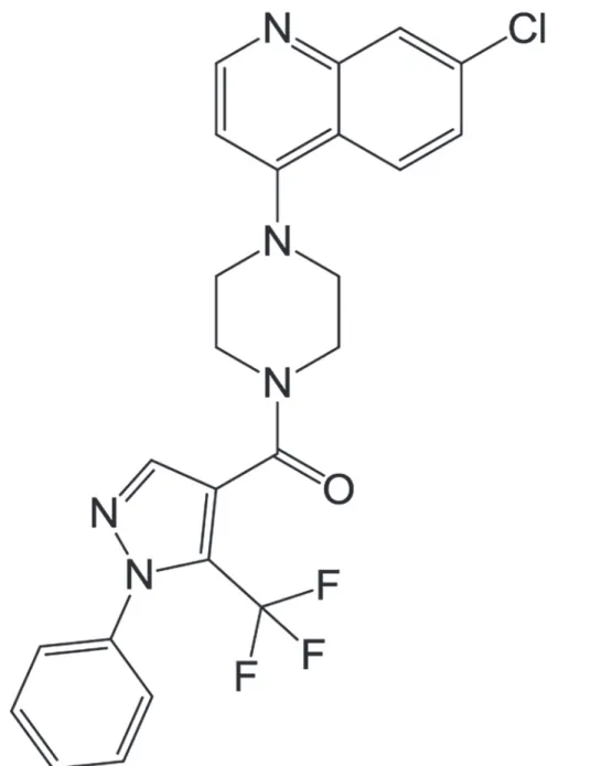
Most agents that are used to inhibit bone loss are anti-resorptive agents, but the develop-ment of anabolic agents for stimulating bone formation is also an area of interest [2,3]. Among FDA-approved anabolic agents, recombinant human bone morphogenetic proteins (rhBMPs) have potential clinical applications in spinal fusion, fracture healing, and dental tissue engi-neering [4–7]. BMPs play crucial roles in bone formation, repair, and regeneration [8–10]. As one of osteogenic BMP family, BMP-2 strongly triggers the commitment of mesenchymal stem cells into pre-osteoblasts for bone formation and mineralization. rhBMP-2 has been approved by the FDA for application in spinal fusion and the treatment of long bone fractures [7,11], but its clinical use is limited due to its comparatively expensive cost and severe side effects, among other reasons. Therefore, recent studies have focused on the identification of new effec-tive anabolic small molecules that are less expensive and simple to use [3]. In the previous study, chemical library in Korea Chemical Bank was screened in order to identify anabolic compounds in the BMP-2-mediated osteoblast differentiation model of bi-potential mesenchy-mal precursor C2C12 cells [12], and finally [4-(7-chloroquinolin-4-yl) piperazino][1-phenyl-5-(trifluoromethyl)-1H-pyrazol-4-yl]methanone (KM11073;Fig. 1) was identified as a BMP-2 enhancer that can accelerate the BMP-2-mediated commitment of C2C12 cells into osteoblasts. Therefore, in the present study, the effect of KM11073 on the commitment of C2C12 cells into osteoblasts was confirmed and potential mechanisms explaining its osteogenic activity investigated.
Materials and Methods
Materials
KM11073 was purchased from Maybridge (MO, USA). In this study, 10 mM KM11073 in DMSO was used as a stock solution and diluted with culture medium. Therefore, 0.2% DMSO was used as a vehicle control in all experiments. Recombinant human BMP-2 (rhBMP-2) and noggin were purchased from PeproTech (Seoul, Korea). Ras inhibitor FTI-277, PI3K inhibitor LY294002, Akt inhibitor, and p38 inhibitors (PD169316, SB203581, and SB202190) were pur-chased from Calbiochem (EMD Biosciences, Inc., La Jolla, CA, USA).
Cell culture
C2C12 cells were maintained in Dulbecco’s Modified Eagle’s Medium (DMEM, Hyclone) con-taining 10% fetal bovine serum (FBS), 100 U/ml of penicillin, and 100μg/ml streptomycin.
Cells were seeded and, after 1 day, differentiated by replacing the medium with differentiation medium (DM; DMEM containing 5% FBS and 100 ng/ml rhBMP-2). The medium was changed every 3 days.
Cell viability assay
Cell Counting Kit-8 (Dojindo Molecular Technologies, ML, USA) according to the manufac-turer’s protocol. Absorbance was measured at 450 nm using the Wallac EnVision microplate reader (PerkinElmer, Finland) and the measured absorbance converted to cell number using the standard curve.
Alkaline phosphatase (ALP) staining and activity assay
ALP, an early biomarker of osteoblastogenesis, was assayed as described previously [13]. Brief-ly, C2C12 cells (4 × 10³cells/well) were seeded in a 96-well plate, and after 24 h, the medium replaced with DM in the absence or presence of KM11073. The medium was changed every 3 days. After 6 days, the cells were washed twice with PBS, fixed with 10% formalin in PBS for
Fig 1. Chemical structure of KM11073.
30 s, rinsed with deionized water, and stained using the Alkaline Phosphatase (ALP) Kit (Sigma). To measure ALP activity, cells were washed twice with PBS and sonicated in lysis buff-er (10 mM of Tris–HCl pH 7.5, 0.5 mM of MgCl2, and 0.1% Triton X-100). After
centrifuga-tion at 10,000 × g for 20 min at 4°C, ALP activity was measured in triplicate in the supernatant using the LabAssay ALP Kit (Wako Pure Chemicals Industries).
Evaluation of mRNA expression
Primers were designed using an online primer design program [14] (Table 1). Total RNA was isolated and cDNA prepared as described previously [13]. Briefly, total RNA was isolated in C2C12 cells (2 × 10⁴cells/well in a 24-well plate) using TRIzol reagent (Life Technologies, MD, USA) and the first strand cDNA synthesized using 2μg of total RNA, 1μM of oligo-dT18
prim-er, and Omniscript Reverse Transcriptase (Qiagen, CA, USA). SYBR green-based quantitative PCR was performed using the Stratagene Mx3000P Real-Time PCR system and Brilliant SYBR Green Master Mix (Stratagene, CA, USA) as described previously [13]. All reactions were run in triplicate and data analyzed using the 2−ΔΔCTmethod [15,16]. GAPDH was used as the
con-trol gene. Significance was determined with GAPDH-normalized 2−ΔΔCTvalues. The mRNA
levels in zebrafish were evaluated as follows. Five days after fertilization, 20 embryos per well were transferred to a 12-well plate with 2 ml of test solution. After 1 day, total RNA was isolat-ed using TRIzol reagent and the first strand cDNA synthesizisolat-ed using 2μg of total RNA, 1μM
of oligo-dT18primer, and Omniscript Reverse Transcriptase (Qiagen, CA, USA). SYBR
green-based quantitative PCR was performed as described above.
Western blot analysis
Western blot analysis was performed as described previously [13]. Briefly, cells were homoge-nized in ice-cold buffer consisting 10 mM Tris–HCl (pH 7.5), 150 mM NaCl, 0.05% (v/v) Tween 20, 1 mM PMSF, and one protease inhibitor cocktail tablet (Roche, Germany) and then centrifuged at 10,000 × g for 15 min. Protein concentrations were determined using the BCA protein assay kit (Pierce, IL, USA) and denatured proteins separated and transferred to PVDF
membrane (Millipore, USA). Membranes were incubated with TBST buffer (10 mM Tris–HCl
pH 7.5, 150 mM NaCl, 0.1% Tween 20) with 5% nonfat dry milk and then incubated with
Table 1. Primer sequences used in this study.
Target Forward (5’–3’) Reverse (5’–3’)
C2C12
BMP-2 GCTCCACAAACGAGAAAAGC AGCAAGGGGAAAAGGACACT
BMP-4 CCTGGTAACCGAATGCTGAT AGCCGGTAAAGATCCCTCAT
BMP-6 TTCTTCAAGGTGAGCGAGGT TAGTTGGCAGCGTAGCCTTT
BMP-7 CGATACCACCATCGGGAGTTC AAGGTCTCGTTGTCAAATCGC
BMP-9 CAGAACTGGGAACAAGCATCC GCCGCTGAGGTTTAGGCTG
ALP ATGGGCGTCTCCACAGTAAC TCACCCGAGTGGTAGTCACA
GAPDH AACTTTGGCATTGTGGAAGG ACACATTGGGGGTAGGAACA
Zebrafish
runx2a TTTGAGCGTCAATTCCCAAG GGTACGGTGGAGGCAGGTAT
BMP-2a CTCCATCCCAGACGAGGAGT GCTCCTGAAGAGAACCGGAC
BMP-2b GTGAGGGTCAGTCGTTCCCT AGCATGTCGCCTACAGTTCG
osteopontin ATGATCTGGAGGACGGGAAC GCTGGGAGAGTCCCTAGCAC
ALP CGCAATTAAGCAGGGAATCA CCTGCGTTTACGGATTTTCA
GAPDH AGAACATCATCCCAGCCTCC TTGGCAGGTTTCTCAAGACG
diluted primary antibodies (1:1,000) overnight at 4°C. The antibodies used in this study were purchased from Cell Signaling (MA, USA) or Santa Cruz Biotechnology, Inc. (Santa Cruz, CA, USA). Following the primary antibody reactions, the membranes were washed with TBST buff-er three times (15 min each) and then probed with diluted secondary antibodies (1:5,000) for 1 h. Next, the membranes were washed three times (15 min each) and developed with Super-Signal West Femto Maximum Sensitivity Substrate (Pierce Biotechnology) using the LAS-3000 luminescent image analyzer (Fuji Photo Film Co., Ltd., Japan). Each experiment was per-formed at least three times, and the figures showed the results from one representative experi-ment. Image J software-based quantification of the detected band was carried out and the relative, normalized ratio between phosphorylated protein and the protein itself was presented in the figures.
Alizarin red S-based zebrafish skeleton development
The development of zebrafish skeletons were visualized by alizarin red S according to the Stan-dard Protocol was approved the Institutional Animal Care and Use Committees of Chungnam National University (CNU-00393). As described previously [17], zebrafish embryos were placed in a 24-well plate (9 embryos per well) 5 days after fertilization and maintained in 1 ml of buffered medium (sea salt, 0.06 mg/l) containing KM11073. The medium was changed every day. Seven days after fertilization, the embryos were fixed in 2% paraformaldehyde for 1 h and washed three times with PBS containing 0.1% Tween 20 (PBST) at 10-min intervals. Next, the embryos were treated with 1 ml of alizarin red S staining buffer (pH 4.2) to stain the formed bone overnight. The embryos were washed two times with 25% glycerol (in 1% KOH) and bleaching solution (1% KOH and 3% H2O2in deionized water) at room temperature until
pig-mented cells removed after approximately 15 min. When the pigpig-mented cell removal was com-plete, the embryos were washed three times with 25% glycerol (in 1% KOH) at 10-min
intervals and the embryos treated successively with KOH solutions containing 25% glycerol, 50% glycerol, and 80% glycerol.
In vivo
murine calvarial bone formation assay
In vivobone formation experiments were carried out according to the Ethics Guidelines of the Korea Research Institute of Chemical Technology (Protocol ID No. 7D-M5). The protocol was approved by the Institutional Committee of the Korea Research Institute of Chemical Technol-ogy (Approval No. 2014-7D-04-05). The surgery forin vivobone formation experiments was performed under anesthesia, and all efforts were made to minimize animal suffering. Briefly,in vivobone-forming activity was evaluated using lyophilized collagen sponges as described previ-ously [13]. After anesthesia with the intraperitoneal injection of 1.2% avertin (600μl/mouse),
collagen sponges loaded with 5μl of DMSO or KM11073 (2.5 or 5 mM) were implanted over
the calvarial bones of 5-week-old male ICR mice (n = 5 per group; Central Lab Animal, Seoul, Korea). When mice were monitored daily, no suffering by operation was observed. Three weeks after drug implantation, the calvariae were harvested after cervical dislocation, fixed in 4% paraformaldehyde, decalcified in 12% EDTA, embedded in paraffin, and sectioned. Sec-tions were deparaffinized through graded xylene washes, dehydrated in a graded series of etha-nol washes, and stained with hematoxylin and eosin (H&E).
Statistical analysis
Results
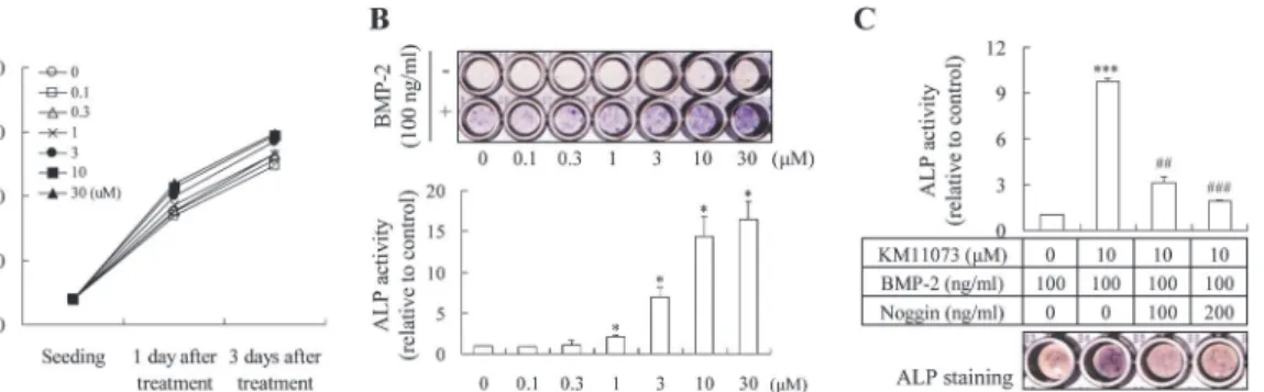
The enhancing effect of KM11073 on the BMP-2-mediated commitment of C2C12 cells into osteoblasts was evaluated by visualizing ALP expression and measuring its activity. At non-cy-totoxic concentrations (30μM;Fig. 2A), KM11073 induced the expression of ALP in a
dose-dependent manner in the presence of BMP-2 (Fig. 2B). Consistent with this result, KM11073 significantly enhanced BMP-2-stimulated ALP activity in a dose-dependent manner (Fig. 2B). In the absence of BMP-2, KM11073 did not induce ALP expression, suggesting that the osteo-genic activity of KM11073 relies on BMP-2 signaling. This finding was confirmed by the post-treatment of noggin; when noggin was post-treated alone on differentiation day 4 after BMP-2 and KM11073 treated on differentiation day 0 and 2, noggin dose-dependently and significant-ly inhibited the ALP activity induced by both BMP-2 and KM11073 (Fig. 2C).
The inhibition of KM11073-enhanced ALP induction by noggin also suggested the involve-ment of endogenous BMP induction in the osteogenic activity of KM11073. Therefore, we eval-uated the effect of KM11073 on the expression of osteogenic BMPs (Table 2). ALP expression and osteogenic BMPs were significantly induced by BMP-2 alone, and KM11073 significantly enhanced the BMP-2-stimulated mRNA levels of BMP-2, BMP-6, and BMP-7, as well as ALP, but not BMP-4 and BMP-9. In the absence of BMP-2, KM11073 did not induce the mRNA ex-pressions of BMP-2, BMP-6, and BMP-7 (S1 Table).
Next, in order to investigate the underlying mechanism explaining the osteogenic activity of KM11073, a pharmacological inhibition study was performed. The KM11073-enhanced ALP induction in the presence of BMP-2 was strongly inhibited by p38 inhibitors, but not inhibitors of Ras, phosphatidylinositol 3-kinase (PI3K), or Akt (Fig. 3).
The involvement of the p38 signaling pathway in KM11073-mediated ALP induction was confirmed by Western blot analysis. BMP-2 induced the phosphorylation of p-38 1 h after its treatment, and this induction was triggered more quickly by the addition of KM11073; 5 min after the addition of KM11073, p38 phosphorylation was observed in C2C12 cells (Fig. 4A). However, the induction was strongly inhibited by p38 inhibitors, suggesting the involvement of p38 activation in the osteogenic action of KM11073 (Fig. 4B). In addition, KM11073-en-hanced expression of BMP-2, BMP-6, and BMP-7 was significantly inhibited by p38 inhibitors (Table 2). In the absence of 2, KM11073 did not induce the mRNA expressions of BMP-2, BMP-6, and BMP-7 (S1 Table).
Fig 2. KM11073 enhanced BMP-2-induced osteoblast differentiation in C2C12 cells.Cell viability was assayed 1 and 3 days after treatment with KM11073 (A). Effect of KM11073 on BMP-2-stimulated ALP induction. Cells (4 × 10³cells/well) were cultured in a 96-well plate for 1 day and then the
medium replaced with DMEM containing 5% FBS and KM11073 in the presence or absence of rhBMP-2 (100 ng/ml). The medium was changed every 3 days. On differentiation day 6, ALP staining and its activity were assayed(B). Effect of noggin on KM11073-mediated enhancement of BMP-2-stimulated ALP induction. Osteogenesis was enhanced by KM11073 in the presence of BMP-2 on differentiation days 0 and 2, and then noggin was treated on differentiation day 4. On differentiation day 6, ALP staining and its activity were assayed (C).***p<0.001 compared to the BMP-2-treated group;##p<0.01,###p<
0.001 compared to the group treated with BMP-2 and KM11073.
The involvement of Smad activation in the anabolic action of KM11073 via p38 activation was evaluated by Western blot analysis. As shown inFig. 4A, BMP-2 induced the phosphoryla-tion of Smad1/5/8, but its inducphosphoryla-tion was not enhanced or accelerated by KM11073. Moreover, the phosphorylation of Smad1/5/8 induced by BMP-2 and KM11073 was not dramatically in-hibited by p38 inhibitors, suggesting that the osteogenic action of KM11073 is Smad-indepen-dent (Fig. 4B). In the absence of BMP-2, the protein expressions of Smad and p38, and their phosphorylations were not changed by KM11073 or inhibitor alone (S1 Fig.).
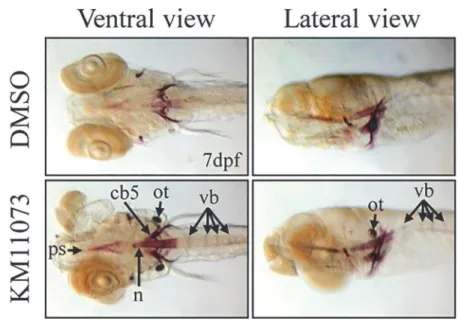
The anabolic activity of KM11073 was further evaluated in twoin vivomodels, the zebrafish
skeletal development model and the mouse calvarial bone formation model. KM11073 (1μM)
enhanced the development of the parasphenoid, notochord, ceratobranchial 5, otolith, and ver-tebrae (Fig. 5A). The bone-forming activity of KM11073 was also confirmed by evaluating the mRNA expression levels of genes related to osteogenesis in zebrafish (Table 3). When larvae at
Table 2. Involvement of p38 inhibitors in the KM11073-mediated enhancement of BMP-2-stimulated induction of osteogenic genes.
mRNA BMP-2 BMP-4 BMP-6 BMP-7 BMP-9 ALP
Control 1.00±0.13 1.00±0.08 1.00±0.13 1.00±0.14 1.00±0.17 1.00±0.43
BMP-2 3.32±0.96* 2.54±0.16** 3.07±0.54* 3.93±0.52* 3.25±0.06* 2.44±0.14 BMP-2 + KM11073 10.44±1.12**,# 2.10±0.31* 12.52±2.62**,# 12.52±2.62**,# 2.36±0.87 9.82±0.45*,## BMP-2 + KM11073 + PD169316 5.19±0.08**,† 1.98±0.39
* 2.73±0.24*,† 3.74±0.31
** 3.80±0.21** 2.42±0.81††
BMP-2 + KM11073 + SB202190 3.48±0.54*,† 1.63±0.14*,# 2.52±0.10*,†† 3.96±0.02* 3.53±0.04**,# 1.55±0.20#,††
BMP-2 + KM11073 + SB203580 6.78±2.62* 2.04±0.36* 3.15±0.63*,†
3.60±1.08* 3.44±0.37* 2.81±0.03††
Cells were treated with each inhibitor for 2 h and then incubated with BMP-2 (100 ng/ml) and KM11073 (10μM) for 3 days. The mRNA expression levels were evaluated by quantitative real-time PCR. Fold changes relative to each gene level in the control are presented as mean±standard deviation
*p<0.05
**p<0.01 (compared to the control) #p<0.05
##p
<0.01 (compared to the group treated with BMP-2) †p
<0.05 ††p
<0.01 (compared to the group treated with BMP-2 + KM11073).
doi:10.1371/journal.pone.0120150.t002
Fig 3. Involvement of p38 in the KM11073-mediated enhancement of BMP-2-stimulated ALP induction.In a 96-well plate, cells (4 × 10³cells/well) were
treated with each inhibitor for 2 h and then treated with BMP-2 and KM11073. After 3 days, the cells were treated with each inhibitor. On differentiation day 6, ALP staining (A) and its activity (B) were assayed.###p<0.001 compared to the BMP-2-treated group;*p<0.05,**p<0.01,***p<0.001 compared to
the group treated with BMP-2 and KM11073.
5 days post fertilization (dpf) were incubated with KM11073 (1μM) for 1 day, the mRNA levels
of Runx2a, BMP-2a, OP, and ALP were significantly induced by KM11073. In addition, the en-hancing effect of KM11073 onin vitromineralization in osteoblast cells was also observed (S2 Fig.).
Thein vivomouse calvarial bone formation assay also revealed the bone-forming activity of KM11073; H&E staining showed that implanted collagen sponges soaked with KM11073 in-duced woven bone formation over the periosteum of the calvarial bones (Fig. 5B). The thick-ness of newly formed woven bone in mice treated with 2.5 and 5 mM of KM11073 was 21.03 and 33.85μm, respectively.
Discussion
An increased risk of complications and adverse events has been suggested for patients receiving rhBMP-2 in spinal fusion, and safety concerns about its clinical application have emerged, in-cluding a greater apparent risk of new malignancy with higher doses of rhBMP-2 [18]. Because BMPs are expensive to produce, small molecules that enhance their potential to induce bone formation would be a cost-effective alternative that reduces the BMP-mediated adverse effects [19,20].
In this study, we found an enhancing effect of KM11073 on the BMP-2-mediated commit-ment of C2C12 cells into osteoblasts. The stimulatory effect of KM11073 on ALP induction was not observed in the absence of BMP-2, suggesting that its osteogenic activity relies on BMP-2 signaling. This suggestion was confirmed by strong inhibition of the KM11073-mediat-ed enhancement of ALP induction in the presence of BMP-2 by noggin post-treatment. Noggin is a well-known BMP antagonist and its functional role in bone formation was identified in a silencing study; the knockdown of noggin has been shown to enhance osteoblastogenesis of BMP-responding cellsin vitroand rhBMP-2-induced new bone formationin vivo[21].
We also identified a BMP-2-dependent osteogenic action of KM11073 via accelerated acti-vation of p38. Recently, the actiacti-vation of Akt and MAP kinases, which are downstream in the Ras-PI3K signaling pathway, has been shown to enhance osteoblastogenesis and bone forma-tion by inducing BMP-2 gene expression [22]. However, in this study, pharmacological inhibi-tion revealed that p38 inhibitors exhibit a dose-dependent inhibitory effect on ALP expression induced by KM11073 in the presence of BMP-2, suggesting the involvement of p38 activation in the osteogenic action of KM11073. The involvement of p38 activation in osteoblast differen-tiation has been reported in several studies [13,23,24].
Fig 4. Effect of KM11073 on the activation of p38.Cells (1 × 10⁵cells/well) were cultured in a 6-well plate for 1 day and then incubated with DMEM containing 5% FBS in the presence or absence of BMP-2 and/or KM11073 (A). Inhibitory effects of p38 inhibitors (1, SB202190; 2, PD169316; 3, SB203580) on the activation of p38 by BMP-2 and KM11073. Western blot analysis was performed with protein samples prepared with cells treated with each inhibitor for 30 min and then incubated with BMP-2 and KM11073 for 30 min (B). The relative, normalized ratio between phosphorylated protein and the protein itself was presented.
BMP-2 triggers osteogenic signaling through the action of its signaling mediator, Smad. The phosphorylation and translocation of Smad has been shown to induce the expression of osteo-blastogenesis-related genes [25–28]. In this study, BMP-2 induced the phosphorylation of
Fig 5. Evaluation of thein vivoosteogenic activity of KM11073 in zebrafish and mouse calvariae.Five days after fertilization, zebrafish were treated with KM11073 (1μM) for 2 days and then fixed and stained with alizarin red S. The parasphenoid (ps), notochord (n), ceratobranchial 5 (cb5), otolith (ot), and vertebrae (vb) are indicated with arrows (A). Collagen sponges soaked in 5μl of 2.5 or 5 mM KM11073 were placed onto mouse calvarial bones. After 3-week implantation, the mice were sacrificed. Calvarial bones were removed, fixed, decalcified, embedded in paraffin, and sectioned. Sections were stained with H&E and photographed at 200 × magnification. Arrows indicate the thickness of newly formed woven bones (B). The thickness of newly formed woven bones was quantified compared to the scale bar.
Smad, but its induction was not enhanced or accelerated by KM11073. Furthermore, the phos-phorylation of Smad was not inhibited by p38 inhibitors. These results suggest that the 2-dependent osteogenic action of KM11073 relies on the activation of p38 but not Smad. BMP-2-induced osteogenic differentiation through a Smad1/5/8 phosphorylation-independent path-way has been reported [29], and conversely, activation of the Smad pathway has been shown to be independent of BMP-2 signaling [30]. Distinct BMP-receptor complexes have been sug-gested to initiate Smad-dependent or Smad-independent signaling [31].
The induction of osteogenic BMPs is also important in the BMP-2-mediated commitment of mesenchymal stem cells into osteoblasts. BMP-2 enhanced the expression of other osteogen-ic BMP genes during osteoblast differentiation [32], and the gene transfer of BMP-2 and -7 in-duced rapid bone formation and increased the expression of endogenous BMP-4 [33]. Here, BMP-2 significantly induced the mRNA expression of all BMPs tested in this study, and KM11073 enhanced the BMP-2-induced mRNA expression of BMP-2, BMP-6, and BMP-7, but not BMP-4 or BMP-9. Although the osteogenic effect differs among the BMPs and among the types of cell exposed to these proteins [34], BMP-6 can also induce osteoblastic differentia-tion of mesenchymal stem cells [35,36]. The osteogenic activity of BMP-7 in C2C12 cells has also been reported [37]. KM11073-enhanced expression of osteogenic BMP-2, BMP-6, and BMP-7 could further augment the osteogenic activity of KM11073. This possibility may also be supported by the results that the post-treatment of noggin strongly inhibited the KM11073-en-hanced induction of ALP. In addition, KM11073-enKM11073-en-hanced expression of BMP-2, BMP-6, and BMP-7 was significantly inhibited by p38 inhibitors, confirming the involvement of p38 activa-tion in the osteogenic acactiva-tion of KM11073.
Furthermore, thein vivobone-forming activity of KM11073 was confirmed in twoin vivo
models, the zebrafish skeletal development model and the mouse calvarial bone formation model [38]. The pathway for bone formation in mammal has been suggested to be conserved during the development of the skeleton in zebrafish [39]. Runx2 (runt-related transcription factor) is essential for osteoblast differentiation during skeletal development in mammals. In an expression analysis of bone markers in zebrafish larvae,runx2a, one of the zebrafish homo-logs of mouseRunx2, was expressed in the early stage of bone development [39,40]. Also, both bmp2a and bmp2b in zebrafish have been cloned and are expressed 10 to 36 h after fertilization [41,42]. Here, in addition to markers of osteogenesis (osteopontin and ALP),runx2aand
bmp2amRNA, but notbmp2b, were significantly induced by KM11073 in zebrafish larvae. In summary, KM11073 exhibited BMP-2-dependentin vitroosteogenic activity via the acti-vation of p38 signaling. In addition, the activities of quinolin analogues to enhance BMP-2-me-diated ALP activity were also found (S2 Table). Therefore, the combination of rhBMP-2 with osteogenic small molecules could reduce the use of expensive rhBMP-2, which mitigates unde-sirable side effects caused by its supra-physiological dose for therapeutic efficacy. Furthermore,
Table 3. Osteogenesis-related gene expression in zebrafish.
mRNA runx2a bmp2a bmp2b osteopontin ALP
Control 1.00±0.34 1.00±0.06 1.00±0.33 1.00±0.15 1.00±0.13
KM11073 11.69±0.88* 4.83±1.63* 3.75±1.52 4.20±0.60* 3.90±0.91*
At 5.0 dpf, larvae were treated with KM11073 (1μM), and after 1 day the mRNA levels were evaluated by quantitative real-time PCR. Fold changes relative to each gene level in the control are presented as mean±standard deviation.
*p<0.05
**p<0.01 (compared to the control).
thein vivoosteogenic activity of KM11073per sealso suggests its potential single use for bone formation. The inherent physical properties of small molecules minimizing the limitations of using biologics suggests that they represent the next generation of regenerative medicine [43].
Supporting Information
S1 Fig. Effect of KM11073 or inhibitor on the expression and activation of Smad and p38 in C2C12 cells.Effect of KM11073 or inhibitor on the expression and activation of Smad and p38 was evaluated by Western blot analysis. Briefly, C2C12 Cells (1 × 10⁵cells/well) were cul-tured in a 6-well plate for 1 day and then incubated with DMEM containing 5% FBS in the presence or absence of KM11073 and each p38 inhibitor for 30 min.
(TIF)
S2 Fig. KM11073 enhanced the mineralization in mouse primary osteoblast cells.(A) The primary calvarial pre-osteoblasts differentiated with ascorbic acid (50μg/ml),β
-glyceropho-sphate (10 mM), and BMP-2 (50 ng/ml) in the absence or presence of KM11073 (0.3μM).
Me-dium was changed every 3 days, and the mineralization was visualized by alizarin red S staining on day 9. (B) Deposited alizarin red S was dissolved with 10% cetylpyridinium (Sigma-Aldrich) for 15 min at room temperature and quantified by a multiplate reader (Envision) at 560 nm.
(TIF)
S1 Supporting Information. Materials and Methods.
(DOCX)
S1 Table. Effect of KM11074 and inhibitors on mRNA expression of BMPs.
(DOCX)
S2 Table. Enhancing effect of quinolin analogues on the BMP-2-induced ALP expression in C2C12 cells.
(DOCX)
Author Contributions
Conceived and designed the experiments: SHK. Performed the experiments: SHB SWC SJP SHL HSC SHK. Analyzed the data: SHB SWC SJP SHL HSC SHK. Contributed reagents/mate-rials/analysis tools: SHB SWC SJP SHL HSC SHK. Wrote the paper: SHB SWC SJP SHK.
References
1. Teitelbaum SL, Ross FP. Genetic regulation of osteoclast development and function. Nat. Rev. Genet. 2003; 4: 638–649. PMID:12897775
2. Rodan GA, Martin TJ. Therapeutic approaches to bone diseases. Science 2000; 289: 1508–1514. PMID:10968781
3. Garrett IR. Anabolic agents and the bone morphogenetic protein pathway. Curr Top Dev Biol. 2007; 78: 127–171. PMID:17338916
4. Valentin-Opran A, Wozney J, Csimma C, Lilly L, Riedel GE. Clinical evaluation of recombinant human bone morphogenetic protein-2. Clin Orthop Relat Res. 2002; 395:110–120. PMID:11937870 5. Nakashima M, Reddi AH. The application of bone morphogenetic proteins to dental tissue engineering.
Nat Biotechnol. 2003; 21: 1025–1032. PMID:12949568
6. Seeherman H, Wozney JM. Delivery of bone morphogenetic proteins for orthopedic tissue regenera-tion. Cytokine Growth Factor Rev. 2005; 16: 329–345. PMID:15936978
8. Hollinger JO, Schmitt JM, Buck DC, Shannon R, Joh SP, Zegzula HD, et al. Recombinant human bone morphogenetic protein-2 and collagen for bone regeneration. J Biomed Mater Res. 1998; 43: 356–364. PMID:9855194
9. Murakami N, Saito N, Takahashi J, Ota H, Horiuchi H, Nawata M, et al. Repair of a proximal femoral bone defect in dogs using a porous surfaced prosthesis in combination with recombinant BMP-2 and a synthetic polymer carrier. Biomaterials. 2003; 24: 2153–2159. PMID:12699651
10. Vögelin E, Jones NF, Huang JI, Brekke JH, Lieberman JR. Healing of a critical-sized defect in the rat femur with use of a vascularized periosteal flap, a biodegradable matrix, and bone morphogenetic pro-tein. J Bone Joint Surg Am. 2005; 87: 1323–1331. PMID:15930543
11. McKay WF, Peckham SM, Badura JM. A comprehensive clinical review of recombinant human bone morphogenetic protein-2 (INFUSE Bone Graft). Int Orthop. 2007; 31: 729–734. PMID:17639384 12. Katagiri T, Yamaguchi A, Komaki M, Abe E, Takahashi N, Ikeda T, et al. Bone morphogenetic protein-2
converts the differentiation pathway of C2C12 myoblasts into the osteoblast lineage. J Cell Biol. 1994; 127(6 Pt 1): 1755–1766. PMID:7798324
13. Kim HJ, Kim SH. Tanshinone IIA enhances BMP-2-stimulated commitment of C2C12 cells into osteo-blasts via p38 activation. Amino Acids. 2010; 39: 1217–1226. doi:10.1007/s00726-010-0557-8PMID: 20300786
14. Rozen S, Skaletsky HJ. Primer3 on the WWW for general users and for biologist programmers. Meth-ods Mol Biol. 2000; 132: 365–386. PMID:10547847
15. Livak KJ, Schmittgen TD. Analysis of relative gene expression data using real-time quantitative PCR and the 2−ΔΔCTmethod. Methods. 2001; 25: 402–408 PMID:11846609
16. Seeherman H, Wozney JM. Delivery of bone morphogenetic proteins for orthopedic tissue regenera-tion. Cytokine Growth Factor Rev. 2005; 16: 329–45. PMID:15936978
17. Westerfield M. The Zebrafish Book. A guide for the laboratory use of zebrafish (Danio rerio). 4th ed. University of Oregon Press: Eugene, OR; 2000.
18. Carragee EJ, Hurwitz EL, Weiner BK. A critical review of recombinant human bone morphogenetic pro-tein-2 trials in spinal surgery: emerging safety concerns and lessons learned. Spine J. 2001; 11: 471–
491.
19. Kato S, Sangadala S, Tomita K, Titus L, Boden SD. A synthetic compound that potentiates bone mor-phogenetic protein-2-induced transdifferentiation of myoblasts into the osteoblastic phenotype. Mol Cell Biochem. 2011; 349: 97–106. doi:10.1007/s11010-010-0664-6PMID:21110071
20. Vrijens K, Lin W, Cui J, Farmer D, Low J, Pronier E, et al. Identification of small molecule activators of BMP signaling. PLoS One. 2013; 8: e59045. doi:10.1371/journal.pone.0059045PMID:23527084 21. Takayama K, Suzuki A, Manaka T, Taguchi S, Hashimoto Y, Imai Y, et al. RNA interference for noggin
enhances the biological activity of bone morphogenetic proteins in vivo and in vitro. J Bone Miner Metab. 2009; 27: 402–411. doi:10.1007/s00774-009-0054-xPMID:19252814
22. Ghosh-Choudhury N, Mandal CC, Choudhury GG. Statin-induced Ras activation integrates the phos-phatidylinositol 3-kinase signal to Akt and MAPK for bone morphogenetic protein-2 expression in osteo-blast differentiation. J Biol Chem. 2007; 282: 4983–4993. PMID:17179158
23. Gallea S, Lallemand F, Atfi A, Rawadi G, Ramez V, Spinella-Jaegle S, et al. Activation of mitogen-acti-vated protein kinase cascades is involved in regulation of bone morphogenetic protein-2-induced oste-oblast differentiation in pluripotent C2C12 cells. Bone. 2001; 28: 491–498. PMID:11344048
24. Lee KS, Hong SH, Bae SC. Both the Smad and p38 MAPK pathways play a crucial role in Runx2 ex-pression following induction by transforming growth factor-beta and bone morphogenetic protein. On-cogene. 2002; 21: 7156–7163. PMID:12370805
25. Nishimura R, Kato Y, Chen D, Harris SE, Mundy GR, Yoneda T. Smad5 and DPC4 are key molecules in mediating BMP-2-induced osteoblastic differentiation of the pluripotent mesenchymal precursor cell line C2C12. J. Biol. Chem. 1998; 273: 1872–1879. PMID:9442019
26. Nöth U, Tuli R, Seghatoleslami R, Howard M, Shah A, Hall DJ, et al. Activation of p38 and Smads medi-ates BMP-2 effects on human trabecular bone-derived osteoblasts. Exp. Cell Res. 2003; 291: 201–
211. PMID:14597420
27. Nakamura Y, Wakitani S, Saito N, Takaoka K. Expression profiles of BMP-related molecules induced by BMP-2 or -4 in muscle-derived primary culture cells. J. Bone Miner. Metab. 2005; 23; 426–434. PMID:16261448
28. Yamachika E, Tsujigiwa H, Shirasu N, Ueno T, Sakata Y, Fukunaga J, et al. Immobilized recombinant human bone morphogenetic protein-2 enhances the phosphorylation of receptor-activated Smads. J. Biomed. Mater. Res. A. 2009; 88: 599–607. doi:10.1002/jbm.a.31833PMID:18314893
of Smad-1/5/8 phosphorylation. J Cell Physiol. 2010; 222: 465–473. doi:10.1002/jcp.21968PMID: 19918795
30. Jadlowiec JA, Zhang X, Li J, Campbell PG, Sfeir C. Extracellular matrix-mediated signaling by dentin phosphophoryn involves activation of the Smad pathway independent of bone morphogenetic protein. J Biol Chem. 2003; 281: 5341–5347.
31. Hassel S, Schmitt S, Hartung A, Roth M, Nohe A, Petersen N, et al. Initiation of Smad-dependent and Smad-independent signaling via distinct BMP-receptor complexes. J Bone Joint Surg Am. 2003; 85(-A Suppl 3): 44–51.
32. Chen D, Harris MA, Rossini G, Dunstan CR, Dallas SL, Feng JQ, et al. Bone morphogenetic protein 2 (BMP-2) enhances BMP-3, BMP-4, and bone cell differentiation marker gene expression during the in-duction of mineralized bone matrix formation in cultures of fetal rat calvarial osteoblasts. Calcif Tissue Int. 1997; 60: 283–290. PMID:9069167
33. Kawai M, Bessho K, Maruyama H, Miyazaki J, Yamamoto T (2006) Simultaneous gene transfer of bone morphogenetic protein (BMP)-2 and BMP-7 by in vivo electroporation induces rapid bone forma-tion and BMP-4 expression. BMC Musculoskelet Disord. 2006 Aug 3. doi:10.1186/1471-2474-7-62. 2006. 7. 62
34. Cheng H, Jiang W, Phillips FM, Haydon RC, Peng Y, Zhou L, et al. Osteogenic activity of the fourteen types of human bone morphogenetic proteins (BMPs). J Bone Joint Surg Am. 2003; 85-A: 1544–1552. PMID:12925636
35. Gitelman SE, Kirk M, Ye JQ, Filvaroff EH, Kahn AJ, Derynck R. Vgr-1/BMP-6 induces osteoblastic dif-ferentiation of pluripotential mesenchymal cells. Cell Growth Differ. 1995; 6: 827–836. PMID:7547504 36. Yamaguchi A, Ishizuya T, Kintou N, Wada Y, Katagiri T, Wozney JM, et al. Effects of BMP-2, BMP-4,
and BMP-6 on osteoblastic differentiation of bone marrow-derived stromal cell lines, ST2 and MC3T3-G2/PA6. Biochem Biophys Res Commun. 1996; 220: 366–371. PMID:8645311
37. Yeh LC, Tsai AD, Lee JC. Osteogenic protein-1 (OP-1, BMP-7) induces osteoblastic cell differentiation of the pluripotent mesenchymal cell line C2C12. J Cell Biochem. 2002; 87: 292–304. PMID:12397611 38. Kim SN, Bae SJ, Kwak HB, Min YK, Jung SH, Kim CH, et al. In vitro and in vivo osteogenic activity of
licochalcone A. Amino Acids. 2012; 42: 1455–1465. doi:10.1007/s00726-011-0901-7PMID: 21468757
39. Li N, Felber K, Elks P, Croucher P, Roehl HH. Tracking gene expression during zebrafish osteoblast dif-ferentiation. Dev Dyn. 2009; 238: 459–466. doi:10.1002/dvdy.21838PMID:19161246
40. Flores MV, Tsang VW, Hu W, Kalev-Zylinska M, Postlethwait J, Crosier P, et al. Duplicate zebrafish runx2 orthologues are expressed in developing skeletal elements. Gene Expr Patterns. 2004; 4: 573–
581. PMID:15261836
41. Martínez-Barberá JP, Toresson H, Da Rocha S, Krauss S. Cloning and expression of three members of the zebrafish Bmp family: Bmp2a, Bmp2b and Bmp4. Gene. 1997; 198: 53–59. PMID:9370264 42. Nikaido M, Tada M, Saji T, Ueno N. Conservation of BMP signaling in zebrafish mesoderm patterning.
Mech Dev. 1997; 61: 75–88. PMID:9076679